

PET-MRI nanoparticles imaging of blood-brain barrier damage and modulation after stroke reperfusion
- PMID: 33305265
- PMCID: PMC7716090
- DOI: 10.1093/braincomms/fcaa193
PET-MRI nanoparticles imaging of blood-brain barrier damage and modulation after stroke reperfusion
Abstract
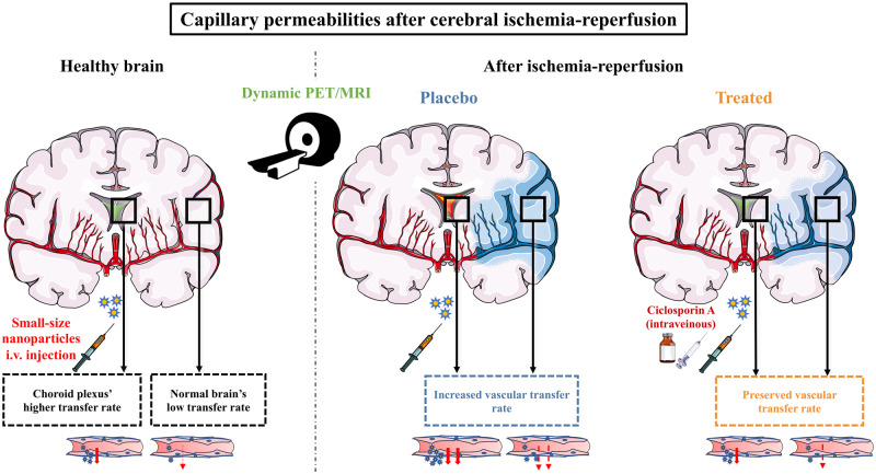
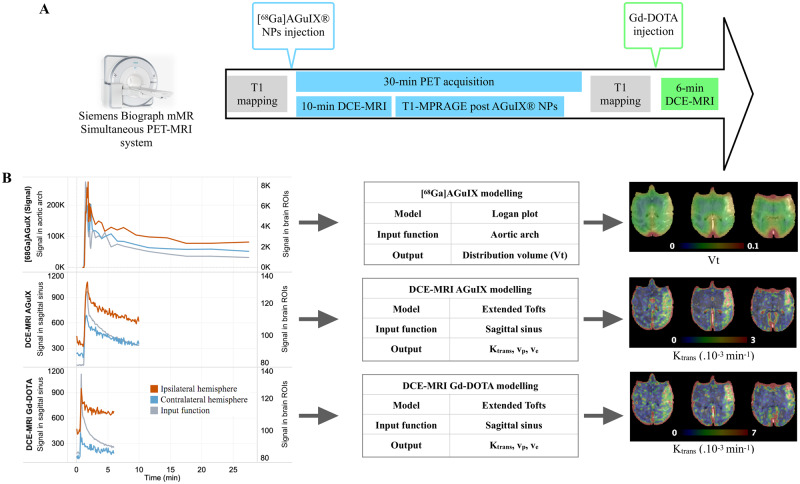
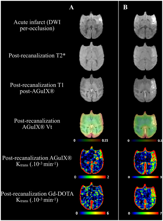
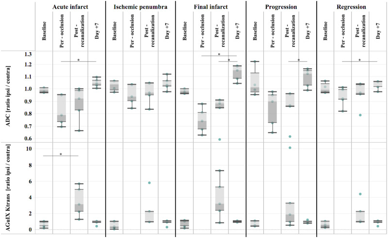
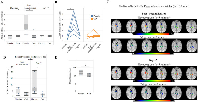
In an acute ischaemic stroke, understanding the dynamics of blood-brain barrier injury is of particular importance for the prevention of symptomatic haemorrhagic transformation. However, the available techniques assessing blood-brain barrier permeability are not quantitative and are little used in the context of acute reperfusion therapy. Nanoparticles cross the healthy or impaired blood-brain barrier through combined passive and active processes. Imaging and quantifying their transfer rate could better characterize blood-brain barrier damage and refine the delivery of neuroprotective agents. We previously developed an original endovascular stroke model of acute ischaemic stroke treated by mechanical thrombectomy followed by positron emission tomography-magnetic resonance imaging. Cerebral capillary permeability was quantified for two molecule sizes: small clinical gadolinium Gd-DOTA (<1 nm) and AGuIX® nanoparticles (∼5 nm) used for brain theranostics. On dynamic contrast-enhanced magnetic resonance imaging, the baseline transfer constant K trans was 0.94 [0.48, 1.72] and 0.16 [0.08, 0.33] ×10-3 min-1, respectively, in the normal brain parenchyma, consistent with their respective sizes, and 1.90 [1.23, 3.95] and 2.86 [1.39, 4.52] ×10-3 min-1 in choroid plexus, confirming higher permeability than brain parenchyma. At early reperfusion, K trans for both Gd-DOTA and AGuIX® nanoparticles was significantly higher within the ischaemic area compared to the contralateral hemisphere; 2.23 [1.17, 4.13] and 0.82 [0.46, 1.87] ×10-3 min-1 for Gd-DOTA and AGuIX® nanoparticles, respectively. With AGuIX® nanoparticles, K trans also increased within the ischaemic growth areas, suggesting added value for AGuIX®. Finally, K trans was significantly lower in both the lesion and the choroid plexus in a drug-treated group (ciclosporin A, n = 7) compared to placebo (n = 5). K trans quantification with AGuIX® nanoparticles can monitor early blood-brain barrier damage and treatment effect in ischaemic stroke after reperfusion.
Keywords: blood–brain barrier; choroid plexus; ischaemia–reperfusion damage; nanoparticles; stroke.
© The Author(s) (2020). Published by Oxford University Press on behalf of the Guarantors of Brain.
Figures
References
-
- Bai J, Lyden PD. Revisiting cerebral postischemic reperfusion injury: New insights in understanding reperfusion failure, hemorrhage, and edema. Int J Stroke 2015; 10: 143–52. - PubMed
-
- Ballanger B, Tremblay L, Sgambato-Faure V, Beaudoin-Gobert M, Lavenne F, Le Bars D, et al. A multi-atlas based method for automated anatomical Macaca fascicularis brain MRI segmentation and PET kinetic extraction. Neuroimage 2013; 77: 26–43. - PubMed
-
- Basseville A, Hall MD, Chau CH, Robey RW, Gottesman M, Figg WD, et al. The ABCG2 multidrug transporter In: George AM, editor. ABC transporters - 40 years on. Cham: Springer International Publishing; 2016. p. 195–226.
-
- Broocks G, Hanning U, Flottmann F, Schönfeld M, Faizy TD, Sporns P, et al. Clinical benefit of thrombectomy in stroke patients with low ASPECTS is mediated by oedema reduction. Brain 2019; 142: 1399–407. - PubMed
LinkOut - more resources
Full Text Sources
Miscellaneous