The histopathological diagnosis of atypical meningioma: glass slide versus whole slide imaging for grading assessment
- PMID: 33305338
- PMCID: PMC7990834
- DOI: 10.1007/s00428-020-02988-1
The histopathological diagnosis of atypical meningioma: glass slide versus whole slide imaging for grading assessment
Abstract
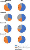
Limited studies on whole slide imaging (WSI) in surgical neuropathology reported a perceived limitation in the recognition of mitoses. This study analyzed and compared the inter- and intra-observer concordance for atypical meningioma, using glass slides and WSI. Two neuropathologists and two residents assessed the histopathological features of 35 meningiomas-originally diagnosed as atypical-in a representative glass slide and corresponding WSI. For each histological parameter and final diagnosis, we calculated the inter- and intra-observer concordance in the two viewing modes and the predictive accuracy on recurrence. The concordance rates for atypical meningioma on glass slides and on WSI were 54% and 60% among four observers and 63% and 74% between two neuropathologists. The inter-observer agreement was higher using WSI than with glass slides for all parameters, with the exception of high mitotic index. For all histological features, we found median intra-observer concordance of ≥ 79% and similar predictive accuracy for recurrence between the two viewing modes. The higher concordance for atypical meningioma using WSI than with glass slides and the similar predictive accuracy for recurrence in the two modalities suggest that atypical meningioma may be safely diagnosed using WSI.
Keywords: Atypical meningioma; Digital; Recurrence; Reproducibility; Whole slide imaging.
Conflict of interest statement
The authors declare that they have no conflict of interest.
Figures


References
-
- Bokhorst JM, Blank A, Lugli A, Zlobec I, Dawson H, Vieth M, Rijstenberg LL, Brockmoeller S, Urbanowicz M, Flejou JF, Kirsch R, Ciompi F, van der Laak J, Nagtegaal ID. Assessment of individual tumor buds using keratin immunohistochemistry: moderate interobserver agreement suggests a role for machine learning. Mod Pathol. 2020;33:825–833. doi: 10.1038/s41379-019-0434-2. - DOI - PMC - PubMed
-
- Administration UFD (2017) FDA news release: FDA allows marketing of first whole slide imaging system for digital pathology. https://www.fda.gov/newsevents/newsroom/pressannouncements/ucm552742.htm.
Publication types
MeSH terms
Grants and funding
LinkOut - more resources
Full Text Sources

