Review
. 2020 Dec 11;477(23):4603-4621.
doi: 10.1042/BCJ20200506.

Functions and regulation of the serine/threonine protein kinase CK1 family: moving beyond promiscuity

Affiliations
Review

Functions and regulation of the serine/threonine protein kinase CK1 family: moving beyond promiscuity

Luke J Fulcher et al. Biochem J. .

Abstract

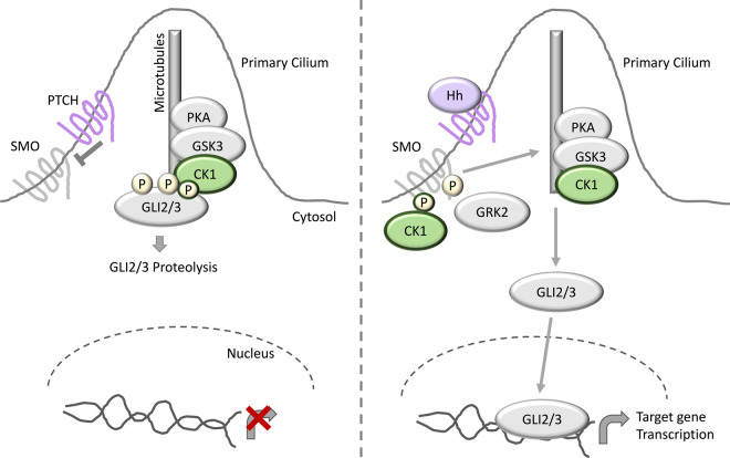
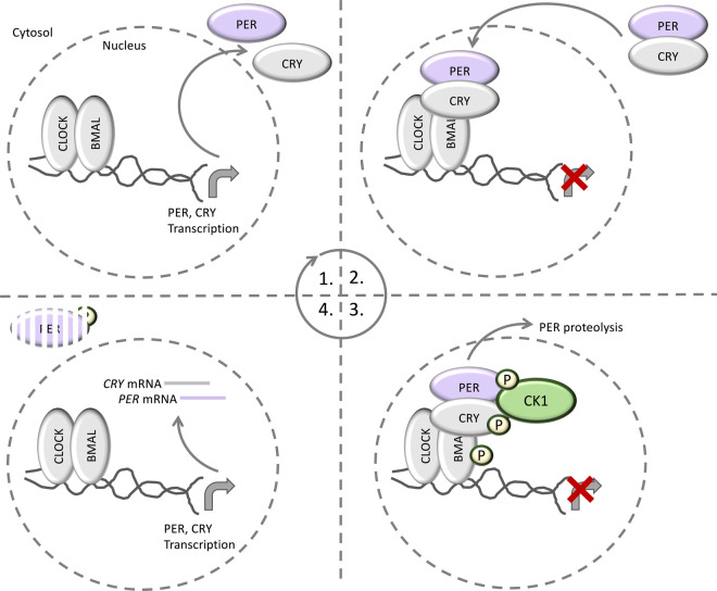
Regarded as constitutively active enzymes, known to participate in many, diverse biological processes, the intracellular regulation bestowed on the CK1 family of serine/threonine protein kinases is critically important, yet poorly understood. Here, we provide an overview of the known CK1-dependent cellular functions and review the emerging roles of CK1-regulating proteins in these processes. We go on to discuss the advances, limitations and pitfalls that CK1 researchers encounter when attempting to define relationships between CK1 isoforms and their substrates, and the challenges associated with ascertaining the correct physiological CK1 isoform for the substrate of interest. With increasing interest in CK1 isoforms as therapeutic targets, methods of selectively inhibiting CK1 isoform-specific processes is warranted, yet challenging to achieve given their participation in such a vast plethora of signalling pathways. Here, we discuss how one might shut down CK1-specific processes, without impacting other aspects of CK1 biology.

Keywords: FAM83; Wnt proteins; casein kinase; cell cycle; circadian clock.


Conflict of interest statement

The authors declare that there are no competing interests associated with the manuscript.

Figures

Figure 1.
Figure 1.. Domain architecture of CK1 isoforms.
(A) Phylogenetic tree of the CK1 family of protein kinases (based on [7]). (B) Schematic detailing the domain architecture for the CK1 isoforms. Percentage sequence similarity of the kinase domains for each CK1 isoform, relative to the CK1α kinase domain, is shown.
Figure 2.
Figure 2.. Reported regulators of CK1.
(A) CG-NAP localises CK1δ and ɛ to centrosomes. (B) CK1ɛ is sequestered and bound by Axin, which competes with Dvl for CK1ɛ binding. Following Wnt stimulation, CK1ɛ dissociates from Axin and binds to DDX3. DDX3 increases the catalytic activity of CK1ɛ to facilitate the phosphorylation of Dvl by CK1ɛ. (C) The FAM83 family of proteins co-ordinate the localisation of distinct subsets of CK1 isoforms to various subcellular sites as shown. A question mark (?) denotes cases where the physiological function of the FAM83–CK1 interaction is still to be determined.
Figure 3.
Figure 3.. CK1 in Wnt signalling.
In the absence of Wnt ligands, β-catenin is held by the so-called destruction complex composed of Axin, APC, the E3 ubiquitin ligase substrate receptor β-TrCP, and the kinases CK1 and GSK3. Sequential phosphorylation of β-catenin by CK1 and GSK3 triggers β-catenin ubiquitination by the β-TrCP-complex, and its subsequent proteasomal degradation. In the nucleus, Groucho binds TCF to block the transcription of Wnt target genes. Following binding of Wnt ligands to the receptors LRP5/6 and FZD, LRP5/6 becomes phosphorylated, and triggers the formation of the Wnt signalosome, largely composed of Axin and Dvl. This recruits the destruction complex to the membrane. β-catenin is stabilised in the cytosol and is free to enter the nucleus where it displaces Groucho from TCF, and together with LEF, activates transcription of Wnt target genes. CK1 has also been reported to phosphorylate the LRP5/6 and Dvl in the presence of Wnt ligands.
Figure 4.
Figure 4.. CK1 in Hippo signalling.
Under conditions of low cell density, the YAP/TAZ transcriptional co-activator complex cycles between the cytosol and nucleus, and in the nucleus YAP/TAZ bind to their cognate transcription factor targets, such as SMADs to trigger transcription of Hippo target genes to promote cell growth and differentiation. CK1 phosphorylates Dvl to promote Wnt signalling under these conditions. Under conditions of cell stress, such as contact inhibition, the kinase MST1/2 becomes activated and phosphorylates LATS1/2, which promotes LATS1/2-mediated phosphorylation of YAP/TAZ. YAP/TAZ phosphorylation acts as a phosphodegron signal to promote YAP/TAZ ubiquitination by β-TrCP, and subsequent YAP/TAZ proteolysis. As YAP/TAZ cannot enter the nucleus, Hippo target gene transcription is suppressed, and cells cease to grow. CK1 has been reported to phosphorylate YAP/TAZ after priming phosphorylation from LATS1/2, and following CK1 binding to MST1/2, CK1 phosphorylation of Dvl is reduced, leading to inhibition of Wnt signalling.
Figure 5.
Figure 5.. CK1 in hedgehog signalling.
In the absence of hedgehog (Hh) ligands, the Hh receptor PTCH associates with SMO to inhibit SMO function. The GLI2/3 transcription factors are held in complex by a primary cilium-localized inhibitory complex consisting of the kinases CK1, GSK3 and PKA. Following GLI2/3 phosphorylation by CK1, GSK3 and PKA, GLI2/3 is marked for proteasomal degradation. In the presence of Hh ligands, SMO is released from PTCH and is free to mediate the release of GLI2/3 from the inhibitory kinase complex. GLI2/3 can now translocate to the nucleus and induce Hh target gene transcription. CK1 and GRK2 have been reported to phosphorylate SMO to promote full SMO activation and maximal release of GLI2/3 from the inhibitory complex.
Figure 6.
Figure 6.. CK1 in circadian rhythm.
The circadian cycle begins with the CLOCK–BMAL complex, which acts to activate the transcription of PER and CRY proteins. Following their translation, PER and CRY proteins heterodimerize and translocate to the nucleus, where they act to inhibit CLOCK–BMAL, thereby preventing their own transcription. Subsequently, CK1 phosphorylates PER to trigger PER proteolysis and relieve the CLOCK–BMAL inhibition. The cycle then begins again. CK1 has also been reported to phosphorylate CRY and BMAL, but the relevance of these phosphorylation events remains to be elucidated.

References

    1. Cohen P. (2002) The origins of protein phosphorylation. Nat. Cell Biol. 4, E127–E130 10.1038/ncb0502-e127 - DOI - PubMed
    1. Pawson T. and Scott J.D. (2005) Protein phosphorylation in signaling–50 years and counting. Trends Biochem. Sci. 30, 286–290 10.1016/j.tibs.2005.04.013 - DOI - PubMed
    1. Cohen P. (2009) Targeting protein kinases for the development of anti-inflammatory drugs. Curr. Opin. Cell Biol. 21, 317–324 10.1016/j.ceb.2009.01.015 - DOI - PubMed
    1. Cohen P. (2002) Protein kinases–the major drug targets of the twenty-first century? Nat. Rev. Drug Discov. 1, 309–315 10.1038/nrd773 - DOI - PubMed
    1. Cohen P. (1999) The development and therapeutic potential of protein kinase inhibitors. Curr. Opin. Chem. Biol. 3, 459–465 10.1016/S1367-5931(99)80067-2 - DOI - PubMed

Publication types