. 2020 Dec 11;18(12):e3000975.
doi: 10.1371/journal.pbio.3000975. eCollection 2020 Dec.

In silico APC/C substrate discovery reveals cell cycle-dependent degradation of UHRF1 and other chromatin regulators

Affiliations

In silico APC/C substrate discovery reveals cell cycle-dependent degradation of UHRF1 and other chromatin regulators

Jennifer L Franks et al. PLoS Biol. .

Abstract

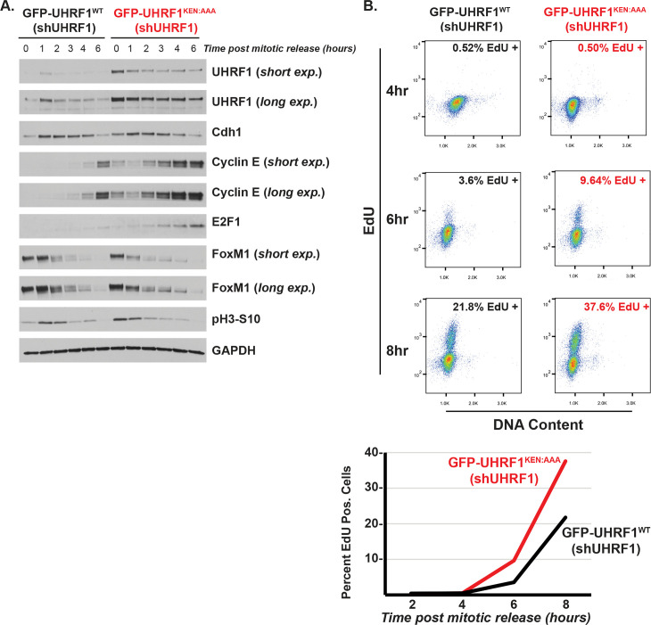
The anaphase-promoting complex/cyclosome (APC/C) is an E3 ubiquitin ligase and critical regulator of cell cycle progression. Despite its vital role, it has remained challenging to globally map APC/C substrates. By combining orthogonal features of known substrates, we predicted APC/C substrates in silico. This analysis identified many known substrates and suggested numerous candidates. Unexpectedly, chromatin regulatory proteins are enriched among putative substrates, and we show experimentally that several chromatin proteins bind APC/C, oscillate during the cell cycle, and are degraded following APC/C activation, consistent with being direct APC/C substrates. Additional analysis revealed detailed mechanisms of ubiquitylation for UHRF1, a key chromatin regulator involved in histone ubiquitylation and DNA methylation maintenance. Disrupting UHRF1 degradation at mitotic exit accelerates G1-phase cell cycle progression and perturbs global DNA methylation patterning in the genome. We conclude that APC/C coordinates crosstalk between cell cycle and chromatin regulatory proteins. This has potential consequences in normal cell physiology, where the chromatin environment changes depending on proliferative state, as well as in disease.


Conflict of interest statement

I have read the journal's policy and the authors of this manuscript have the following competing interests: NGB is a consultant for Cullgen, Inc.

Figures

Fig 1
Fig 1. In silico analysis reveals a high confidence set of APC/C substrates involved in mitosis.
(A) KEN-box containing human proteins were identified and cross-referenced against a set of 651 genes whose expression is cell cycle regulated based on multiple, independent studies. This revealed a set of 145 KEN-box containing proteins whose mRNA expression is cell cycle regulated. (B) Analysis of the enrichment of bona fide KEN-dependent substrates among these 3 datasets (blue, KEN box only set (2,206); black, cell cycle regulated mRNAs (651); red, the overlapping set of 145 proteins compared against a curated set of bona fide, KEN-dependent APC/C substrates [16]). Enrichment was calculated based on the expected number of substrates, which would be captured by chance based on the size of the dataset. (S1 Data) (C) Analysis of putative substrates recovered in the indicated studies. (S1 Data) (D) GO analysis for indicated studies (blue, KEN box only set (2,206); black, cell cycle–regulated mRNAs (651); red, the overlapping set of 145 proteins). (S1 Data) (E) The set of 145 putative substrates was manually curated and analyzed for roles in various aspects of cell cycle progression. Seventy proteins, involved in cell cycle activities, are shown. The ones labeled in magenta signify that there is evidence in the literature of their regulation by APC/C. (Note that AURORA B, a mitotic kinase that phosphorylates histone H3, is listed here and in Fig 2A). APC/C, anaphase-promoting complex/cyclosome; GO, gene ontology.
Fig 2
Fig 2. Putative APC/C substrates are enriched for roles in chromatin regulation.
(A) The set of 145 known and putative APC/C substrates is enriched for proteins involved in various chromatin-related process. This includes chromatin readers and writers, chaperones, RNA regulation and processing, DNA damage repair, and others. (Note that AURORA B, a mitotic kinase that phosphorylates histone H3, is listed here and in Fig 1E) (B) GO analysis of the overlapping KEN-box containing cell cycle–regulated transcripts. This set is enriched for the indicated biological process, including DNA metabolism, protein-DNA complex assembly, DNA packaging, and DNA conformation. (S1 Data) (C) APC/C activation assay to monitor substrate degradation. Following synchronization in mitosis, cells were washed 1 time and treated with CDK inhibitors to remove inhibitory phosphorylation marks that hinder the formation of APC/CCdh1 needed for the M/G1 phase transition. Protein degradation was monitored by immunoblot. CHAF1B and PCAF are putative APC/C substrates, and FoxM1 and Cyclin B are known targets. Data representative of n = 3 experiments. (D) coIP of HA-Cdh1 with Myc-CHAF1B in transiently transfected HEK-293T cells treated with proteasome inhibitors prior to harvesting. The underline indicates which protein or tag was blotted for in a particular panel (here and below). Input equal to 1% of IP, here and below. Data representative of n = 2 experiments. (E) coIP of HA-Cdh1 with FLAG-PCAF in transiently transfected 293T cells treated with proteasome inhibitors prior to harvesting. Data representative of n = 2 experiments. (F) coIP of HA-Cdh1 with Myc-NCOA3 in transiently transfected 293T cells treated with proteasome inhibitors prior to harvesting. Data representative of n = 3 experiments. (G) coIP of HA-Cdh1 with Myc-TTF2 in transiently transfected 293T cells treated with proteasome inhibitors prior to harvesting. Data representative of n = 4 experiments. (H) Mitotic shake-off of synchronized U2OS cells collected after release at the indicated timepoints. Immunoblotting for select endogenous proteins that are putative APC/C substrates or the positive control cyclin B. Data representative of n = 3 experiments. APC/C, anaphase-promoting complex/cyclosome; coIP, co-immunoprecipitation; GO, gene ontology.
Fig 3
Fig 3. UHRF1 levels are controlled by APC/CCdh1.
(A) HeLa S3 cells were synchronized in mitosis and released into the cell cycle. Time points were taken at the indicated time points and analyzed by immunoblot. Data representative of n = 3 experiments. (B) U2OS cells were synchronized in prometaphase with 250-ng/mL nocodazole for 16 h prior to mitotic shake-off. Cells were released into fresh media containing 10-μM RO-3306 CDK inhibitor (used as described in Fig 2C) with or without addition of 20 μM of proteasomal inhibitor MG-132 and harvested 1 h later. Cyclin B is a positive control for a known APC/C substrate that is degraded at mitotic exit. Data representative of n = 3 experiments. (C) HCT116 cells were transfected with siRNA targeting Cdh1 (Fzr1 mRNA) or firefly luciferase as a control and harvested after 24 h for immunoblotting. Data representative of n = 3 experiments. (D) Myc-UHRF1 was transiently expressed in HEK-293T cells with increasing concentrations of FLAG-Cdh1 for 24 h before analysis by immunoblot. Data representative of n = 3 experiments. (E) HeLa S3 cells transfected with siRNA targeting FF or FZR1 for 8 h prior to synchronization in mitosis for 14 h and then released into the cell cycle. Time points were taken at the indicated time points and analyzed by immunoblot. Line indicates blots from multiple gels. Data representative of n = 3 experiments. (F) UHRF1 degradation assay in G1 phase-synchronized and UBE2S-depleted HeLa S3 cell extracts supplemented with ATP and Ub. Aliquots were collected at the indicated time points and analyzed by immunoblot. Data representative of n = 2 experiments. APC/C, anaphase-promoting complex/cyclosome; siRNA, small interfering RNA; Ub, ubiquitin; UHRF1, ubiquitin-like with PHD and RING finger domains 1.
Fig 4
Fig 4. UHRF1 binding and ubiquitylation by APC/CCdh1 depends on KEN degron.
(A) Schematic of UHRF1 domain structure with location of KEN degron in both FL and truncated LPS UHRF1. (B) Endogenous IP of UHRF1 with Cdh1 in cells at pro-metaphase arrest and during mitotic exit. HeLa S3 cells were synchronized by nocodazole block and release as described previously. Cells were collected at 0 h and 1 h after release, flash frozen prior to α-Cdh1 IP, and analyzed by immunoblot. Data representative of n = 5 experiments. (C) coIP of HA-Cdh1 with Myc-UHRF1 in transiently transfected HEK-293T cells treated with proteasome inhibitors prior to harvesting and α-Myc IP. Input equal to 1% of IP, here and below. Data representative of n = 4 experiments. (D) coIP of Myc-UHRF1 with HA-Cdh1 in transiently transfected HEK-293T cells treated with proteasome inhibitors prior to harvesting and α-HA IP. Data representative of n = 3 experiments. (E) Ubiquitylation reactions with APC/CCdh1, UBE2C, FL UHRF1* or LPS UHRF1*, and WT Ub. UHRF1 was detected by fluorescence scanning (* indicates fluorescently labeled protein). Data representative of n = 3 experiments. (F) Ubiquitylation reactions similar as in (D) but using 2 variants of APC/C: WT and catalytically dead APC/CΔRINGΔWHB, a version of APC/C that can neither recruit nor activate its E2, UBE2C. UHRF1 was detected by fluorescence scanning. Samples were collected at 30 min. Data representative of n = 3 experiments. (G) Representative in vitro ubiquitylation reactions showing UBE2S-dependent chain elongation reactions of LPS UHRF1*. Titration of UBE2S: 0 μM, 0.1 μM (+), and 0.5 μM (++). The addition of Emi1 completely inhibited the reaction. UHRF1 was detected by fluorescence scanning. Samples were collected at 30 min. Data representative of n = 3 experiments. (H) coIP of HA-Cdh1 with Myc-UHRF1WT or Myc-UHRF1KEN:AAA in transiently transfected HEK-293T cells treated with proteasome inhibitors prior to harvesting and α-Myc IP. Data representative of n = 2 experiments. (I) Polyubiquitylation reactions of FL-UHRF1* and LPS-UHRF1* by APC/CCdh1, UBE2C, and UBE2S. UHRF1 ubiquitylation by APC/CCdh1 is dependent on the KEN degron motif (lane 4 in both gels). UHRF1 was detected by fluorescence scanning. Samples were collected at 30 min. Data representative of n = 3 experiments. (J) Dependence of UHRF1 ubiquitylation on phosphorylation state of the APC/C (referred to as pE-APC/C) and subsequent coactivator recruitment. The well-established APC/C substrates, CycBNTD* and Securin*, are ubiquitylated by either APC/CCdc20 or APC/CCdh1, whereas UHRF1 is only ubiquitylated by APC/CCdh1. Reactions were run in parallel. Collections taken at 1 h (for FL and LPS UHRF1*) and 30 min (for CycBNTD* and Securin*). Ubiquitylated proteins were detected by fluorescence scanning. Data representative of n = 3 experiments. APC/C, anaphase-promoting complex/cyclosome; coIP, co-immunoprecipitation; FL, full-length; IP, immunoprecipitations; LPS, Linker, PHD, and SRA domains; Ub, ubiquitin; UHRF1, ubiquitin-like with PHD and RING finger domains 1; WT, wild-type.
Fig 5
Fig 5. UHRF1 nondegradable mutant protein is stable at mitotic exit.
(A) Myc-UHRF1WT or mutant versions harboring alanine substitutions in either its KEN-box (KEN) or the fourth putative D-box motif (D4) (see Fig 4A for location of sequences) were transiently expressed in HEK-293T cells with or without FLAG-Cdh1 for 24 h before analysis by immunoblot. Data representative of n = 2 experiments. (B) HeLa S3 stably expressing GFP-UHRF1WT or GFP-UHRF1KEN:AAA were synchronized in mitosis, released into the cell cycle, and collected for immunoblot analysis at the indicated timepoints. Data representative of n = 3 experiments. UHRF1, ubiquitin-like with PHD and RING finger domains 1.
Fig 6
Fig 6. UHRF1 degradation restrains S-phase entry.
(A) HeLa S3 stably expressing GFP-UHRF1WT or GFP-UHRF1KEN:AAA along with 3′UTR targeting shUHRF1 were synchronized in mitosis as described previously, released into the cell cycle, and collected for immunoblot analysis at the indicated time points, probing for cell cycle proteins as shown. Data representative of n = 3 experiments. (B) HeLa S3 stably expressing GFP-UHRF1WT or GFP-UHRF1KEN:AAA along with 3′UTR targeting shUHRF1 were synchronized in mitosis, released into the cell cycle, and pulsed with 10-μM EdU for 30 min prior to harvest and analysis by flow cytometry. Data representative of n = 3 experiments. (S1 Data). UHRF1, ubiquitin-like with PHD and RING finger domains 1.
Fig 7
Fig 7. A nondegradable form of UHRF1 induces DNA hypermethylation of gene bodies and early replicating regions of the genome.
(A) Global DNA methylation analysis for Parental U2OS and U2OS cells overexpressing GFP-UHRF1WT or GFP-UHRF1KEN:AAA with the Infinium MethylationEPIC BeadChip (Illumina) platform. Each sample group is represented in biological triplicate. All CpG probes that passed quality control analysis (n = 724,622 CpGs) are plotted as β-values population averages from 0 (fully unmethylated) to 1 (fully methylated). The midlines of each box plot represent the median DNA methylation value for all CpG probes in a sample. (S1 Data has information on accessing information for these experiments in Gene Expression Omnibus (GEO)) (B) MDS of the top 50,000 variable CpG probes among samples. (C) Number of CpG probes that were differentially hypermethylated or hypomethylated in the GFP-UHRF1WT and GFP-UHRF1KEN:AAA groups relative to the Parental samples adjusted p-value ≤ 0.05. (D) Overlap analysis of significantly hypermethylated (left) or hypomethylated (right) CpG probes between GFP-UHRF1WT and GFP-UHRF1KEN:AAA sample groups. (E) DNA methylation levels of significantly hypermethylated (left) or hypomethylated (right) probes from (D) that are common between GFP-UHRF1WT and GFP-UHRF1KEN:AAA sample groups, unique to GFP-UHRF1KEN:AAA (KEN only) or unique to GFP-UHRF1WT (WT only). Color code from Fig 7A applies. Outliers removed to simplify visualization. (F) Enrichment bias analysis of significantly hypermethylated (left) or hypomethylated (right) CpG probes among genomic annotations and U2OS replication timing data. *p-value ≤ 1E−300 for positive enrichment of the feature by hypergeometric testing. GEO, accession GSE137913; MDS, multidimensional scaling; UHRF1, ubiquitin-like with PHD and RING finger domains 1; WT, wild-type.

References

    1. Oh E, Akopian D, Rape M. Principles of Ubiquitin-Dependent Signaling. Annu Rev Cell Dev Biol. 2018;34:137–62. 10.1146/annurev-cellbio-100617-062802 - DOI - PubMed
    1. Alfieri C, Zhang S, Barford D. Visualizing the complex functions and mechanisms of the anaphase promoting complex/cyclosome (APC/C). Open Biol. 2017. 10.1098/rsob.170204 - DOI - PMC - PubMed
    1. Sivakumar S, Gorbsky GJ. Spatiotemporal regulation of the anaphase-promoting complex in mitosis. Nat Rev Mol Cell Biol. 2015;16:82–94. 10.1038/nrm3934 - DOI - PMC - PubMed
    1. García-Higuera I, Manchado E, Dubus P, Cañamero M, Méndez J, Moreno S, et al. Genomic stability and tumour suppression by the APC/C cofactor Cdh1. Nat Cell Biol. 2008;10:802–11. 10.1038/ncb1742 - DOI - PubMed
    1. The I, Ruijtenberg S, Bouchet BP, Cristobal A, Prinsen MBW, van Mourik T, et al. Rb and FZR1/Cdh1 determine CDK4/6-cyclin D requirement in C. elegans and human cancer cells. Nat Commun. 2015;6:5906 10.1038/ncomms6906 - DOI - PMC - PubMed

Publication types

MeSH terms