. 2020 Dec 11;15(12):e0232101.
doi: 10.1371/journal.pone.0232101. eCollection 2020.

Ancestry of the AUTS2 family-A novel group of polycomb-complex proteins involved in human neurological disease

Affiliations

Ancestry of the AUTS2 family-A novel group of polycomb-complex proteins involved in human neurological disease

Robert A Sellers et al. PLoS One. .

Abstract

Autism susceptibility candidate 2 (AUTS2) is a neurodevelopmental regulator associated with an autosomal dominant intellectual disability syndrome, AUTS2 syndrome, and is implicated as an important gene in human-specific evolution. AUTS2 exists as part of a tripartite gene family, the AUTS2 family, which includes two relatively undefined proteins, Fibrosin (FBRS) and Fibrosin-like protein 1 (FBRSL1). Evolutionary ancestors of AUTS2 have not been formally identified outside of the Animalia clade. A Drosophila melanogaster protein, Tay bridge, with a role in neurodevelopment, has been shown to display limited similarity to the C-terminal of AUTS2, suggesting that evolutionary ancestors of the AUTS2 family may exist within other Protostome lineages. Here we present an evolutionary analysis of the AUTS2 family, which highlights ancestral homologs of AUTS2 in multiple Protostome species, implicates AUTS2 as the closest human relative to the progenitor of the AUTS2 family, and demonstrates that Tay bridge is a divergent ortholog of the ancestral AUTS2 progenitor gene. We also define regions of high relative sequence identity, with potential functional significance, shared by the extended AUTS2 protein family. Using structural predictions coupled with sequence conservation and human variant data from 15,708 individuals, a putative domain structure for AUTS2 was produced that can be used to aid interpretation of the consequences of nucleotide variation on protein structure and function in human disease. To assess the role of AUTS2 in human-specific evolution, we recalculated allele frequencies at previously identified human derived sites using large population genome data, and show a high prevalence of ancestral alleles, suggesting that AUTS2 may not be a rapidly evolving gene, as previously thought.


Conflict of interest statement

The authors have declared that no competing interests exist.

Figures

Fig 1
Fig 1. Evolutionary history of AUTS2-related proteins.
Phylogenetic tree produced using MEGA6. Drawn to scale; branch lengths measured in the number of substitutions per site; 558 amino acid positions in the final dataset. 1000 bootstraps were applied; bootstrap agreement figures are displayed for clades with >70% bootstrap support. Labelling format: [Given Protein Name]_[Species: Latinised]_[Species: Common]_[Length]. Branch colour: teal: aAUTS2p; black: Tay homologs; blue: FBRS; red: FBRSL1; green: AUTS2.
Fig 2
Fig 2. Schematic diagram of AUTS2 displaying previously annotated regions.
Regions, motifs and predicted sites were attained through literature and database searches; only conserved predicted sites are displayed, non-conserved (interacting residue is not present in 100% of a 30-way AUTS2 multiple sequence alignment) sites were omitted. Conservation values for regions and motifs are displayed in S7 Table.
Fig 3
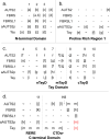
Fig 3. Shared regions of conservation between AUTS2-related proteins (R1-11).
Heat map colour coding displays the average pairwise identity of each protein region between AUTS2-related proteins (107 sequences) as calculated by MView [34]. Connecting lines represent the arrangement of conserved regions in Tay bridge compared to aAUTS2p (C. floridanus). Amino acid positions are displayed above each protein. PR1: Proline rich region 1. Percentage identity values are displayed in Table 2.
Fig 4
Fig 4. Internally conserved regions in AUTS2-related proteins.
Internally conserved regions are displayed above the midpoint and those regions identified as conserved across all AUTS2-related proteins are displayed below; an exon-by-exon conservation schematic is displayed above each primary isoform. Average conservation of each region within the respective ortholog grouping is represented by a heat map. ATG1 and ATG2: start codons used by AUTS2 Variants 1 and 2 respectively. a. AUTS2-001: canonical isoform containing all exons; regions critical to both the Nuclear and Cytosolic are displayed with the blue dashed and black solid brackets respectively. AUTS2-003: N-terminal isoform containing exons 1–4 of the canonical transcript and an alternate exon 5 containing a premature stop codon (displayed in yellow); b. FBRS-001: long form of FBRS containing 18 exons, no analogous exon analogous to AUTS2 Exon 3 is present. FBRS-201: predominant C-terminal isoform of FBRS (ENST00000287468.5) analogous to AUTS2 Variant 2 and contains the majority of the Tay. c. FBRSL1-001: canonical FBRSL1 isoform. FBRSL1-201: C-terminal isoform containing exons 8–19; validated in zebrafish and is analogous to Auts2 Variant 1 (AUTS2-201). d. Schematic of Tay bridge (D. melanogaster) and aAUTS2p (C. floridanus).
Fig 5
Fig 5. Alignment of internally conserved regions within AUTS2-related proteins.
Regions aligned to R1-11 (shared conservation) are displayed above each matrix. []: internally conserved regions; divergent regions of conservation are in red; ~: not highly conserved;—: no alignment or alignment to a region of low conservation. H8: octahistidine tract/trinucleotide repeat specific to AUTS2 orthologs. RERE: RERE repeat region; CTDvr: C-terminal domain variable region.
Fig 6
Fig 6
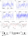
Putative domain structure for AUTS2. a-d: hydrophobicity plots. a. AUTS2; 3 hydrophobic cores: TayH2 is present in all AUTS2-related proteins; X14 (Exon 14) is the most conserved region of AUTS2 and displays a peak in hydrophobicity in all AUTS2-related proteins; CTDc (C-terminal Domain Core) is not conserved in FBRS, FBRSL1 and Tay. b. FBRS. c. FBRSL1; a peak is also visible at ~400–410 which is located between Regions 4 and 5. d. Tay bridge; the distance between both cores in comparison to the AUTS2 family proteins should be noted. e. Line graph displaying conservation across AUTS2 along with disorder potential. Regions of high conservation and low disorder potential: TayH2 (Region 2), 36R (Region 3), X10 (Exon 10; Region 6), X14 (Region 8) and CTDc. Regions of low conservation and high disorder potential: PR1 and RERE repeat region. f. Revised three domain structure for AUTS2.

References

    1. Beunders G, van de Kamp J, Vasudevan P, Morton J, Smets K, Kleefstra T, et al. A detailed clinical analysis of 13 patients with AUTS2 syndrome further delineates the phenotypic spectrum and underscores the behavioural phenotype. Journal of Medical Genetics. 2016;53(8):523 10.1136/jmedgenet-2015-103601 - DOI - PubMed
    1. Beunders G, Voorhoeve E, Golzio C, Pardo Luba M, Rosenfeld Jill A, Talkowski Michael E, et al. Exonic Deletions in AUTS2 Cause a Syndromic Form of Intellectual Disability and Suggest a Critical Role for the C Terminus. American Journal of Human Genetics. 2013;92(2):210–20. 10.1016/j.ajhg.2012.12.011 - DOI - PMC - PubMed
    1. Hori K, Nagai T, Shan W, Sakamoto A, Abe M, Yamazaki M, et al. Heterozygous Disruption of Autism susceptibility candidate 2 Causes Impaired Emotional Control and Cognitive Memory. PLOS ONE. 2016;10(12):e0145979. - PMC - PubMed
    1. Gao Z, Lee P, Stafford JM, von Schimmelmann M, Schaefer A, Reinberg D. AUTS2 confers gene activation to Polycomb group proteins in the CNS. Nature. 2014;516(7531):349–54. 10.1038/nature13921 - DOI - PMC - PubMed
    1. Hori K, Nagai T, Shan W, Sakamoto A, Taya S, Hashimoto R, et al. Cytoskeletal Regulation by AUTS2 in Neuronal Migration and Neuritogenesis. Cell Reports. 2014;9(6):2166–79. 10.1016/j.celrep.2014.11.045 - DOI - PubMed

LinkOut - more resources