

Anti-inflammatory, wound healing and antioxidant potential of compounds from Dioscorea bulbifera L. bulbils
- PMID: 33306733
- PMCID: PMC7732089
- DOI: 10.1371/journal.pone.0243632
Anti-inflammatory, wound healing and antioxidant potential of compounds from Dioscorea bulbifera L. bulbils
Abstract
Background: Dioscorea bulbifera L. (Dioscoreaceae) has been traditionally used in Thai folk medicine as a diuretic and anthelmintic, for longevity preparations, and for wound and inflammation treatment. This plant is also commonly used in traditional Indian and Chinese medicines in the treatment of sore throat, gastric cancer, rectal carcinoma and goiters. However, the wound healing effects of the active compounds in this plant have not been investigated.
Objective: This study aimed to identify compounds responsible for the wound healing activity of D. bulbifera and determine their potential anti-inflammatory and antioxidant activities.
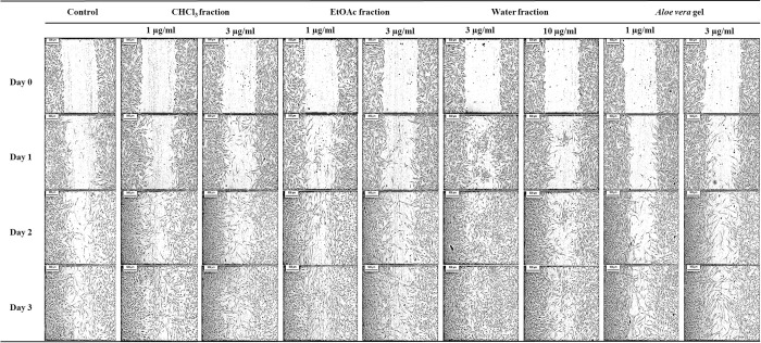
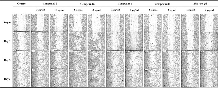
Methods: Crude extracts of D. bulbifera bulbils, their derived fractions and eleven purified compounds were tested for anti-inflammatory activity against LPS-induced NO production in RAW264.7 macrophages. The wound healing effects were evaluated via cell proliferation and migration assays using human dermal fibroblasts (HDFs), and the antioxidant effects were determined using 2,2-diphenyl-1-picrylhydrazyl (DPPH) and hydroxyl radical (•OH) scavenging activity assays.
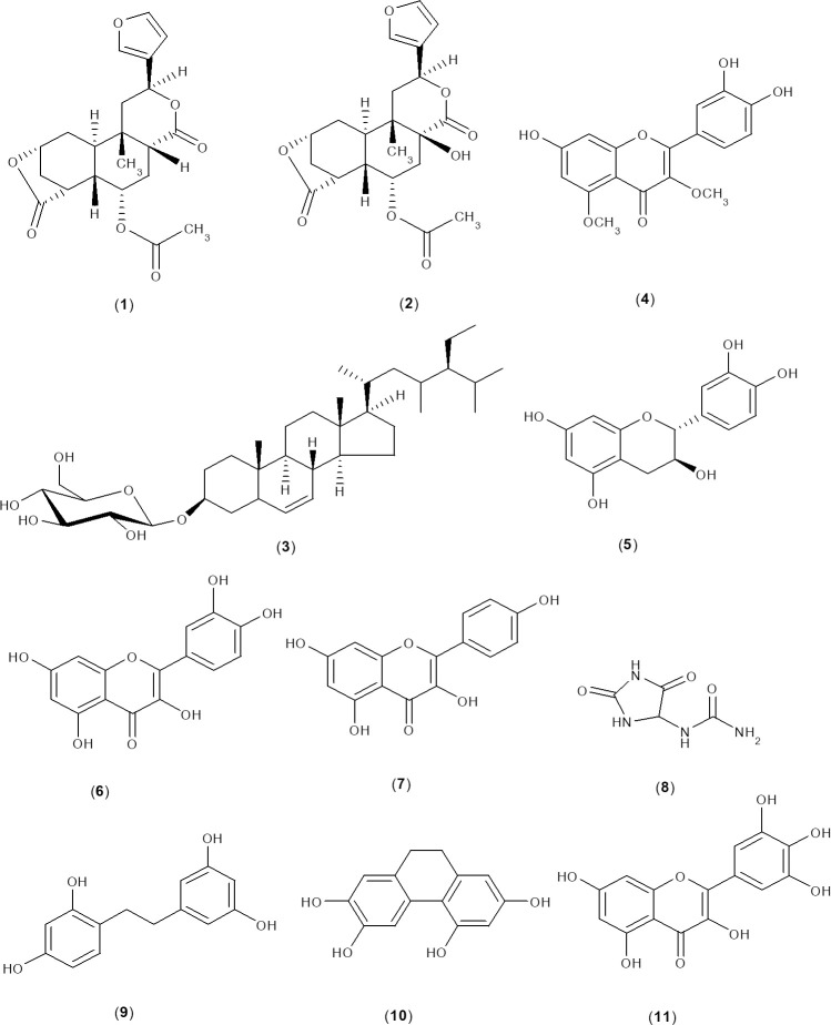
Results: 15,16-Epoxy-6α-O-acetyl-8β-hydroxy-19-nor-clero-13(16),14-diene-17,12;18,2-diolide (2), (+)-catechin (5), quercetin (6) and myricetin (11) exhibited significantly potent wound healing effects and promoted marked cell proliferation, resulting in % viabilities of 107.4-137.6, 121.1-151.9, 98.0-131.9, 90.9-115.9, respectively. Among them, (+)-catechin produced the highest % cell migration, resulting in 100.0% wound closure sooner (at day 2) than the other compounds. In addition, 1 μg/ml (+)-catechin significantly increased fibroblast migration by 2.4-fold compared to that in the control after 24 h. Regarding anti-inflammatory properties, kaempferol (7) and quercetin (6) decreased (p < 0.005) NO production, with IC50 values of 46.6 and 56.2 μM, respectively. In addition, the crude extracts, solvent fractions and flavonoid compounds were also found to possess marked antioxidant activity in both DPPH and •OH radical scavenging assays.
Conclusions: These findings provide more evidence to support the traditional use of D. bulbifera for the treatment of wounds and inflammation.
Conflict of interest statement
The authors have declared that no competing interests exist.
Figures
References
-
- Harper D, Young A, McNaught C-E. The physiology of wound healing. Surgery (Oxford). 2014;32(9):445–50.
Publication types
MeSH terms
Substances
LinkOut - more resources
Full Text Sources
Medical
