Case Reports
. 2021 Jan;9(1):e1566.
doi: 10.1002/mgg3.1566. Epub 2020 Dec 11.

Defective INPP5E distribution in NPHP1-related Senior-Loken syndrome

Affiliations
Case Reports

Defective INPP5E distribution in NPHP1-related Senior-Loken syndrome

Ke Ning et al. Mol Genet Genomic Med. 2021 Jan.

Abstract

Background: Senior-Loken syndrome is a rare genetic disorder that presents with nephronophthisis and retinal degeneration, leading to end-stage renal disease and progressive blindness. The most frequent cause of juvenile nephronophthisis is a mutation in the nephronophthisis type 1 (NPHP1) gene. NPHP1 encodes the protein nephrocystin-1, which functions at the transition zone (TZ) of primary cilia.

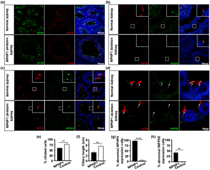
Methods: We report a 9-year-old Senior-Loken syndrome boy with NPHP1 deletion, who presents with bilateral vision decrease and cystic renal disease. Renal function deteriorated to require bilateral nephrectomy and renal transplant. We performed immunohistochemistry, H&E staining, and electron microscopy on the renal sample to determine the subcellular distribution of ciliary proteins in the absence of NPHP1.

Results: Immunohistochemistry and electron microscopy of the resected kidney showed disorganized cystic structures with loss of cilia in renal tubules. Phosphoinositides have been recently recognized as critical components of the ciliary membrane and immunostaining of kidney sections for phosphoinositide 5-phosphatase, INPP5E, showed loss of staining compared to healthy control. Ophthalmic examination showed decreased electroretinogram consistent with early retinal degeneration.

Conclusion: The decreased expression of INPP5E specifically in the primary cilium, coupled with disorganized cilia morphology, suggests a novel role of NPHP1 that it is involved in regulating ciliary phosphoinositide composition in the ciliary membrane of renal tubular cells.

Keywords: INPP5E; NPHP1; Senior-Loken syndrome; primary cilia; transition zone.


Conflict of interest statement

The authors declare no financial/non‐financial competing interest.

Figures

FIGURE 1
FIGURE 1
Early retinal degeneration and kidney cyst in a patient with NPHP1 deletion. (a) Representative color fundus photographs of the patient with NPHP1 deletion showing normal fundus in both eyes without optic disc pallor or retinal dystrophy. (b) Visual field examination demonstrating bilateral visual field loss in this patient. The grayscale plots present as a cluster of paracentral points with decreased sensitivity and scotoma. (c) Full‐field standard ERG of OD showing increased implicit time and reduced amplitude in scotopic 0.01 ERG, and in photopic 3.0 ERG. (d) Representative hematoxylin and eosin (H&E)‐stained kidney sections showing the histological pattern in normal human and the patient with NPHP1 deletion. Kidney specimen is shown at low (left) and high (right) magnification. Kidney histology of the NPHP1 patient illustrates the characteristic triad of corticomedullary cysts, tubular basement membrane disruption, and tubulointerstitial nephropathy. Scale bars = (left) 200 μm; (right) 50 μm. Transmission electron micrographs of primary cilia and TBM in kidney epithelial cells from the NPHP1 deletion patient. TEM reveals a thickened tubular membrane and decreased cilia. Scale bars = 2 μm
FIGURE 2
FIGURE 2
Disorganized cilia with abnormal ciliary protein expression in renal tubular cells of a patient with NPHP1 deletion. (a) Disorganized cilia in renal tubular cells of a patient with NPHP1 deletion. Confocal immunomicroscopy of normal and patient kidney sections stained with Arl13b and Ac‐tubulin antibodies to detect primary cilia and with IFT88 to detect basal body. Primary cilia appear shorter and disorganized in epithelial cells of patient kidney sections. (e and f) Quantification of ciliation and ciliary length (n = 200). (b) Normal expression of NPHP8 and abnormal expression of (c) NPHP4 in cilia of renal tubular cells from the NPHP1 deletion patient. Confocal immunomicroscopy of patient kidney sections stained for NPHP8 and NPHP4. (g) Quantification of cells with abnormal expression of NPHP4 (n = 50). (d) Aberrant expression of phosphoinositide 5‐phosphatases in cilia of renal tubular cells from the NPHP1 deletion patient. Confocal immunomicroscopy of patient kidney sections stained for INPP5E. (h) Quantification of cells with abnormal expression (n = 50). Statistical analysis in E‐H was performed using Student's test, p < 0.05 was considered statistically significant. Scale bars: (a–c) 10 μm; (d) magnified images 40 μm. (Arrow, marks primary cilia)

Similar articles

Cited by

References

    1. Aggarwal, H. K. , Jain, D. , Yadav, S. , Kaverappa, V. , & Gupta, A. (2013). Senior‐loken syndrome with rare manifestations: A case report. The Eurasian Journal of Medicine, 45(2), 128–131. 10.5152/eajm.2013.25 - DOI - PMC - PubMed
    1. Anderson, C. T. , Castillo, A. B. , Brugmann, S. A. , Helms, J. A. , Jacobs, C. R. , & Stearns, T. (2008). Primary cilia: Cellular sensors for the skeleton. The Anatomical Record: Advances in Integrative Anatomy and Evolutionary Biology, 291(9), 1074–1078. 10.1002/ar.20754 - DOI - PMC - PubMed
    1. Awata, J. , Takada, S. , Standley, C. , Lechtreck, K. F. , Bellve, K. D. , Pazour, G. J. , Fogarty, K. E. , & Witman, G. B. (2014). NPHP4 controls ciliary trafficking of membrane proteins and large soluble proteins at the transition zone. Journal of Cell Science, 127(Pt 21), 4714–4727. 10.1242/jcs.155275 - DOI - PMC - PubMed
    1. Berbari, N. F. , O'Connor, A. K. , Haycraft, C. J. , & Yoder, B. K. (2009). The primary cilium as a complex signaling center. Current Biology, 19(13), R526–R535. 10.1016/j.cub.2009.05.025 - DOI - PMC - PubMed
    1. Chavez, M. , Ena, S. , Van Sande, J. , de Kerchove d'Exaerde, A. , Schurmans, S. , & Schiffmann, S. N. (2015). Modulation of ciliary phosphoinositide content regulates trafficking and sonic hedgehog signaling output. Developmental Cell, 34(3), 338–350. 10.1016/j.devcel.2015.06.016 - DOI - PubMed

Publication types

MeSH terms

Substances

Supplementary concepts