. 2021 May 15;203(10):1230-1244.
doi: 10.1164/rccm.202006-2403OC.

Human Lung-Resident Macrophages Colocalize with and Provide Costimulation to PD1hi Tissue-Resident Memory T Cells

Affiliations

Human Lung-Resident Macrophages Colocalize with and Provide Costimulation to PD1hi Tissue-Resident Memory T Cells

Mark E Snyder et al. Am J Respir Crit Care Med. .

Abstract

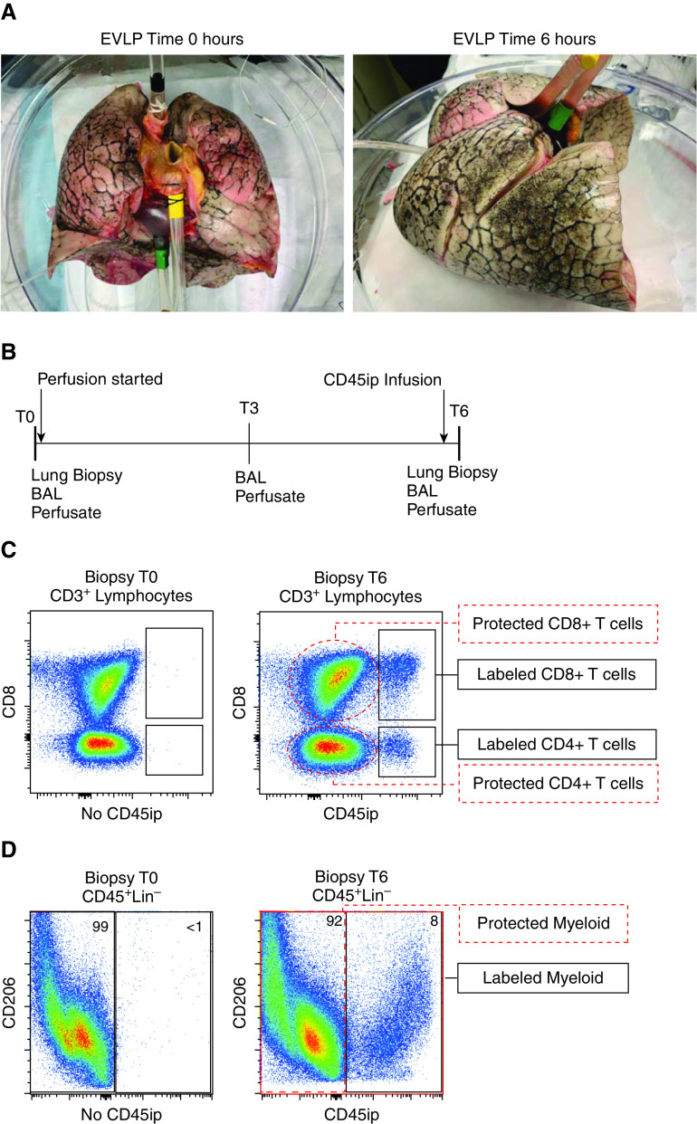
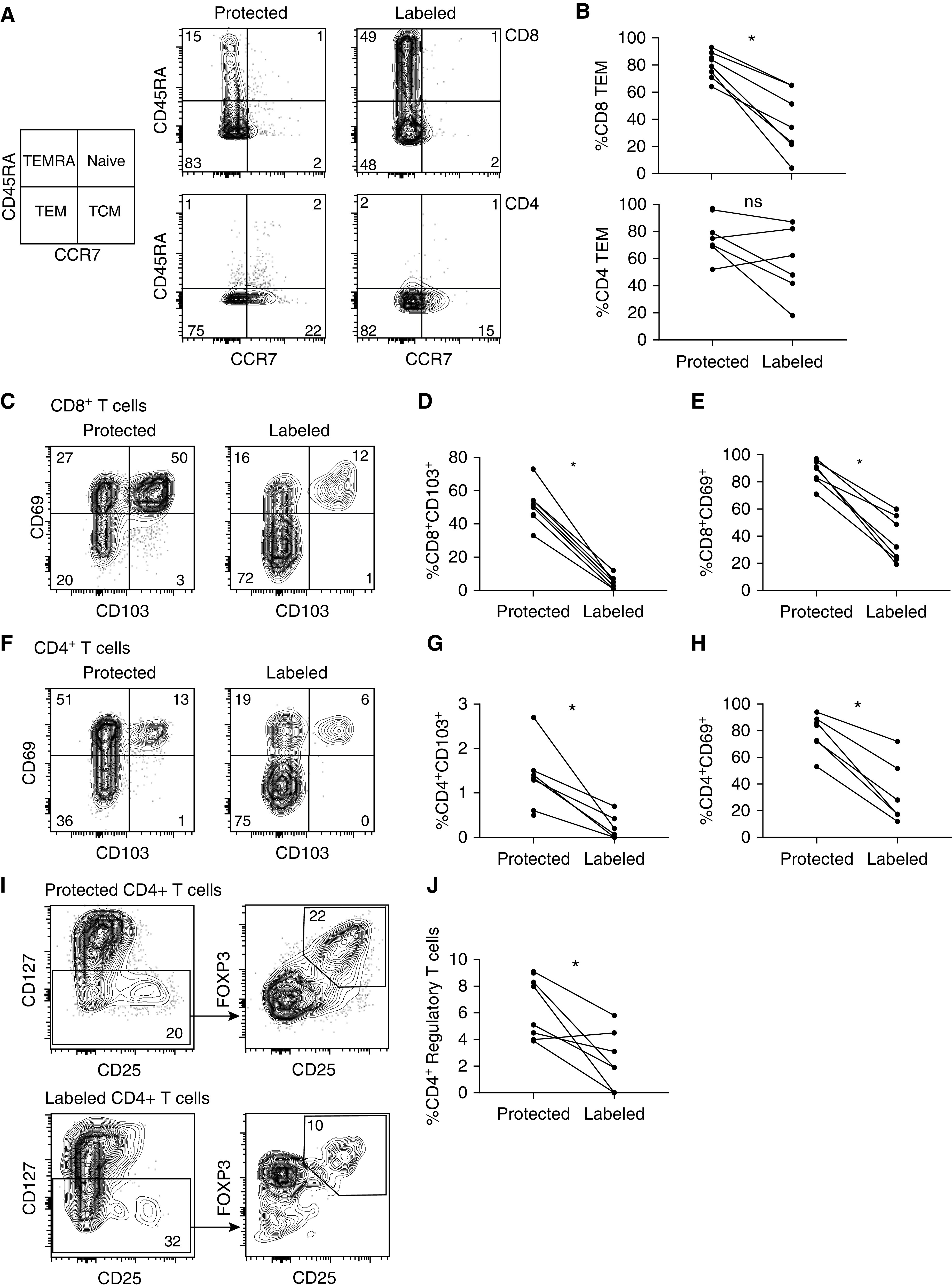
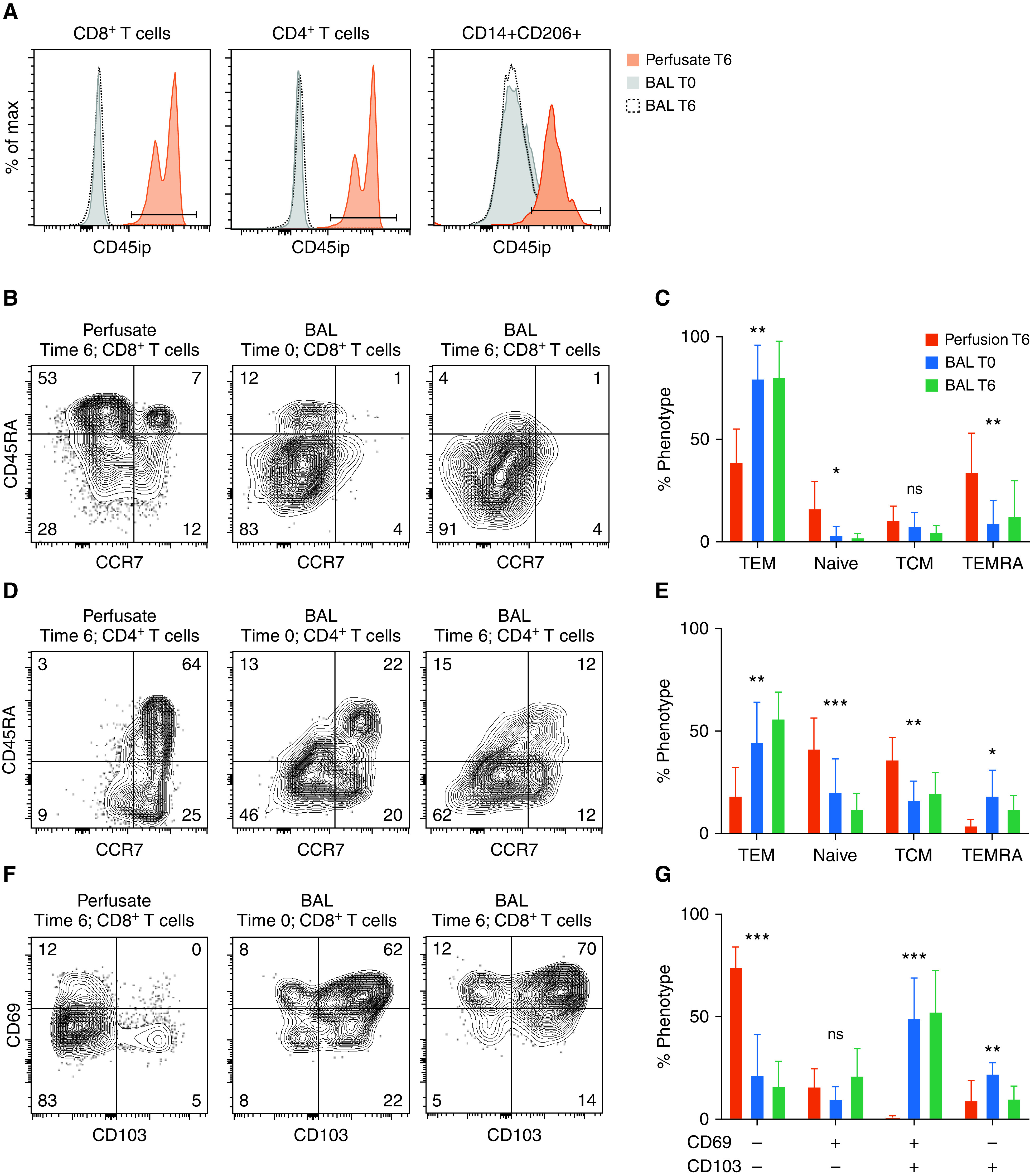
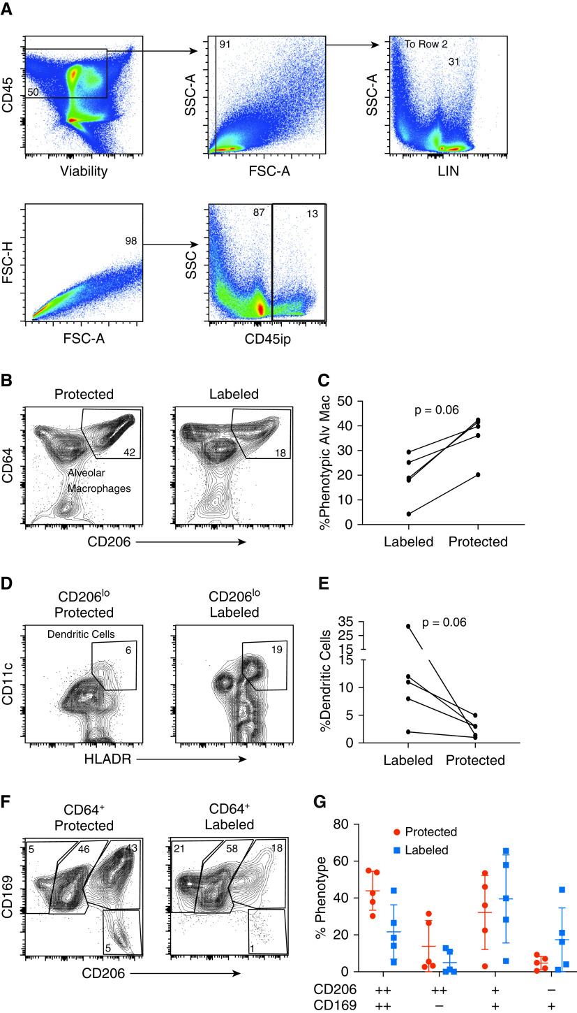
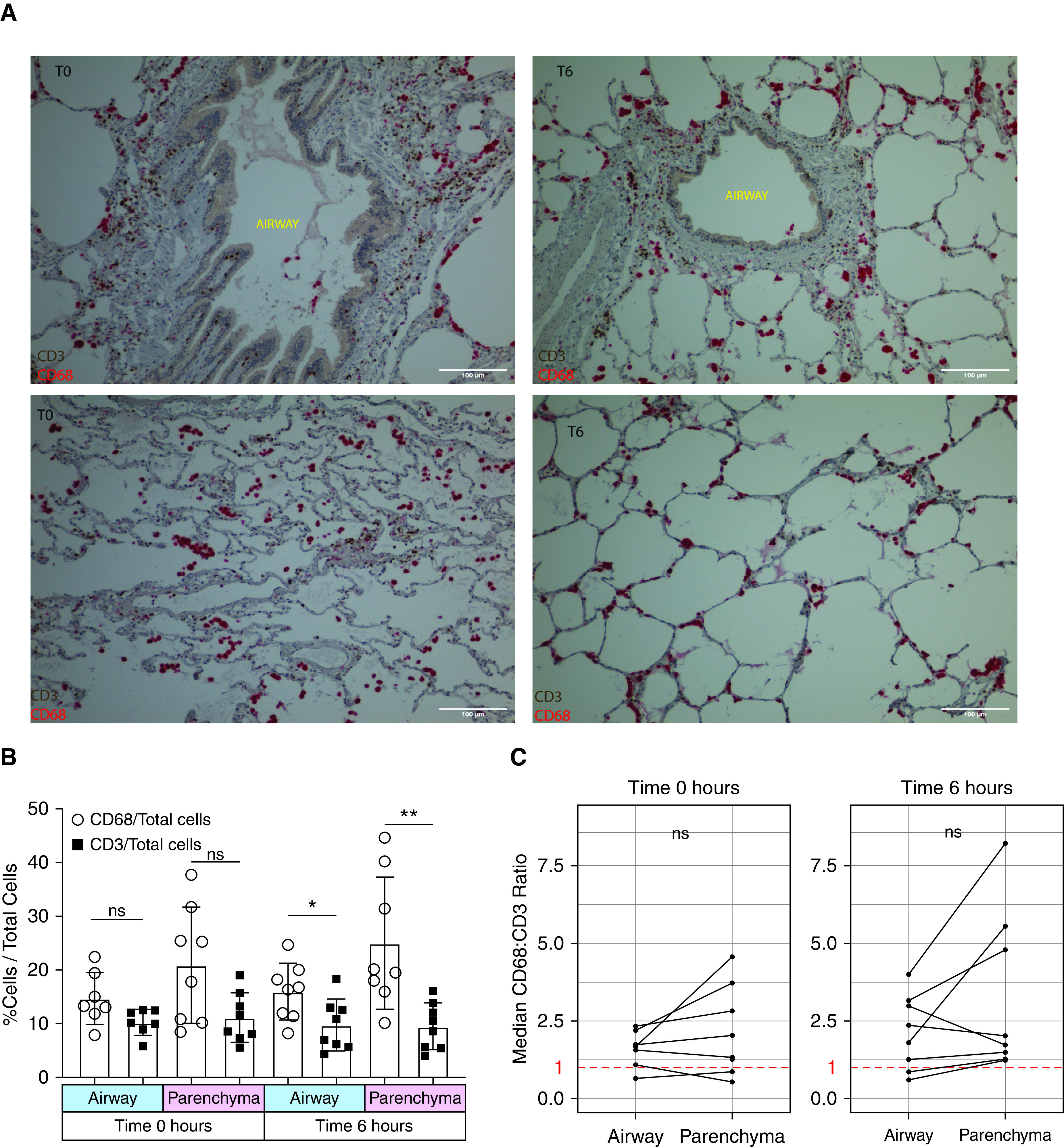
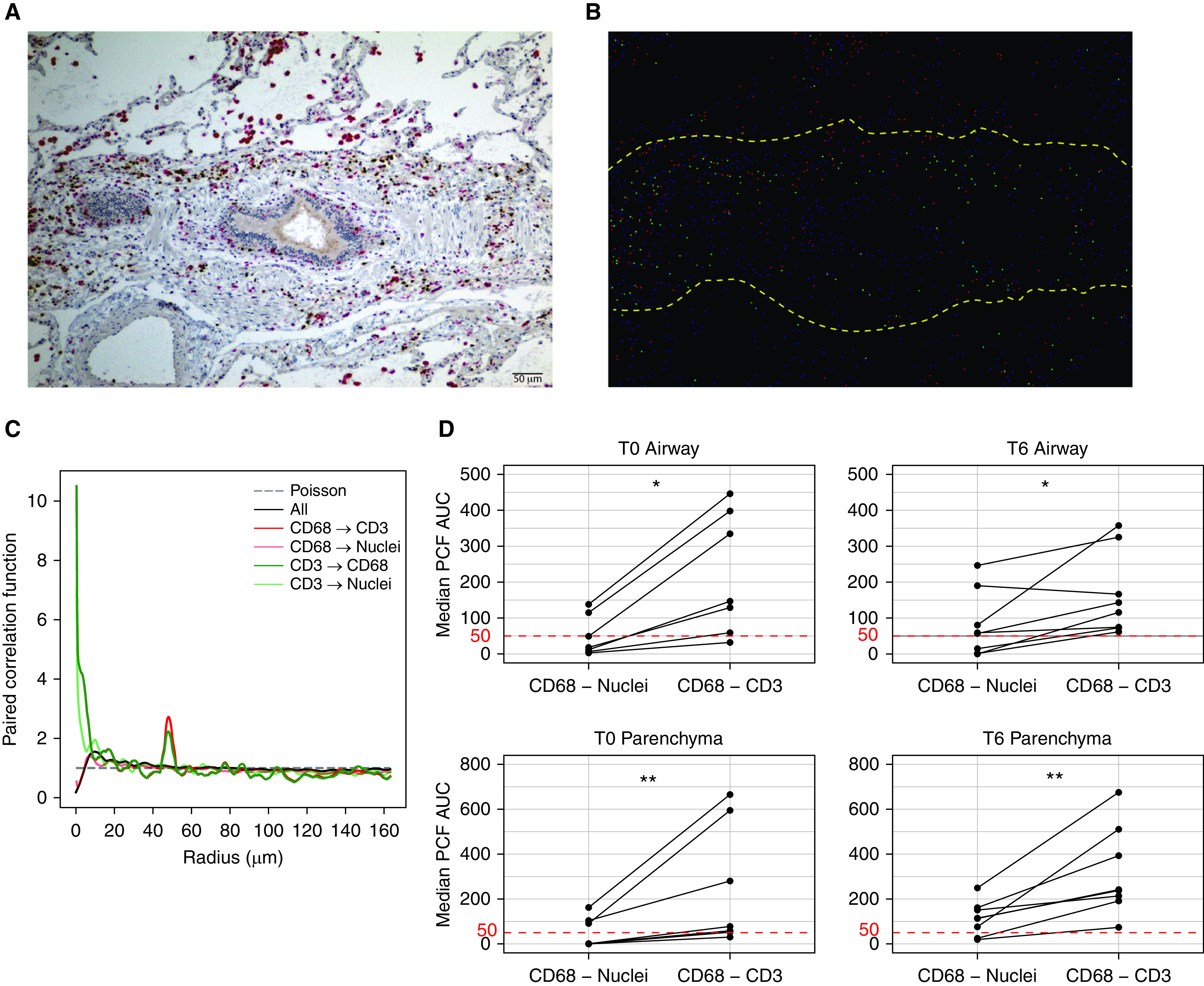
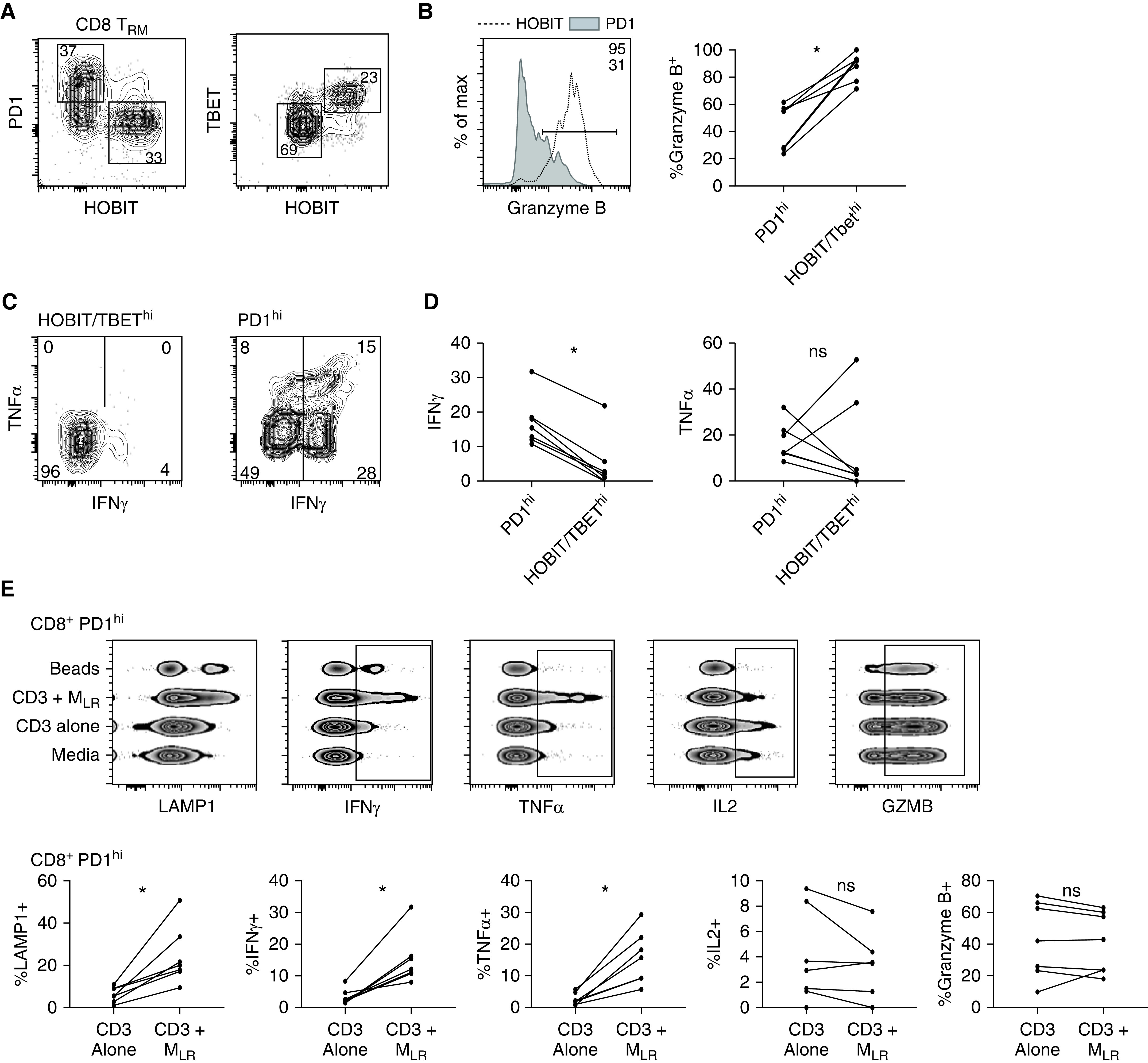
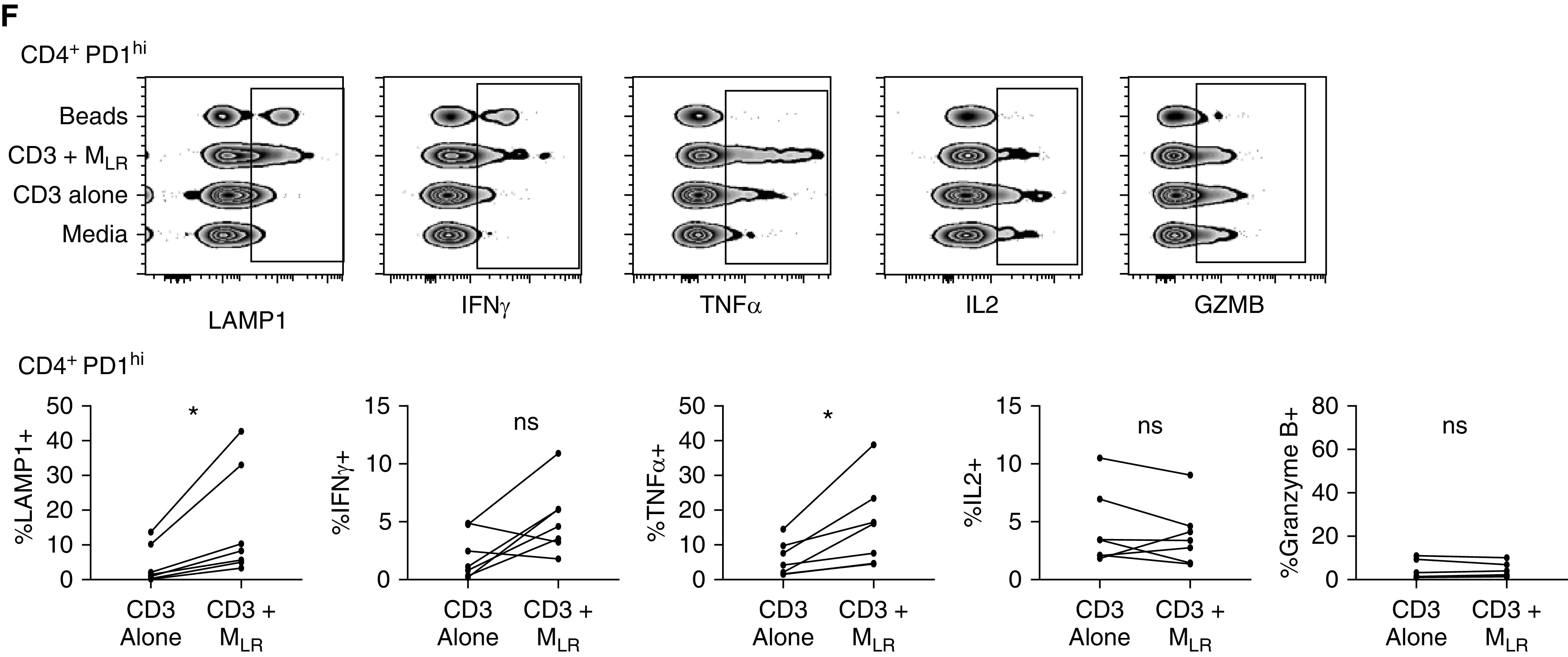
Rationale: Tissue-resident memory T cells (TRM) play a critical role in the defense against inhaled pathogens. The isolation and study of human lung tissue-resident memory T cells and lung-resident macrophages (MLR) are limited by experimental constraints. Objectives: To characterize the spatial and functional relationship between MLR and human lung tissue-resident memory T cells using ex vivo lung perfusion (EVLP). Methods: TRM and MLR were isolated using EVLP and intraperfusate-labeled CD45 antibody. Cells isolated after 6 hours of EVLP were analyzed using spectral flow cytometry. Spatial relationships between CD3+ and CD68+ cells were explored with multiplexed immunohistochemistry. Functional relationships were determined by using coculture and T-cell-receptor complex signal transduction. Measurements and Main Results: Lungs from 8 research-consenting organ donors underwent EVLP for 6 hours. We show that human lung TRM and MLR colocalize within the human lung, preferentially around the airways. Furthermore, we found that human lung CD8+ TRM are composed of two functionally distinct populations on the basis of PD1 (programed cell death receptor 1) and ZNF683 (HOBIT) protein expression. We show that MLR provide costimulatory signaling to PD1hi CD4+ and CD8+ lung TRM,, augmenting the effector cytokine production and degranulation of TRM. Conclusions: EVLP provides an innovative technique to study resident immune populations in humans. Human MLR colocalize with and provide costimulation signaling to TRM, augmenting their effector function.

Keywords: ex vivo lung perfusion; human lung immunology; innate and adaptive immune interaction; lung-resident macrophage; tissue-resident memory T cell.


Figures

Figure 1.
Figure 1.
Ex vivo lung perfusion (EVLP) to isolate tissue-resident memory T cells (TRM) and lung-resident macrophages (MLR). Experimental model of isolating human lung TRM and MLR using 6 hours of EVLP and an intraperfusate-labeled CD45 (CD45ip) antibody: (A) human lungs before (left) and after (right) 6 hours of EVLP. (B) Study timeframe showing timing of sample collection and labeled antibody administration 20 minutes before the 6-hour time point (T6) of EVLP. (C) Representative flow-cytometric plot of live CD3+ lymphocytes taken from lung biopsy specimens at the 0-hour time point (T0; left) where no CD45ip antibody is present and at T6 (after 20 min of intraperfusate antibody circulation). T cells in communication with the perfusate are positive for the antibody (black rectangles), defined as “labeled” T cells, whereas the majority of CD4+ and CD8+ T cells are protected from intraperfusate labeling (red dashed ovals) and labeled “protected.” (D) Representative flow-cytometric plot of live, CD45+, Lin singlets taken from lung biopsy specimens at T0 (left) and T6 (right). Those cells positive for CD45ip antibody are identified as labeled myeloid cells, and those absent of CD45ip antibody are identified as protected myeloid cells. Lin = CD3, CD19, and CD56 lineage–negative.
Figure 2.
Figure 2.
Protected T cells have increased canonical markers of tissue residency and include CD4+ regulatory T cells. (A) Subset composition of representative samples using flow cytometry to identify the phenotype on the basis of CCR7 and CD45RA cell-surface expression as TEM (CCR7CD45RA), TCM (CCR7+CD45RA), naive T cells (CCR7+, CD45RA+), and TEMRA (CCR7CD45RA+) from lung biopsy specimens taken at 6 hours of ex vivo lung perfusion. (B) Compiled data showing paired proportion of TEM for both CD8+ (top) and CD4+ (bottom) T cells
Figure 3.
Figure 3.
Differential phenotype of BAL-fluid and perfusate compartments. (A) Representative histograms displaying intraperfusate CD45 labeling by sample location and time for CD8+ T cells (far left), CD4+ T cells (middle), and alveolar macrophages (far right, defined as lineage-negative CD14+CD206+). (B and D) Representative flow-cytometric plots of CD8+ (B) and CD4+ (D) T-cell phenotypes based on CD45RA and CCR7 expression for samples obtained from perfusate at the 6-hour time point (T6; far left), from BAL fluid at the 0-hour time point (T0; middle), and from BAL fluid at T6 (far right). (C and E) Cumulative proportions of phenotypes for CD8+ (C) and CD4+ (E) T cells (bars represent means with SDs shown; N = 8; with *P < 0.05, **P < 0.001, and ***P < 0.0001 by Kruskal-Wallis test). (F and H) Representative flow-cytometric plots of canonical tissue-resident memory T-cell markers CD69 and CD103 for CD8+ (F) and CD4+ (H) T cells obtained from perfusate at 6 hours (far left), BAL fluid at 0 hours (middle), and BAL fluid at 6 hours (far right). (G and I) Cumulative proportions of CD69 and CD103 expression from CD8+ (G) and CD4+ (I) T-cell phenotypes (bars represent means with SDs shown; N = 8; *P < 0.05, **P < 0.001, and ***P < 0.0001 by Kruskal-Wallis test). + = positive; − = negative; CD45ip = intraperfusate-labeled CD45; max = maximum; ns = no statistically significant difference; TCM = central memory T cells; TEM = effector memory T cells; TEMRA = terminally differentiated effector T cells.
Figure 3.
Figure 3.
Differential phenotype of BAL-fluid and perfusate compartments. (A) Representative histograms displaying intraperfusate CD45 labeling by sample location and time for CD8+ T cells (far left), CD4+ T cells (middle), and alveolar macrophages (far right, defined as lineage-negative CD14+CD206+). (B and D) Representative flow-cytometric plots of CD8+ (B) and CD4+ (D) T-cell phenotypes based on CD45RA and CCR7 expression for samples obtained from perfusate at the 6-hour time point (T6; far left), from BAL fluid at the 0-hour time point (T0; middle), and from BAL fluid at T6 (far right). (C and E) Cumulative proportions of phenotypes for CD8+ (C) and CD4+ (E) T cells (bars represent means with SDs shown; N = 8; with *P < 0.05, **P < 0.001, and ***P < 0.0001 by Kruskal-Wallis test). (F and H) Representative flow-cytometric plots of canonical tissue-resident memory T-cell markers CD69 and CD103 for CD8+ (F) and CD4+ (H) T cells obtained from perfusate at 6 hours (far left), BAL fluid at 0 hours (middle), and BAL fluid at 6 hours (far right). (G and I) Cumulative proportions of CD69 and CD103 expression from CD8+ (G) and CD4+ (I) T-cell phenotypes (bars represent means with SDs shown; N = 8; *P < 0.05, **P < 0.001, and ***P < 0.0001 by Kruskal-Wallis test). + = positive; − = negative; CD45ip = intraperfusate-labeled CD45; max = maximum; ns = no statistically significant difference; TCM = central memory T cells; TEM = effector memory T cells; TEMRA = terminally differentiated effector T cells.
Figure 4.
Figure 4.
Differential compartmentalization of alveolar macrophages (Alv Mac) and lung dendritic cells. (A) Representative flow-cytometric plot isolating cells with a myeloid lineage (LIN). (B) Representative flow-cytometric plot identifying Alv Mac (defined as LIN, CD64+CD206+) from lung biopsy specimens taken at 6 hours on the basis of the presence (right) or absence (left) of intraperfusate CD45 labeling (CD45ip). (C) Paired comparison of the proportion of Alv Mac from each cellular compartment (P = 0.06, n = 5). (D) Representative flow-cytometric plot identifying dendritic cells (defined as LIN CD206lo, HLADR+CD11c+) from lung biopsy specimens taken at 6 hours on the basis of the presence (right) or absence (left) of CD45ip. (E) Paired comparison of the proportion of Alv Mac from each cellular compartment (P = 0.06, n = 5). (F) Representative flow-cytometric plots of macrophage phenotypes based on CD169 and CD206 expression for protected (left) and labeled (right) CD64+ cells obtained from lung biopsy specimens at 6 hours. (G) Cumulative data (n = 5). ++ = strongly positive; + = weakly positive; − = negative; FSC-A = forward scatter area; FSC-H = forward scatter height; SSC = side scatter; SSC-A = side scatter area.
Figure 5.
Figure 5.
Tissue-resident memory T cells are abundant in the lung and are mainly airway centered. Multiplexed immunohistochemistry of CD3 (brown) and CD68 (red) was performed on sections of lung obtained at the 0-hour time point (T0) and at the 6-hour time point (T6) of ex vivo lung perfusion (EVLP). (A) Representative immunohistochemistry of the airway (top row) and lung parenchyma (bottom row) at T0 (left column) and after T6 of EVLP (right column). (B) Cumulative data showing the proportion of CD3+ and CD68+ cells of all nucleated cells by time of lung biopsy and area captured on imaging. “Airway” refers to small airways, and “parenchyma” refers to the adjacent lung parenchyma section devoid of airway or blood vessels. The median value per lung is included in each graph, with 8 lungs included (*P = 0.02 and **P = 0.008). (C) Cumulative data of CD68/CD3 ratio by anatomic location for T0 (left) and T6 (right) of EVLP. The median value of each lung is included, 8 lungs were included in the analysis, the orphaned parenchyma dot at 0 hours is due to the lack of an identifiable airway in one biopsy specimen at 0 hours, and the red dashed line equals a ratio of 1. Scale bars, 100 μm. ns = no statistically significant difference.
Figure 6.
Figure 6.
Tissue-resident memory T cells and lung-resident macrophages colocalize within the lung, predominantly around the airways. (A) Representative multiplexed immunohistochemistry showing CD3 (brown) and CD68 (red) cells colocalizing around the airways. (B) Image converted to centroids (dots) using a machine-learning algorithm; yellow dashed lines outline the border of the airway. (C) Representative graph showing PCF (y-axis) across distances between centroids (x-axis) in micrometers for all cellular spatial relationships in one image; deviations above the Poisson distribution suggest colocalization at that radius (for reference, the diameter of a T cell is roughly 7 µm [49], and the diameter of a macrophage is variable but is on average 21 µm [50]). (D) Cumulative data comparing proximity between CD68+ cells and either nuclei or CD3+ cells within and around the small airways (top row) and lung parenchyma (bottom row) at T0 (left column) and T6 (right column) of ex vivo lung perfusion; for reference, an AUC value of 50 µm would represent a random Poisson distribution (*P = 0.02 and **P = 0.008; median value of 3–6 random airway and parenchyma sections included from 8 lungs; no T0 airway identified for one sample). Scale bar, 50 μm. AUC = area under the curve; PCF = pair-correlation function; T0 = 0-hour time point; T6 = 6-hour time point.
Figure 7.
Figure 7.
Lung-resident macrophages (MLR) provide costimulatory signaling to tissue-resident memory T cells (TRM). CD69+, CD4+, and CD8+ T cells were isolated from lung biopsy specimens after 6 hours of ex vivo lung perfusion using an influx cell sorter and were cultured with media alone (media), with monoclonal CD3 (CD3), with monoclonal CD3 plus MLR at an MLR/TRM ratio of 1:1 (CD3 + MLR), and with CD2/CD3/CD28 beads (beads). (A) Representative flow-cytometric plot of PD1 (programed cell death receptor 1) and HOBIT (left) and TBET (T-box transcription factor TBX21) and HOBIT (right) expression of CD8+ TRM cultured in media. (B) Representative histogram (left; horizontal line denotes positive gate) and paired cumulative frequency of intracellular GZMB (granzyme B) expression between PD1hi and HOBIThi or TBEThi CD8+ TRM (*P = 0.02, includes samples from 7 lungs). (C) Representative flow-cytometric plot of TNFα (tumor necrosis factor α) and IFNγ production among HOBIT or TBEThi (left) and PD1hi (right) CD8+ TRM isolated from lung biopsy specimens at 6 hours and cocultured with CD3 + MLR. (D) Composite data comparing the proportion of CD8+ TRM producing IFNγ (left) and TNFα (right) on the basis of expression of PD1 and either HOBIT or TBET (*P = 0.02, includes data from 7 lungs). (E) Representative comparison of cytokine production and LAMP1 expression for CD8+ PD1hi lung TRM under different stimulation conditions displayed as concatenated files from a single lung (top; rectangle outlines positive cells) and cumulative data (bottom; *P = 0.02; includes data from 7 lungs). (F) Representative comparison of cytokine production and LAMP1 expression for CD4+ PD1hi lung TRM under different stimulation conditions displayed as concatenated files from a single lung (top; rectangle outlines positive cells) and cumulative data (bottom; *P = 0.02; includes data from 7 lungs). max = maximum; ns = no statistically significant difference.
Figure 7.
Figure 7.
Lung-resident macrophages (MLR) provide costimulatory signaling to tissue-resident memory T cells (TRM). CD69+, CD4+, and CD8+ T cells were isolated from lung biopsy specimens after 6 hours of ex vivo lung perfusion using an influx cell sorter and were cultured with media alone (media), with monoclonal CD3 (CD3), with monoclonal CD3 plus MLR at an MLR/TRM ratio of 1:1 (CD3 + MLR), and with CD2/CD3/CD28 beads (beads). (A) Representative flow-cytometric plot of PD1 (programed cell death receptor 1) and HOBIT (left) and TBET (T-box transcription factor TBX21) and HOBIT (right) expression of CD8+ TRM cultured in media. (B) Representative histogram (left; horizontal line denotes positive gate) and paired cumulative frequency of intracellular GZMB (granzyme B) expression between PD1hi and HOBIThi or TBEThi CD8+ TRM (*P = 0.02, includes samples from 7 lungs). (C) Representative flow-cytometric plot of TNFα (tumor necrosis factor α) and IFNγ production among HOBIT or TBEThi (left) and PD1hi (right) CD8+ TRM isolated from lung biopsy specimens at 6 hours and cocultured with CD3 + MLR. (D) Composite data comparing the proportion of CD8+ TRM producing IFNγ (left) and TNFα (right) on the basis of expression of PD1 and either HOBIT or TBET (*P = 0.02, includes data from 7 lungs). (E) Representative comparison of cytokine production and LAMP1 expression for CD8+ PD1hi lung TRM under different stimulation conditions displayed as concatenated files from a single lung (top; rectangle outlines positive cells) and cumulative data (bottom; *P = 0.02; includes data from 7 lungs). (F) Representative comparison of cytokine production and LAMP1 expression for CD4+ PD1hi lung TRM under different stimulation conditions displayed as concatenated files from a single lung (top; rectangle outlines positive cells) and cumulative data (bottom; *P = 0.02; includes data from 7 lungs). max = maximum; ns = no statistically significant difference.

Comment in

  • doi: 10.1164/rccm.202012-4358ED

References

    1. Teijaro JR, Turner D, Pham Q, Wherry EJ, Lefrançois L, Farber DL. Cutting edge: tissue-retentive lung memory CD4 T cells mediate optimal protection to respiratory virus infection. J Immunol. 2011;187:5510–5514. - PMC - PubMed
    1. Wu T, Hu Y, Lee YT, Bouchard KR, Benechet A, Khanna K, et al. Lung-resident memory CD8 T cells (TRM) are indispensable for optimal cross-protection against pulmonary virus infection. J Leukoc Biol. 2014;95:215–224. - PMC - PubMed
    1. Turner DL, Bickham KL, Thome JJ, Kim CY, D’Ovidio F, Wherry EJ, et al. Lung niches for the generation and maintenance of tissue-resident memory T cells. Mucosal Immunol. 2014;7:501–510. - PMC - PubMed
    1. Cho A, Bradley B, Kauffman R, Priyamvada L, Kovalenkov Y, Feldman R, et al. Robust memory responses against influenza vaccination in pemphigus patients previously treated with rituximab. JCI Insight. 2017;2:e93222. - PMC - PubMed
    1. McMaster SR, Wilson JJ, Wang H, Kohlmeier JE. Airway-resident memory CD8 T cells provide antigen-specific protection against respiratory virus challenge through rapid IFN-γ production. J Immunol. 2015;195:203–209. - PMC - PubMed

Publication types