Review
. 2021 Jan;1864(1):194666.
doi: 10.1016/j.bbagrm.2020.194666. Epub 2020 Dec 8.

Close to the edge: Heterochromatin at the nucleolar and nuclear peripheries

Affiliations
Review

Close to the edge: Heterochromatin at the nucleolar and nuclear peripheries

Aizhan Bizhanova et al. Biochim Biophys Acta Gene Regul Mech. 2021 Jan.

Abstract

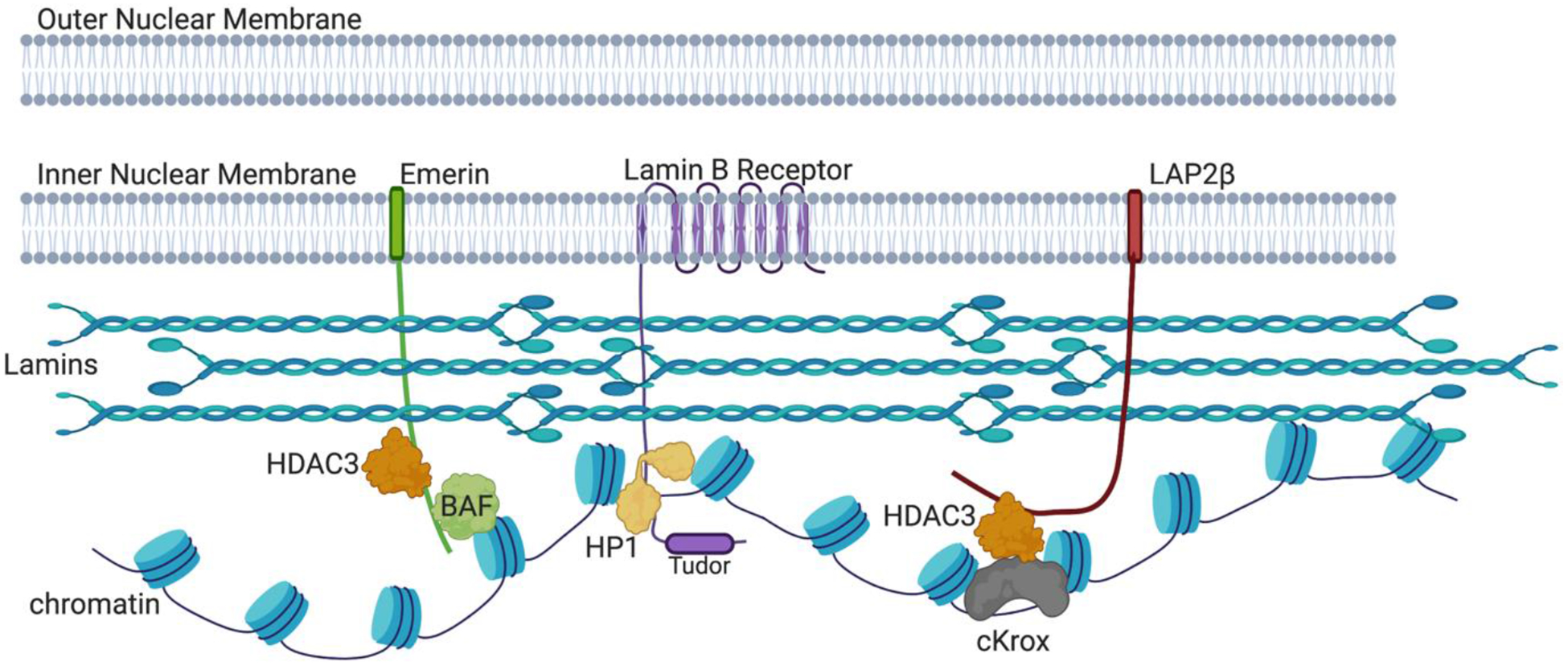
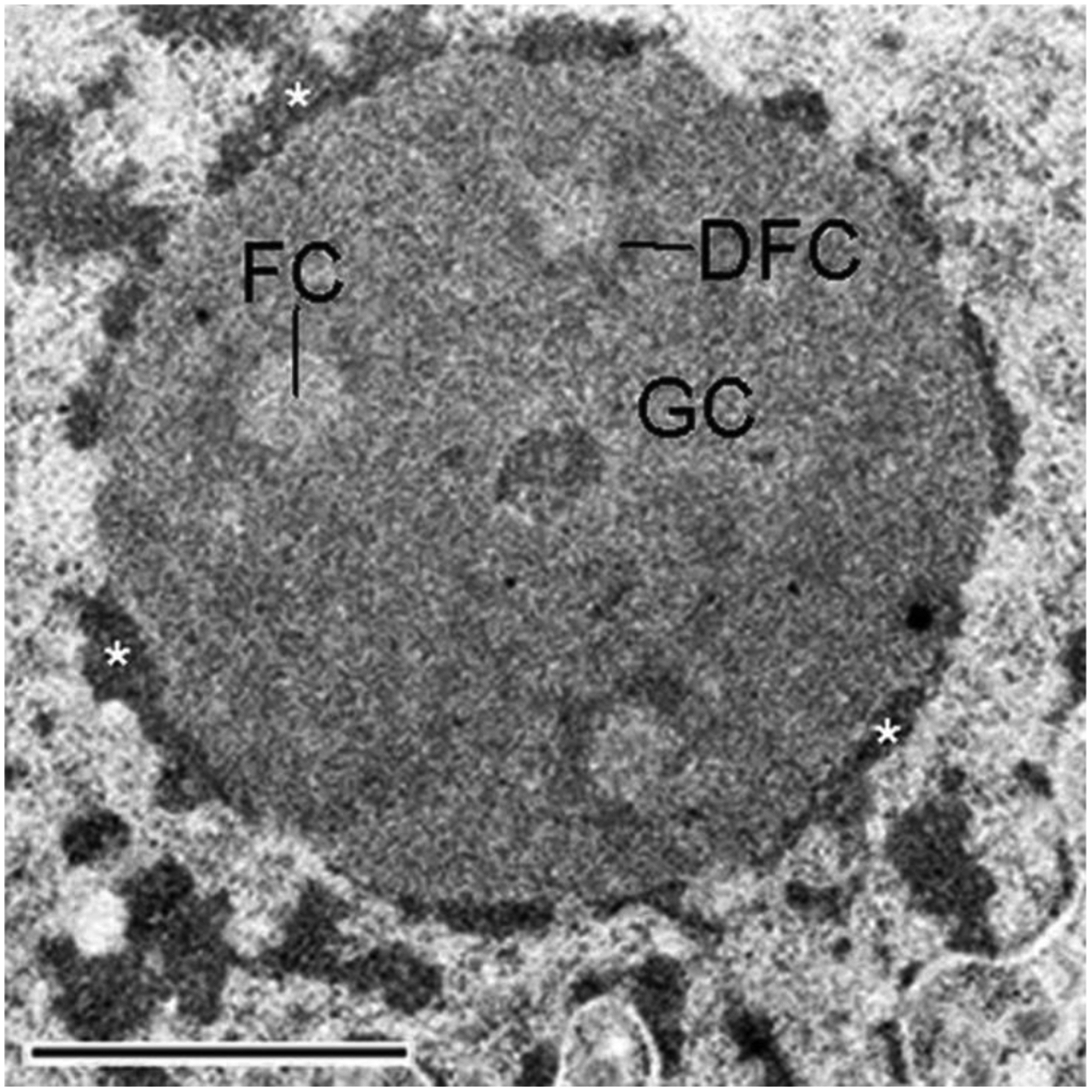
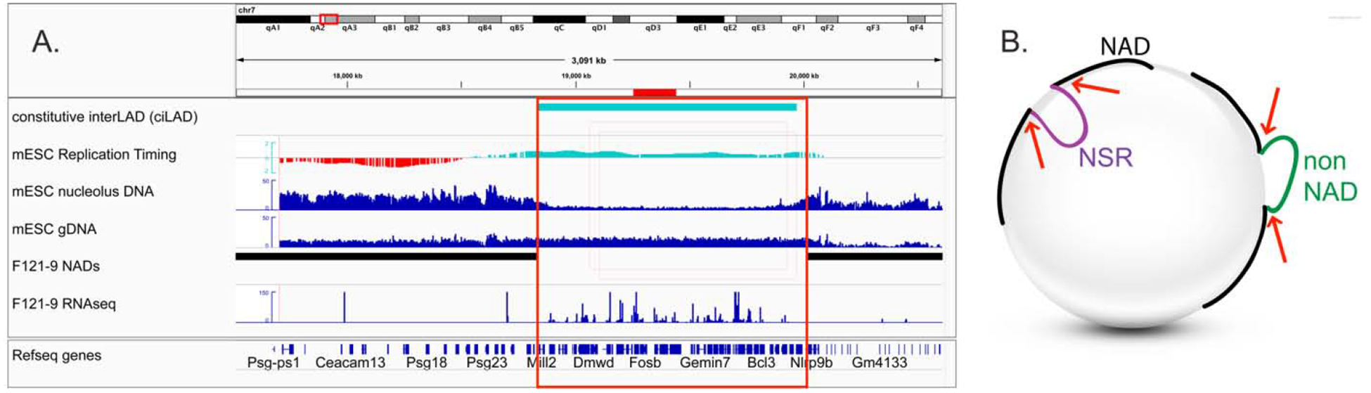
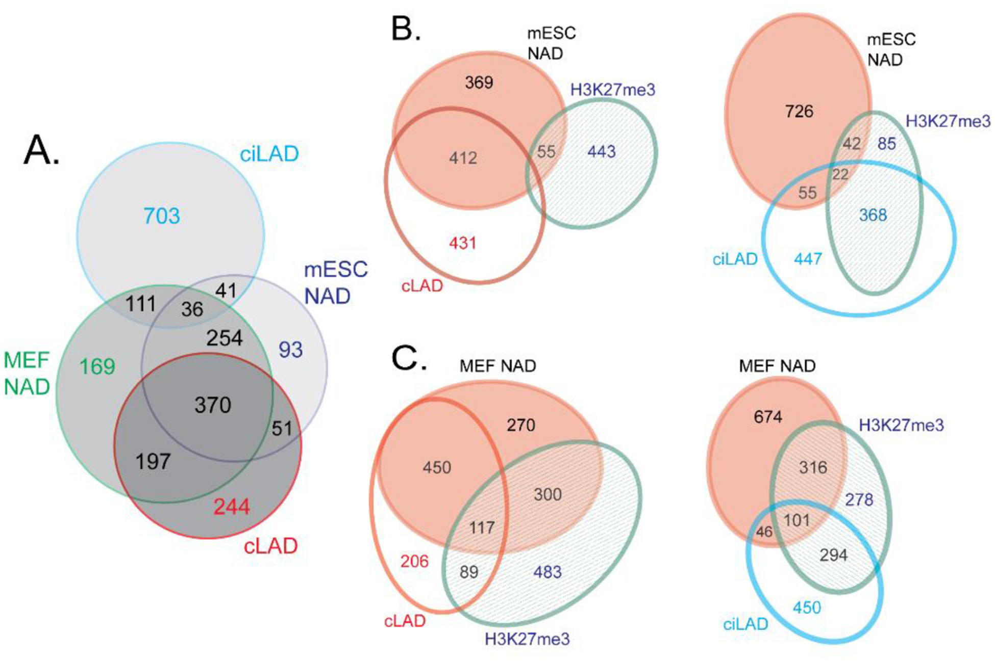
Chromatin is a dynamic structure composed of DNA, RNA, and proteins, regulating storage and expression of the genetic material in the nucleus. Heterochromatin plays a crucial role in driving the three-dimensional arrangement of the interphase genome, and in preserving genome stability by maintaining a subset of the genome in a silent state. Spatial genome organization contributes to normal patterns of gene function and expression, and is therefore of broad interest. Mammalian heterochromatin, the focus of this review, mainly localizes at the nuclear periphery, forming Lamina-associated domains (LADs), and at the nucleolar periphery, forming Nucleolus-associated domains (NADs). Together, these regions comprise approximately one-half of mammalian genomes, and most but not all loci within these domains are stochastically placed at either of these two locations after exit from mitosis at each cell cycle. Excitement about the role of these heterochromatic domains in early development has recently been heightened by the discovery that LADs appear at some loci in the preimplantation mouse embryo prior to other chromosomal features like compartmental identity and topologically-associated domains (TADs). While LADs have been extensively studied and mapped during cellular differentiation and early embryonic development, NADs have been less thoroughly studied. Here, we summarize pioneering studies of NADs and LADs, more recent advances in our understanding of cis/trans-acting factors that mediate these localizations, and discuss the functional significance of these associations.

Keywords: Chromosome; Development; Gene expression; Lamina-associated domain; Nucleolus-associated domain.


Conflict of interest statement

Declaration of interests

The authors declare that they have no known competing financial interests or personal relationships that could have appeared to influence the work reported in this paper.

Figures

Figure 1.
Figure 1.. Molecular interactions at the Inner Nuclear Membrane (INM).
We emphasize here molecules discussed in this review, omitting many interactions at the nuclear lamina for clarity, including lamin-DNA interactions [72,75,79]. Individual lamin protein molecules form dimeric coiled coils, which in turn form polar head-to-tail polymers. These polymers make antiparallel interactions to form a network of intermediate filaments [–82]. The INM proteins Emerin and LAP2β [83] both have “LEM” (LAP2-Emerin-MAN1) domains (not depicted), and bind lamins and HDAC3 (histone deacetylase 3). Emerin also contacts chromatin via its interaction with Barrier-to-Autointegration Factor (BAF) [84]. LAP2β binds to HDAC3 together with cKrox, a DNA-binding protein that contacts LAS elements [56]. The Lamin B receptor (LBR) binds not only to Lamin B, but also to DNA, histones H3/H4 and Heterochromatin Protein 1 (HP1) [85], which binds methylated histone H3K9 residues [–19]. Additionally, the LBR Tudor domain binds H4K20me3-marked heterochromatin [86]. The contributions of H3K9 methylation by Suv3–9 enzymes and H3K27 methylation by Ezh2 are discussed in the text but not depicted here. Created with BioRender.com.
Figure 2:
Figure 2:. Nucleolar tripartite structure.
Shown is an electron microscopy image of a nucleolus. Nucleoli possess a tripartite structure consisting of Fibrillar Center (FC), Dense Fibrillar Component (DFC) and Granular Component (GC). The FC appears as a less-well labeled region surrounded by the more darkly stained DFC. The GC comprises the area outside of the DFC. Ribosomal DNA transcription occurs in the FC or at the interface between FC and DFC, this is where the rRNA-encoding Nucleolar Organizer Regions (NORs) are located. Processing of ribosomal RNA takes place in the DFC, and ribosomal subunit assembly occurs in the GC [105]. On the periphery of this nucleolus is darkly stained heterochromatin, illustrated by asterisks. These regions are where NAD loci are located. This image is from Schöfer and Weipoltshammer, Histochemistry and Cell Biology 150:209–225 (2018) [106], under the terms of the Creative Commons Attribution 4.0 International License (http://creativecommons.org/licenses/by/4.0/).
Figure 3:
Figure 3:. Genomic locations of BACs that correspond to NAD Splitting Regions (NSRs).
“NAD-splitting regions” (NSRs) have distinct attributes: early replication timing, increased transcriptional activity, and low numbers of reads in nucleolus DNA samples from NAD-seq experiments. These features led us to speculate that NSRs are not associated with nucleoli [119]. However, one example of an NSR which is nucleolus-associated in F121–9 mESCs in immuno-FISH assays is illustrated here [120]. These data suggested that NSRs can be systematically lost during the sonication-based purification during the NADseq protocol. (A) A genome browser view of the NSR region around BAC probe pPK1007. This overlaps a constitutive interLAD (ciLAD, cyan), which is early-replicating (cyan portion of replication timing track) and displays elevated transcript levels (RNA-seq) compared to the neighboring NADs (black). The region between the two NADs was designated an NSR based on the low read numbers in the nucleolus DNA sample. The BAC location is indicated with a red horizontal bar above the top track, and the red box higlights the region between the neighboring NADs. ciLAD and mESC LAD tracks are from [30]. mESC DNA replication timing data are from [157]. mESC NAD-seq analyses of nucleolus and genomic DNA (gDNA), and also RNA-seq, are from [120]. (B) Graphical hypothesis. NSRs (purple) and non-NAD regions (green) are released from nucleoli during sonication steps of NAD-seq protocols (sonication-mediated breaks indicated by red arrows). However, in intact cells, NADs (black) and NSRs, but not non-NADs, are closely associated with the nucleolar periphery.
Figure 4.
Figure 4.. Comparison of NADs in mouse embryonic stem cells (mESCs) and embryonic fibroblasts (MEFs).
Numbers indicate the size of regions in Mb. A) Venn diagram showing the overlaps between mESC NADs [120], MEF NADs [119] and constitutive LADs (cLADs) and constitutive interLADs (ciLADs) [30]. B) Venn diagrams illustrating the overlaps among mESC NADs [120], cLADs, ciLADs [30] and mESC H3K27me3-enriched domains [161]. C) As in B, except here the indicated overlaps are between crosslinked MEF NADs [119] and MEF H3K27me3-enriched domains [162]. Note that the overlap between H3K27me3-rich loci is much larger with the NADs from MEFs (panel C) than with the NADs from mESCs (panel B). License 4882511241435. Reprinted by permission from [the Licensor]: [Springer Nature] [Chromosoma] (Distinct features of nucleolus-associated domains in mouse embryonic stem cells. Bizhanova, Yan, Yu, Zhu, Kaufman) [120] [Copyright] (2020)

References

    1. Heitz E, Das Heterochromatin der Moose., Jahrbücher Für Wissenschaftliche Bot. (1928).
    1. Politz JCR, Scalzo D, Groudine M, Something Silent This Way Forms: The Functional Organization of the Repressive Nuclear Compartment, Annu. Rev. Cell Dev. Biol 29 (2013) 241–270. 10.1146/annurev-cellbio-101512-122317. - DOI - PMC - PubMed
    1. Weintraub H, Groudine M, Chromosomal subunits in active genes have an altered conformation, Science (80-.). 193 (1976) 848–856. 10.1126/science.948749. - DOI - PubMed
    1. O’Keefe RT, Henderson SC, Spector DL, Dynamic organization of DNA replication in mammalian cell nuclei: spatially and temporally defined replication of chromosome-specific alpha-satellite DNA sequences, J. Cell Biol 116 (1992) 1095–1110. 10.1083/jcb.116.5.1095. - DOI - PMC - PubMed
    1. Wu R, Terry AV, Singh PB, Gilbert DM, Differential Subnuclear Localization and Replication Timing of Histone H3 Lysine 9 Methylation States, Mol. Biol. Cell 16 (2005) 2872–2881. 10.1091/mbc.e04-11-0997. - DOI - PMC - PubMed

Publication types

Substances

LinkOut - more resources