. 2022 Jan 1;91(1):129-140.
doi: 10.1016/j.biopsych.2020.09.022. Epub 2020 Oct 5.

Sex Differences in Nociceptor Translatomes Contribute to Divergent Prostaglandin Signaling in Male and Female Mice

Affiliations

Sex Differences in Nociceptor Translatomes Contribute to Divergent Prostaglandin Signaling in Male and Female Mice

Diana Tavares-Ferreira et al. Biol Psychiatry. .

Abstract

Background: There are clinically relevant sex differences in acute and chronic pain mechanisms, but we are only beginning to understand their mechanistic basis. Transcriptome analyses of rodent whole dorsal root ganglion (DRG) have revealed sex differences, mostly in immune cells. We examined the transcriptome and translatome of the mouse DRG with the goal of identifying sex differences.

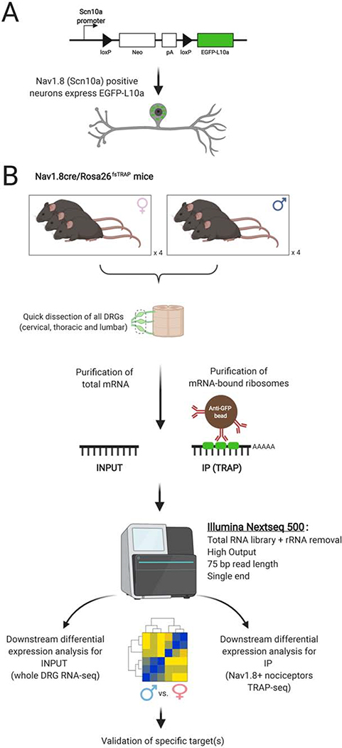
Methods: We used translating ribosome affinity purification sequencing and behavioral pharmacology to test the hypothesis that in Nav1.8-positive neurons, most of which are nociceptors, translatomes would differ by sex.

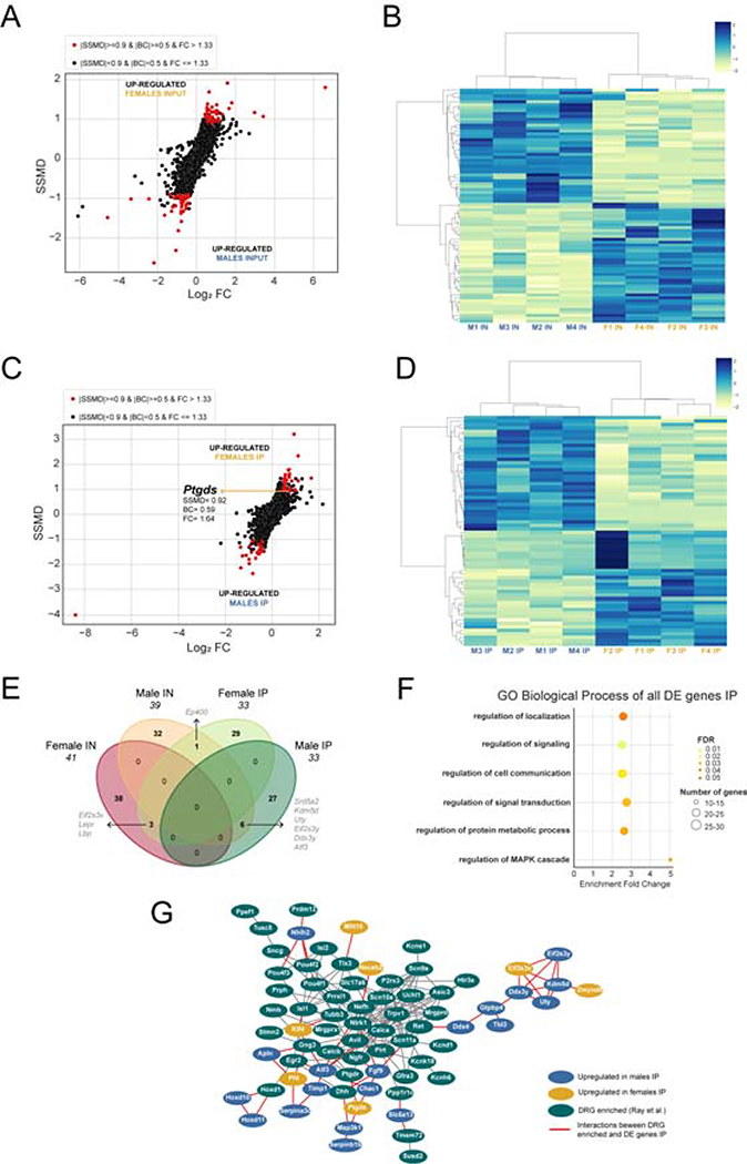
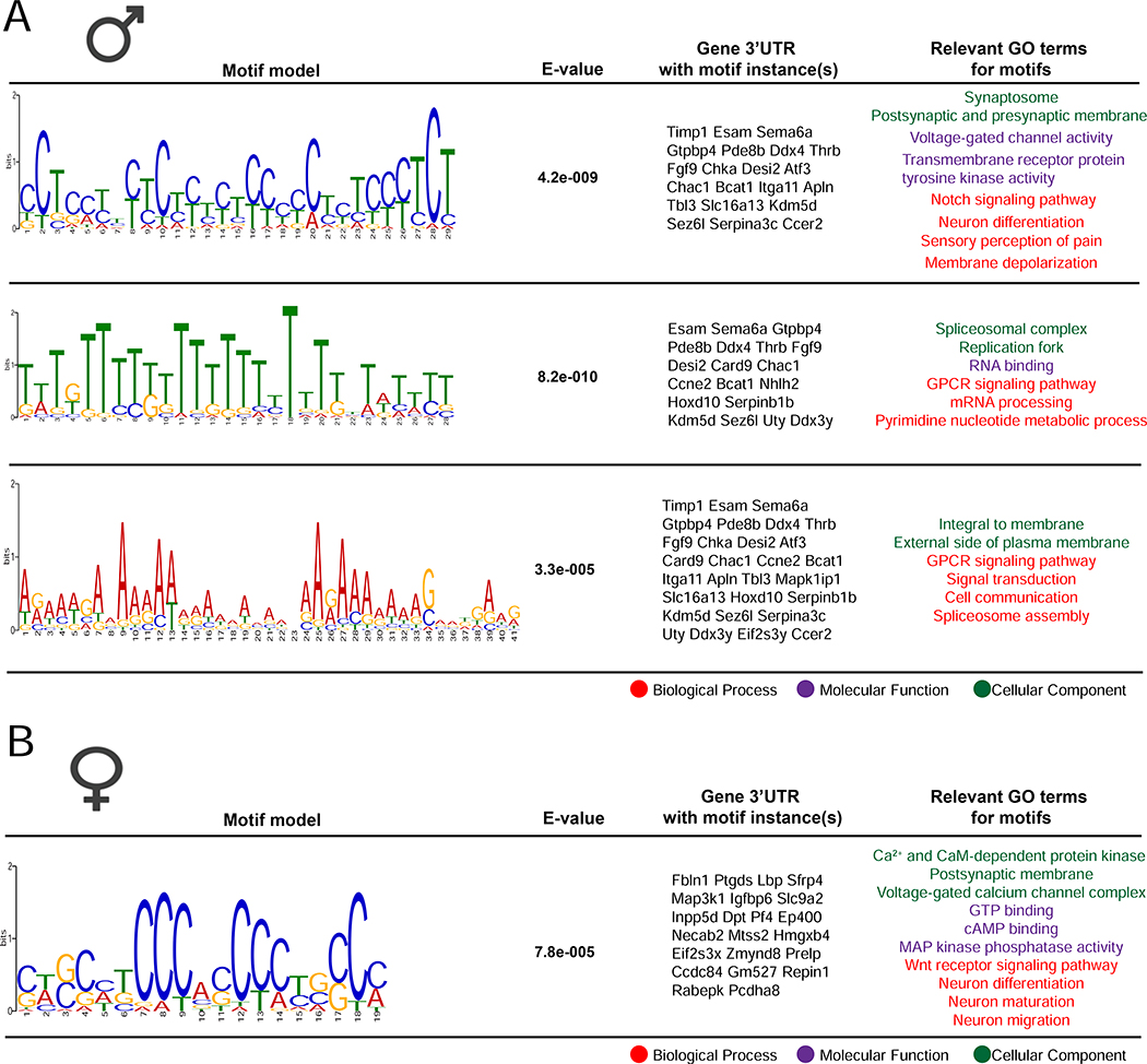
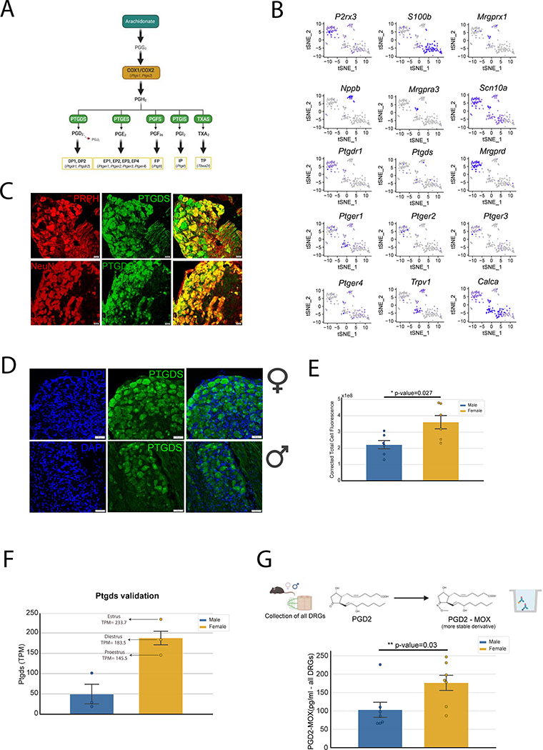
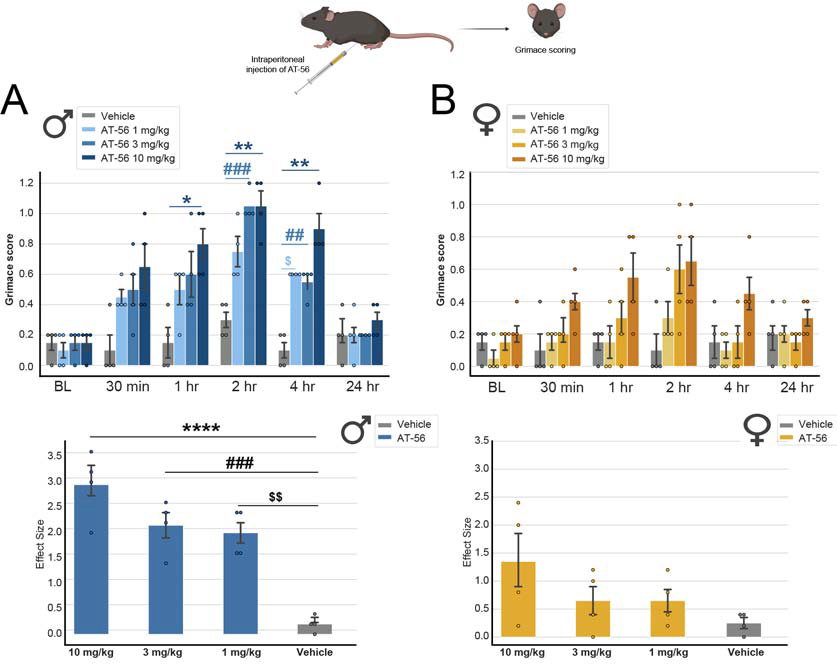
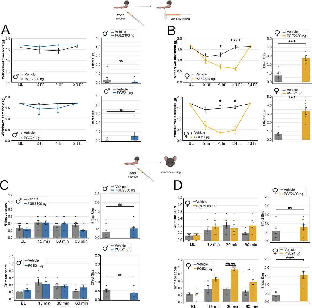
Results: We found 80 genes with sex differential expression in the whole DRG transcriptome and 66 genes whose messenger RNAs were sex differentially actively translated (translatome). We also identified different motifs in the 3' untranslated region of messenger RNAs that were sex differentially translated. In further validation studies, we focused on Ptgds, which was increased in the translatome of female mice. The messenger RNA encodes the prostaglandin PGD2 synthesizing enzyme. We observed increased PTGDS protein and PGD2 in female mouse DRG. The PTGDS inhibitor AT-56 caused intense pain behaviors in male mice but was only effective at high doses in female mice. Conversely, female mice responded more robustly to another major prostaglandin, PGE2, than did male mice. PTGDS protein expression was also higher in female cortical neurons, suggesting that DRG findings may be generalizable to other nervous system structures.

Conclusions: Our results demonstrate sex differences in nociceptor-enriched translatomes and reveal unexpected sex differences in one of the oldest known nociceptive signaling molecule families, the prostaglandins.

Keywords: Nociceptor; PGD2; PGE2; PTGDS; Pain; Prostaglandins; Sex differences; Translating ribosome affinity purification.


Figures

Figure 1:
Figure 1:. Outline of workflow for TRAP sequencing to reveal sex differences in nociceptor translatomes.
A, eGFP-L10a protein is expressed in Nav1.8-positive nociceptors. B, Schematic representation of the methodology shows dissection of all DRGs (cervical, thoracic and lumbar) from Nav1.8cre/Rosa26fsTRAP mice followed by isolation of total RNA (INPUT), and mRNA-bound to the ribosome (IP) using anti-eGFP-coated beads; Samples were sequenced and processed for downstream analysis of differentially expressed genes as shown.
Figure 2:
Figure 2:. Nociceptor TRAP sequencing quality control.
A, Hierarchical clustering analysis and B, Heatmap of the correlation coefficient show clear separation between TRAP-seq and bulk RNA-seq. However, we did not observe a clear distinction between male and female samples. C, Linear correlation plots shows high correlation coefficients of gene TPMs within biological replicates for the INPUT and IP fractions (shown for 2 replicates in each sex and assay), suggesting high reproducibility between replicates. D, Neuronal markers were enriched in IP fractions, such as Calca (encoding CGRP) or Prph (peripherin), while glial markers such as Mbp, Mpz, and Gfap were depleted (based on fold changes of median TPMs in each assay). E, F, The empirically estimated probability density of the raw TPMs and quantile normalized TPM (qTPM) distributions for the INPUT and IP fractions of all samples are shown. For INPUT samples, TPM distributions are shown for all coding genes and qTPMs shown for systematically transcriptome-expressed genes. For IP samples, TPM distributions were plotted for all coding genes, and qTPMs are shown for systematically translatome-expressed genes.
Figure 3:
Figure 3:. Differentially translated mRNAs in male and female DRG nociceptors.
A, Dual-flashlight plot of INPUT samples showing SSMD and Log2 fold change values for all autosomal genes on or above the 30th percentile. B, Heatmap shows the z-scores of the differentially expressed genes in INPUT (IN) samples. Labels represent sex and biological replicate number. C, Dual-flashlight plot of IP samples showing SSMD and Log2 fold change values for all autosomal genes on or above the 15th percentile. D, Heatmap shows the z-scores of the differentially translated mRNAs in IP samples. E, Venn diagram comparing the genes identified as differentially expressed. There were few overlaps between INPUT and IP autosomal genes. F, GO terms enriched for all genes differentially expressed in IP. G, Network of interactions between genes differentially translated between males and females in IP and genes enriched in DRG neurons (Network generated using String database and Cytoscape).
Figure 4:
Figure 4:. Enriched motifs identified in the 3’ UTRs of mRNAs differentially translated in males or females.
The motif analysis was conducted on the list of up-regulated mRNAs in both male and female IP fractions. A, We found 3 motifs significantly enriched in the 3’ UTR of up-regulated male mRNAs compared to the female mRNAs. B, We identified one motif significantly enriched in the 3’ UTR of up-regulated female mRNAs compared to the male mRNAs. This motif is involved in several neuronal functions and it is present in multiple mRNAs, such as Ptgds.
Figure 5:
Figure 5:. Ptgds expression is higher in female DRGs and leads to higher production of PGD2.
A, Ptgds converts PGH2 to PGD2, which has a very short half-life and is rapidly metabolized to PGJ2. B, Single DRG neuron-sequencing shows that Ptgds and PGD2 receptor DP1 (Ptgdr1) are expressed in neurons in the DRG. Ptgds is co-expressed with most neuronal markers, suggesting that it is expressed in all neurons; Ptgdr1 is mostly expressed in non-peptidergic neurons (co-expressed with P2rx3); PGE2 receptors (Ptger1, Ptger2, Ptger3, Ptger4) were expressed by most neuronal subtypes. C, We confirmed using IHC that Ptgds is expressed in neurons. D, E, We found that Ptgds has higher expression in female DRG neurons compared to male DRGs, at the protein level (Unpaired t-test, t = 2.584, df = 10, p-value = 0.0272). F, In a separate TRAP experiment, we monitored the female estrous cycle and validated that Ptgds is higher in females compared to females regardless of estrous cycle. G, PGD2-MOX ELISA demonstrated that PGD2 levels are higher in female DRGs (Unpaired t-test, t = 2.381, df = 12, p-value = 0.0347). Panel C scale bar = 20 μm; Panel D scale bar = 50 μm.
Figure 6:
Figure 6:. Inhibition of Ptgds produces robust grimacing behavior in mice that is greater in males.
A, Intraperitoneal injection of AT-56, a selective inhibitor of Ptgds, led to grimacing behavior in male mice (Two-way ANOVA, F = 4.279, p-value<0.0001, post-hoc Sidak’s, * Vehicle vs. AT-56 10 mg/kg male at 1h, p-value= 0.0314; ** Vehicle vs. AT-56 10 mg/kg male at 2h, p-value= 0.0071; *** Vehicle vs. AT-56 10 mg/kg male at 4h, p-value = 0.0068; ### Vehicle vs. AT-56 3 mg/kg male at 2h, p-value = 0.0004; ## Vehicle vs. AT-56 3 mg/kg male at 4h, p-value = 0.0068; $ Vehicle vs. AT-56 1 mg/kg male at 4h, p-value = 0.0193). We also calculated the effect size (difference from the baseline) and observed a significant difference for the 3 doses of AT-56 compared to vehicle in males (One way-ANOVA, F=22.00, p-value<0.0001, post-hoc Tukey’s; **** 10 mg/kg vs. Vehicle, p-value = <0.0001; **** 10 mg/kg vs. Vehicle p-value = <0.0001; ### 3 mg/kg vs. Vehicle p-value = 0.0006; $ $ 1 mg/kg vs. Vehicle p-value= 0.0012). B, Grimacing behavior in female mice following AT-56 injection was not different from vehicle (Two-way ANOVA, F = 1.136, p-value = 0.3462). When calculating the effect size (difference from the baseline), we did not observe any significant differences between groups in female mice (Two-way ANOVA, F = 2.109, p-value = 0.1524).
Figure 7:
Figure 7:. Intraplantar administration of PGE2 produces greater mechanical allodynia and grimacing in female mice.
A, Male mice did not respond to von Frey filaments after injection of 300 ng (Two-way ANOVA RM, F = 1.030, p-value = 0 .3900) or 1 μg (Two-way ANOVA RM, F = 0.7260, p-value = 0.5444) of PGE2. When calculating the effect size we did not observe any statistical significant differences between groups in males (Effect size 300 ng: Unpaired t-test, t = 0.8756, df = 13, p-value = 0.3971; Effect size 1 μg: Unpaired t-test, t = 0.8709, df = 10, p-value = 0.4042). B, Female mice showed mechanical allodynia up to 24 hours after injection of both 300 ng of PGE2 (Two-way ANOVA, F = 12.35, p-value <0.0001, post-hoc Sidak’s: * Vehicle - 300ng PGE2 at 4h, p-value = 0.0161, **** Vehicle - 300ng PGE2 at 24h, p-value <0.0001) and 1 μg of PGE2 (Two-way ANOVA RM, F = 10.78, p-value <0.0001, post-hoc Sidak’s: *** Vehicle - 1 μg PGE2 at 4h, p-value = 0.0005, ** Vehicle - 1 μg PGE2 at 24h, p-value = 0.0026). We also observed an effect size difference between groups in female mice (*** Effect size 300 ng: Unpaired t-test, t = 4.980, df = 13, p-value = 0.0003; *** Effect size 1 μg: Unpaired t-test, t = 5.569, df = 10, p-value = 0.0002). C, Male mice did not show any significant grimacing behaviors following administration of 300 ng of PGE2 (Two-way ANOVA RM, F = 1.996, p-value = 0.1305). At 1 μg of PGE2 we also did not observe any significant grimacing in male mice (Two-way ANOVA RM, F = 0.5376, p-value = 0.6601). Similarly, we also do not observe any effect size between groups in grimacing of male mice (Effect size 300 ng: Unpaired t-test, t = 0.9225, df = 13, p-value = 0.3731; Effect size 1 μg: Unpaired t-test, t = 0.8305, df = 10, p-value = 0.4257). D, Female mice exhibit robust grimacing after 1 μg PGE2 injection (Two-way ANOVA RM, F = 11.34, p-value <0.0001, post-hoc Sidak’s: **** Vehicle - 1ug PGE2 at 30 min, p-value<0.0001, * Vehicle - 1ug PGE2 at 60 min, p-value = 0.0257) but not at 300 ng (Two-way ANOVA RM, F = 1.197, p-value = 0.1303). We also observed an effect size in the grimacing scores between 1 μg PGE2 and vehicle grimacing in female mice (Effect size 300 ng: Unpaired t-test, t = 0.9458, df = 13, p-value = 0.3615; *** Effect size 1 μg: Unpaired t-test, t = 4.669, df = 10, p-value = 0.0009).

References

    1. Beery AK, Zucker I (2011): Sex bias in neuroscience and biomedical research. Neuroscience & Biobehavioral Reviews. 35:565–572. - PMC - PubMed
    1. Mendrek A, Mancini-Marïe A (2016): Sex/gender differences in the brain and cognition in schizophrenia. Neurosci Biobehav Rev. 67:57–78. - PubMed
    1. Nawka A, Kalisova L, Raboch J, Giacco D, Cihal L, Onchev G, et al. (2013): Gender differences in coerced patients with schizophrenia. BMC Psychiatry. 13:257–257. - PMC - PubMed
    1. Baldereschi M, Di Carlo A, Rocca WA, Vanni P, Maggi S, Perissinotto E, et al. (2000): Parkinson’s disease and parkinsonism in a longitudinal study: two-fold higher incidence in men. ILSA Working Group. Italian Longitudinal Study on Aging. Neurology. 55:1358–1363. - PubMed
    1. Georgiev D, Hamberg K, Hariz M, Forsgren L, Hariz GM (2017): Gender differences in Parkinson’s disease: A clinical perspective. Acta Neurol Scand. 136:570–584. - PubMed

Publication types