Review
. 2021 Apr;27(4):314-331.
doi: 10.1016/j.molmed.2020.11.006. Epub 2020 Dec 10.

Permeability of the Endothelial Barrier: Identifying and Reconciling Controversies

Affiliations
Review

Permeability of the Endothelial Barrier: Identifying and Reconciling Controversies

Lena Claesson-Welsh et al. Trends Mol Med. 2021 Apr.

Abstract

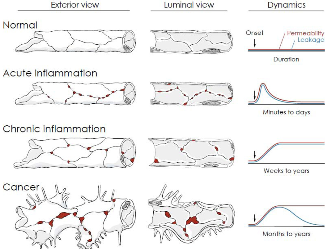
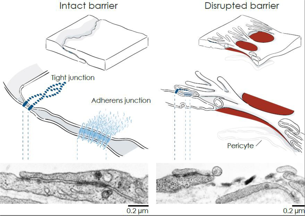
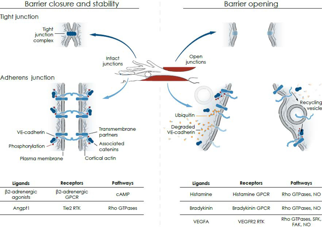
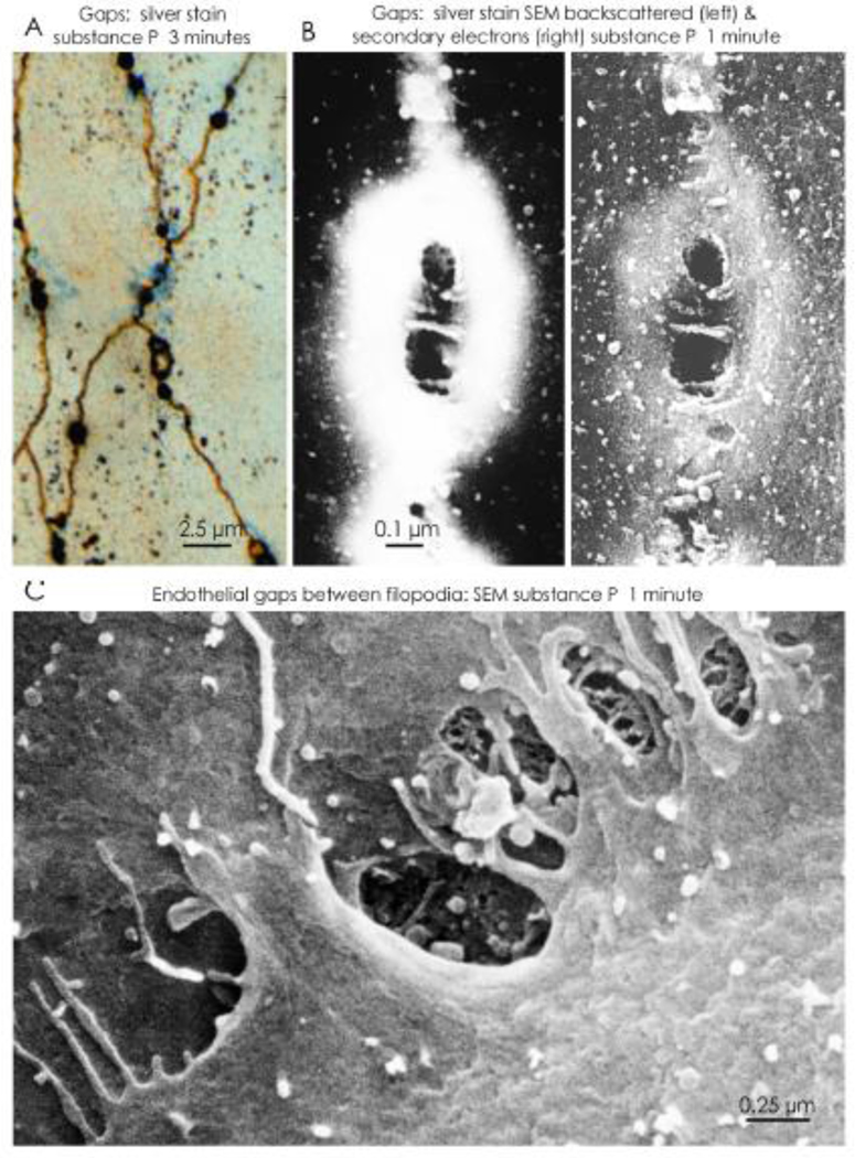
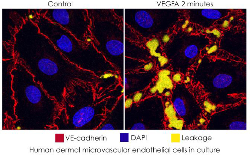
Leakage from blood vessels into tissues is governed by mechanisms that control endothelial barrier function to maintain homeostasis. Dysregulated endothelial permeability contributes to many conditions and can influence disease morbidity and treatment. Diverse approaches used to study endothelial permeability have yielded a wealth of valuable insights. Yet, ongoing questions, technical challenges, and unresolved controversies relating to the mechanisms and relative contributions of barrier regulation, transendothelial sieving, and transport of fluid, solutes, and particulates complicate interpretations in the context of vascular physiology and pathophysiology. Here, we describe recent in vivo findings and other advances in understanding endothelial barrier function with the goal of identifying and reconciling controversies over cellular and molecular processes that regulate the vascular barrier in health and disease.

Keywords: G protein-coupled receptors; Rho GTPases; VEGF receptors; endothelial barrier function; endothelial cell junctions; gap formation; postcapillary venules.


Conflict of interest statement

Conflicts of interest: The authors have no conflicting financial interests.

Figures

Figure 1.
Figure 1.. Vascular permeability and leakage in health and disease.
Comparison of exterior and luminal views of blood vessels of the microcirculation and corresponding dynamics of permeability and leakage over time under normal conditions, inflammation, and cancer. Durations are approximate values reflecting the vasculature of the entire organ rather than individual vessels or gaps. In normal blood vessels, junctions between endothelial cells form a uniform barrier that restricts extravasation of plasma. Gaps are not present between endothelial cells, and permeability and leakage are stable and low. In acute inflammation, leakage occurs as permeability increases rapidly through formation of focal endothelial gaps (red) in postcapillary venules. The gaps are transient, and vessels return to normal after the inflammatory stimulus ends or is inactivated. In chronic inflammation, vessels undergo structural remodeling, reflected by enlargement, proliferation (angiogenesis), increased mediator sensitivity, and sustained gap formation and leakage. The vascular changes do not resolve spontaneously but can be reversed by treatment of the chronic condition. In cancer, blood vessels that supply growing tumors undergo distinctive changes promoted by the abnormal tumor microenvironment, including sprouting angiogenesis, abnormal growth and gene expression, and defective endothelial junctions that result in leakage of plasma and even erythrocytes in some tumors. Increased endothelial permeability is sustained, but leakage decreases as interstitial pressure rises because of impaired lymphatic drainage.
Figure 2.
Figure 2.. Comparison of endothelial junctions at intact and disrupted barriers.
Left: Drawings of the border of normal overlapping endothelial cells (upper) showing the relative locations of the tight junction and adherens junction (middle). Corresponding transmission electron micrograph (TEM) of a normal venule showing overlapping endothelial cell borders with electron-dense junctions and underlying basement membrane (lower). Locations of the tight junction near the lumen (apical) and adherens junction (basolateral) are marked by dashed lines. Right: Disrupted endothelial barrier with focal gaps (red) after exposure to a mediator that increased vascular permeability (upper). Cell connections are maintained by junctions at the tip of finger-like filopodia that extend from one endothelial cell to the adjacent cell (middle). Corresponding TEM of an endothelial gap (lower). Endothelial cells bordering the gap have filopodia on the luminal surface, some with intact intercellular junctions. Extravasated particles of the tracer Monastral blue (black crystals in the center) are trapped in basement membrane exposed by the gap. Basement membrane underlies the endothelial cells and surrounds a pericyte process on the right. TEM images from rat trachea at baseline (left) and after electrical stimulation of vagus nerve for 5 minutes to activate substance P release from sensory axons (right) [131].
Figure 3.
Figure 3.. Mechanisms of barrier opening and closure.
Center: Drawing based on Figure 2 showing intact junctions (blue) between finger-like endothelial filopodia and an adjacent endothelial cell (left, Intact junctions) and a gap (red) between the same endothelial cells (right, Open junctions). Left arrows point to corresponding regions of intact tight junctions and adherens junctions; right arrows point to open junctions. Left: Intact endothelial cell barrier formed by homophilic interactions of tight junction complexes (upper) and adherens junction proteins (lower) at endothelial cell borders. At tight junctions, the fused outer leaflets of plasma membranes of adjacent cells create a diffusion barrier. At adherens junctions, VE-cadherin is in complex with catenins and other transmembrane proteins that provide structural attachments. Tyrosine residues of the intracellular domain of VE-cadherin have a low level of flow-dependent phosphorylation (few red dots) in endothelial cells of postcapillary venules at baseline. Right: Open barrier at endothelial gap resulting from changes in tight junctions (upper), which must still be defined, and adherens junctions (lower), where tyrosine phosphorylation of VE-cadherin increases (many red dots) followed by ubiquitination (orange dots), dismantling of complexes and degradation or internalization into vesicles and recycling back to the plasma membrane where complexes and tyrosine phosphorylation are restored. Bottom table: Ligands, receptors, and pathways implicated in barrier closure and stability (left) and barrier opening (right): Angpt1, angiopoietin 1; cAMP, cyclic adenosine monophosphate; FAK, focal adhesion kinase; GPCR, GTPase protein coupled receptor; NO, nitric oxide; RTK, receptor tyrosine kinase; SFK, Src family kinase; VEGFA, vascular endothelial growth factor.
Figure 4.
Figure 4.. Endothelial gap location and structure.
Distribution and appearance of endothelial gaps in tracheal venules viewed by light microscopy and scanning electron microscopy (SEM) soon after intravenous (iv) injection of the proinflammatory peptide substance P. A. Endothelial gaps are seen as black dots along intercellular junctions (brown lines) in the endothelium of a postcapillary venule stained with silver nitrate 3 minutes after substance P. Leakage of the particulate tracer Monastral blue appears as faint blue staining (from [134]). B. SEM views of endothelial gaps stained with silver nitrate 1 minute after substance P. Silver nitrate appears as a silver annulus (white) around the central gaps (black), which are bridged by endothelial cell filopodia. The gap is visualized by detection of backscattered electrons (left) and secondary electrons (right) (from [136]). C. SEM view of gaps bordered by filopodia at the luminal rim of endothelial cells in a venule 1 minute after iv injection of substance P. Tips of filopodia, reflecting changes in the cortical actin cytoskeleton, maintain contacts between the adjacent endothelial cells. Fibrous basement membrane is visible through some gaps (from [137]).
Figure I box 1
Figure I box 1
compares control and VEGFA-stimulated microvascular endothelial cells, where leakage is detected by fluorescent streptavidin (yellow) bound to biotin in the substrate, in an in vitro vascular permeability imaging assay (Merck KGaA, Darmstadt). Separations of junctions in cultured endothelial cells exposed to VEGFA are larger and more numerous than occur in vivo.

References

    1. Stan RV (2006) Channels across endothelial cells. In Cell-Cell Channels (Baluska F. et al. eds), pp. 271–278, Landes Bioscience, Georgetown, TX, USA.
    1. Ayloo S. and Gu C. (2019) Transcytosis at the blood-brain barrier. Curr Opin Neurobiol 57, 32–38. - PMC - PubMed
    1. Michel CC (1997) Starling: the formulation of his hypothesis of microvascular fluid exchange and its significance after 100 years. Exp Physiol 82 (1), 1–30. - PubMed
    1. Palade GE et al. (1979) Structural aspects of the permeability of the microvascular endothelium. Acta Physiol Scand Suppl 463, 11–32. - PubMed
    1. Simionescu M. and Simionescu N. (1984) Ultrastructure of the microvascular wall: functional correlations. Chapter 3. In Handbook of Physiology (Renkin EM and Michel CC eds), pp. 41–101, American Physiological Society.

Publication types

MeSH terms

Substances