. 2021 Jan 31:168:272-278.
doi: 10.1016/j.ijbiomac.2020.12.038. Epub 2020 Dec 9.

Ritonavir may inhibit exoribonuclease activity of nsp14 from the SARS-CoV-2 virus and potentiate the activity of chain terminating drugs

Affiliations

Ritonavir may inhibit exoribonuclease activity of nsp14 from the SARS-CoV-2 virus and potentiate the activity of chain terminating drugs

Naveen Narayanan et al. Int J Biol Macromol. .

Abstract

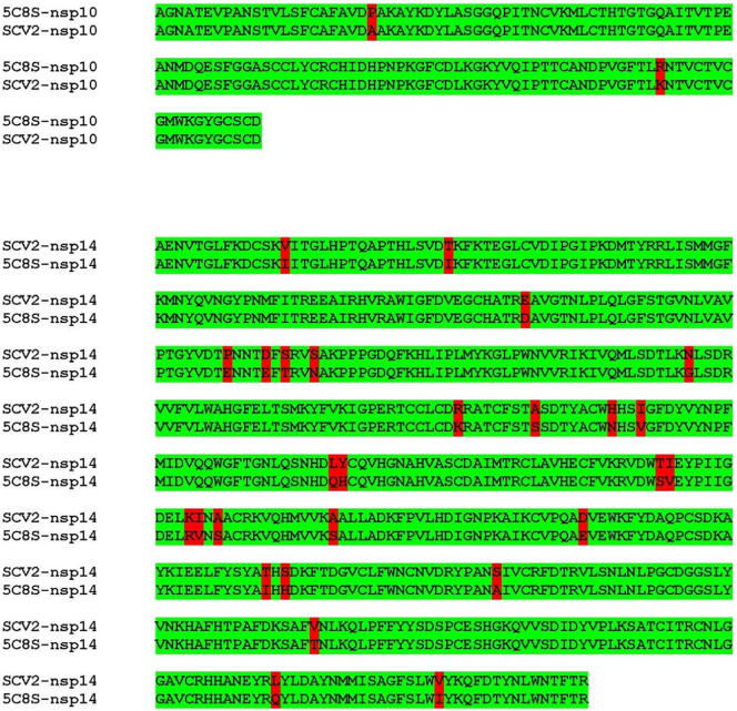
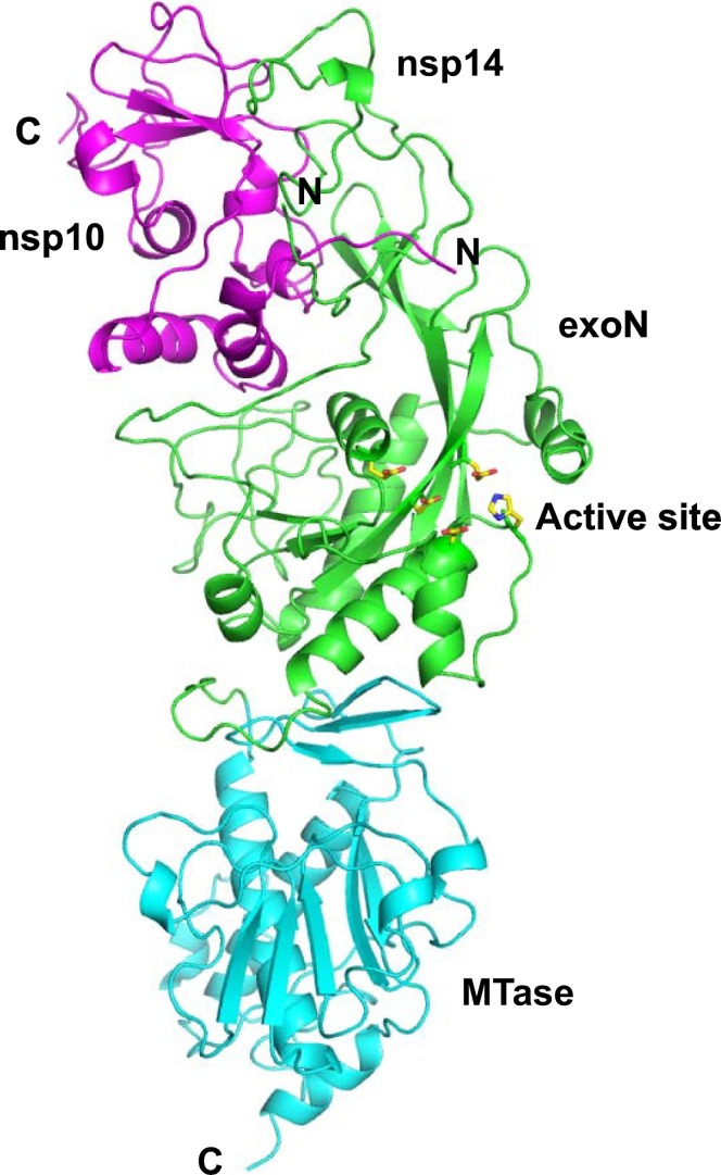
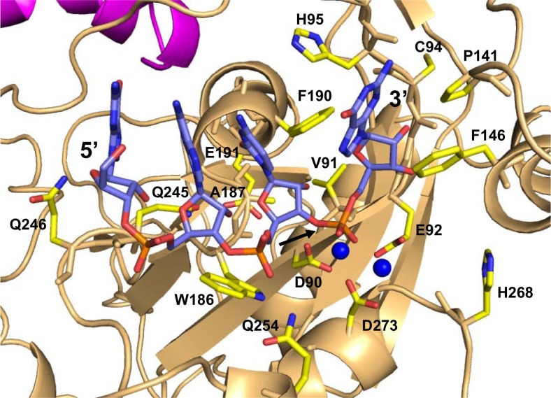
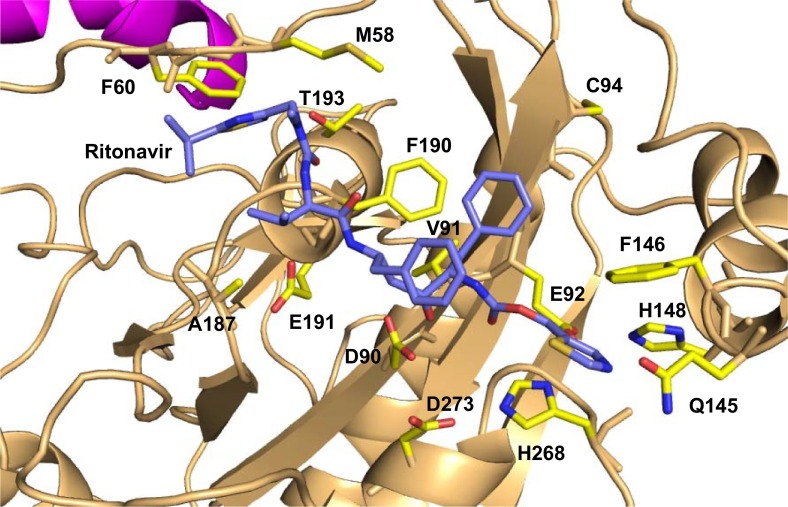
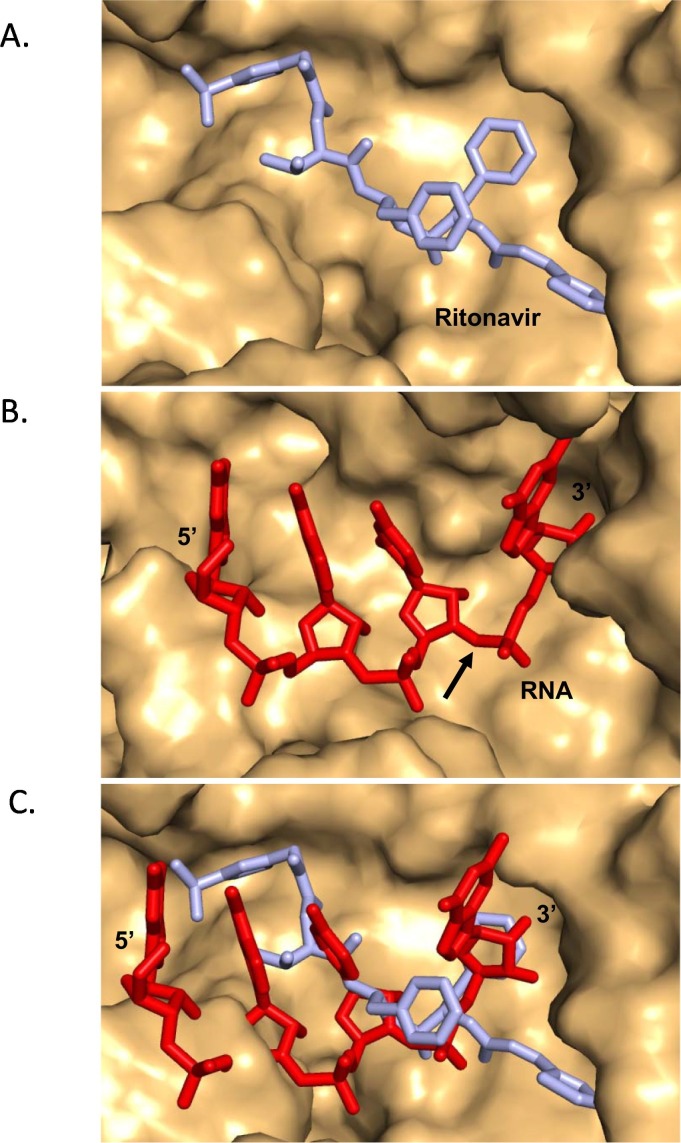
SARS-CoV-2is the causative agent for the ongoing COVID19 pandemic, and this virus belongs to the Coronaviridae family. The nsp14 protein of SARS-CoV-2 houses a 3' to 5' exoribonuclease activity responsible for removing mismatches that arise during genome duplication. A homology model of nsp10-nsp14 complex was used to carry out in silico screening to identify molecules among natural products, or FDA approved drugs that can potentially inhibit the activity of nsp14. This exercise showed that ritonavir might bind to the exoribonuclease active site of the nsp14 protein. A model of the SARS-CoV-2-nsp10-nsp14 complex bound to substrate RNA showed that the ritonavir binding site overlaps with that of the 3' nucleotide of substrate RNA. A comparison of the calculated energies of binding for RNA and ritonavir suggested that the drug may bind to the active site of nsp14 with significant affinity. It is, therefore, possible that ritonavir may prevent association with substrate RNA and thus inhibit the exoribonuclease activity of nsp14. Overall, our computational studies suggest that ritonavir may serve as an effective inhibitor of the nsp14 protein. nsp14 is known to attenuate the inhibitory effect of drugs that function through premature termination of viral genome replication. Hence, ritonavir may potentiate the therapeutic properties of drugs such as remdesivir, favipiravir and ribavirin.

Keywords: Exoribonuclease; Inhibitor; Ritonavir; SARS-CoV-2; nsp14.


Figures

Fig. 1
Fig. 1
Alignment of the sequence of SARS-CoV-2-nsp10 and SARS-CoV-2-nsp14 with the sequence of the available structure of SARS-nsp10-nsp14 complex (5C8S). The sequence of the two proteins from SARS-CoV-2sequence exhibits about 97% identity with the corresponding sequences from SARS. The differences in the alignment are highlighted in red.
Fig. 2
Fig. 2
Model of SARS-CoV-2-nsp10-nsp14 complex. The homology model is displayed here and the nsp10 chain is shown in magenta. The exoribonuclease (exoN) domain of nsp14 is coloured green and the methyltransferase (MTase) region is displayed in cyan. The N- and C-termini of the two chains are highlighted. The active site residues responsible for the exoribonuclease activity are shown in stick representation and coloured according to element.
Fig. 3
Fig. 3
Model of the SARS-CoV-2-nsp10-nsp14 complex bound to RNA. nsp10 and nsp14 are coloured magenta and light orange, respectively. The RNA molecule and the interacting residues from nsp14 are shown in stick representation and coloured according to element. The cofactor ions are shown in the form of blue spheres. The site of cleavage is marked by an arrow.
Fig. 4
Fig. 4
Model of the SARS-CoV-2-nsp10-nsp14 complex bound to ritonavir. nsp10 and nsp14 are coloured magenta and light orange, respectively. The ritonavir molecule is shown in stick representation and coloured according to element (carbon = blue). The residues of nsp14 that interact with ritonavir are shown in stick representation (carbon = yellow).
Fig. 5
Fig. 5
Binding site of ritonavir overlaps with that of substrate RNA. (A) SARS-CoV-2-nsp10-nsp14:ritonavir where the surface of protein molecule is displayed in light orange and ritonavir is displayed in stick representation and coloured cyan. (B) The surface of the protein molecule is displayed and the RNA, is displayed in stick representation and coloured red. The site of cleavage on RNA is marked by an arrow. (C) Superimposition of the models of functional ternary complex (red) and that of SARS-CoV-2-nsp10-nsp14:ritonavir (cyan) is displayed. The comparison suggests that the presence of ritonavir may prevent binding of the natural RNA substrate.

References

    1. Johns Hopkins Coronavirus Resource Center, (n.d.).
    1. Vanden Eynde J.J. 2020. Covid-19: a brief overview of the discovery clinical trial, Pharmaceuticals. - DOI - PMC - PubMed
    1. Wu F., Zhao S., Yu B., Chen Y.-M., Wang W., Song Z.-G., Hu Y., Tao Z.-W., Tian J.-H., Pei Y.-Y., Yuan M.-L., Zhang Y.-L., Dai F.-H., Liu Y., Wang Q.-M., Zheng J.-J., Xu L., Holmes E.C., Zhang Y.-Z. 2020. A new coronavirus associated with human respiratory disease in China, Nature. - DOI - PMC - PubMed
    1. Shang W., Yang Y., Rao Y., Rao X. The outbreak of SARS-CoV-2 pneumonia calls for viral vaccines. Npj Vaccines. 2020;5:18. doi: 10.1038/s41541-020-0170-0. - DOI - PMC - PubMed
    1. Michel C.J., Mayer C., Poch O., Thompson J.D. Characterization of accessory genes in coronavirus genomes. Virol. J. 2020;17:131. doi: 10.1186/s12985-020-01402-1. - DOI - PMC - PubMed

MeSH terms

LinkOut - more resources