Multicenter Study
. 2021 May:225:95-107.
doi: 10.1016/j.ajo.2020.11.022. Epub 2020 Dec 11.

KCNV2-Associated Retinopathy: Genetics, Electrophysiology, and Clinical Course-KCNV2 Study Group Report 1

Affiliations
Multicenter Study

KCNV2-Associated Retinopathy: Genetics, Electrophysiology, and Clinical Course-KCNV2 Study Group Report 1

Michalis Georgiou et al. Am J Ophthalmol. 2021 May.

Abstract

Purpose: To investigate genetics, electrophysiology, and clinical course of KCNV2-associated retinopathy in a cohort of children and adults.

Study design: This was a multicenter international clinical cohort study.

Methods: Review of clinical notes and molecular genetic testing. Full-field electroretinography (ERG) recordings, incorporating the international standards, were reviewed and quantified and compared with age and recordings from control subjects.

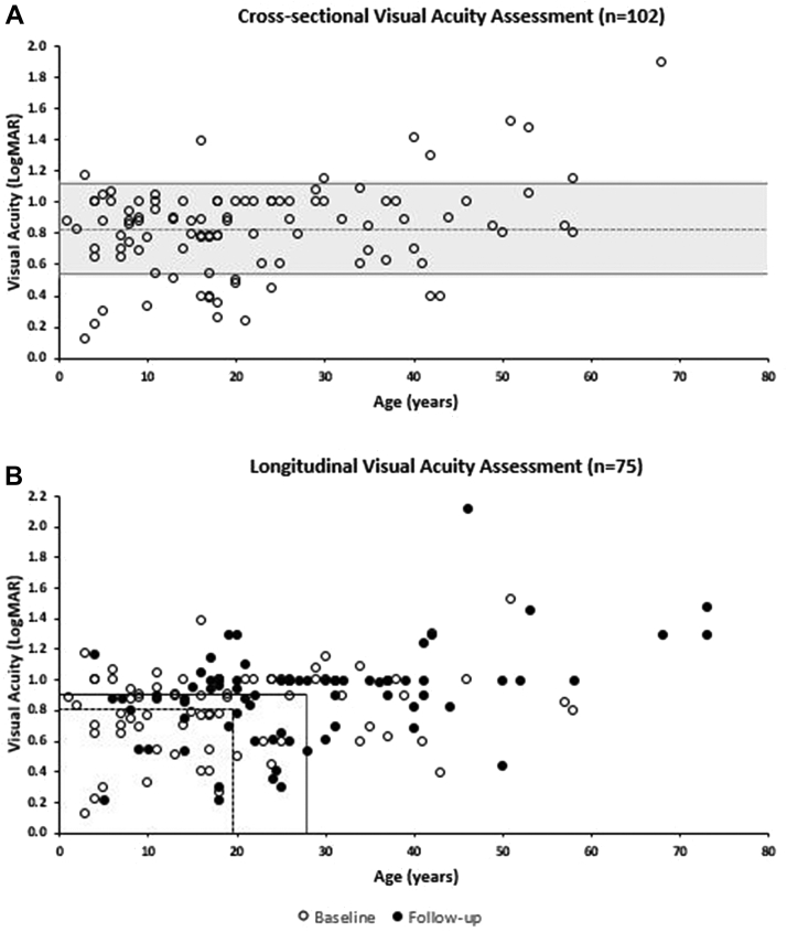
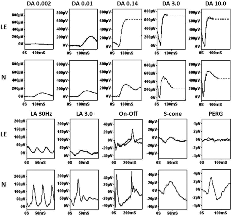
Results: In total, 230 disease-associated alleles were identified from 117 patients, corresponding to 75 different KCNV2 variants, with 28 being novel. The mean age of onset was 3.9 years old. All patients were symptomatic before 12 years of age (range, 0-11 years). Decreased visual acuity was present in all patients, and 4 other symptoms were common: reduced color vision (78.6%), photophobia (53.5%), nyctalopia (43.6%), and nystagmus (38.6%). After a mean follow-up of 8.4 years, the mean best-corrected visual acuity (BCVA ± SD) decreased from 0.81 ± 0.27 to 0.90 ± 0.31 logarithm of minimal angle of resolution. Full-field ERGs showed pathognomonic waveform features. Quantitative assessment revealed a wide range of ERG amplitudes and peak times, with a mean rate of age-associated reduction indistinguishable from the control group. Mean amplitude reductions for the dark-adapted 0.01 ERG, dark-adapted 10 ERG a-wave, and LA 3.0 30 Hz and LA3 ERG b-waves were 55%, 21%, 48%, and 74%, respectively compared with control values. Peak times showed stability across 6 decades.

Conclusion: In KCNV2-associated retinopathy, full-field ERGs are diagnostic and consistent with largely stable peripheral retinal dysfunction. Report 1 highlights the severity of the clinical phenotype and established a large cohort of patients, emphasizing the unmet need for trials of novel therapeutics.


Figures

Figure 1
Figure 1
KCNV2 protein and domains. The schematic diagram shows the KCNV2 encoded protein structure, the alpha-subunit of the potassium channel (Kv8.2), and its domains. It consists of 1) a highly conserved tetramerization domain; N-terminal A and B box (NAB); 2) 6 transmembrane domains (S1-S6); 3) extracellular and intracellular loop segments; and 4) an ultraconserved potassium selective motif in the pore-forming loop between S5 and S6 (P loop). The distribution of the missense variants identified are detailed in the results section.
Figure 2
Figure 2
Best-corrected visual acuity (BCVA) assessment. A. Cross-sectional assessment of BCVA based on data from 101 patients. The dashed line marks the mean of the cohort (0.83 logarithm of minimal angle of resolution [logMAR]). More than two-thirds of the patients (68.2%) had a BCVA between 0.54 and 1.12 logMAR (shadowed area between lines marking ± SD). B. Longitudinal assessment of BCVA on data from 75 patients. The dashed lines mark the mean age and the mean BCVA at baseline—19.5 years and 0.81 logMAR, respectively. The continuous lines mark the mean age and mean BCVA at follow-up—27.9 years and 0.90 logMAR, respectively. After a mean follow-up of 8.4 years, the mean BCVA (±SD) decreased by 0.09 logMAR. The annual rate for the cohort was 0.011 logMAR per year.
Figure 3
Figure 3
Full-field electroretinography (ERG) and pattern ERG recordings in a case of KCNV2-associated retinopathy. The dark-adapted (DA) responses (top panels) show the pathognomonic features. To the dimmest flash the (DA 0.002) ERG was undetectable and the DA0.01 ERG delayed and subnormal. As flash strength increased up to 3 cd.s.m−2, there was abnormal increased enlargement of the ERG. The DA 10 (strong flash) ERG a-wave trough had a characteristic broad shape with a late negative component and the b-wave was disproportionately large relative to the attenuated dim flash responses. Light-adapted (LA) 30-Hz flicker (LA 30 Hz) and single flash cone (LA3) ERGs were delayed and subnormal (bottom panels). The photopic on-off ERG (stimulus duration, 200 ms) showed a delayed and markedly reduced b-wave (an electronegative on-response) and delay and mild reduction of the d-wave (the off-response). The S-cone ERG was simplified and reduced. The pattern ERG P50 component was undetectable in keeping with severe macular dysfunction, typical of the disorder. Representative control recordings from an unaffected individual are shown for comparison (N). All patient recordings showed a high degree of interocular symmetry, are shown from 1 eye only, and are superimposed to demonstrate reproducibility, with the exception of the DA 0.14 ERG (single recording). Broken lines replace eye movement artefacts seen after the b-waves for clarity. LE = left eye, N = normal control, PERG = pattern ERG.
Figure 4
Figure 4
Scatter plots for electrophysiologic parameters and age. The major full-field electroretinography (ERG) component peak times and amplitudes in KCNV2 patients (filled circles) and unaffected control subjects (gray circles) are plotted against age. Data are shown for the dark-adapted (DA) 0.01 ERG (A and B), DA10.0 ERG a- and b-waves (C through F), light-adapted (LA) 30-Hz ERG (G and H), and for the amplitude of the LA 3 ERG a- and b-waves (I and J). Regression lines are shown for the KCNV2-associated retinopathy (solid line) and control (gray broken line) data. See text for details.

References

    1. Gouras P., Eggers H.M., MacKay C.J. Cone dystrophy, nyctalopia, and supernormal rod responses. A new retinal degeneration. Arch Ophthalmol. 1983;101(5):718–724. - PubMed
    1. Wu H., Cowing J.A., Michaelides M. Mutations in the gene KCNV2 encoding a voltage-gated potassium channel subunit cause “cone dystrophy with supernormal rod electroretinogram” in humans. Am J Hum Genet. 2006;79(3):574–579. - PMC - PubMed
    1. Czirjak G., Toth Z.E., Enyedi P. Characterization of the heteromeric potassium channel formed by kv2.1 and the retinal subunit kv8.2 in Xenopus oocytes. J Neurophysiol. 2007;98(3):1213–1222. - PubMed
    1. Gayet-Primo J., Yaeger D.B., Khanjian R.A., Puthussery T. Heteromeric KV2/KV8.2 channels mediate delayed rectifier potassium currents in primate photoreceptors. J Neurosci. 2018;38(14):3414–3427. - PMC - PubMed
    1. Gill J.S., Georgiou M., Kalitzeos A. Progressive cone and cone-rod dystrophies: clinical features, molecular genetics and prospects for therapy. Br J Ophthalmol. 2019;103(5):711–720. - PMC - PubMed

Publication types

MeSH terms

Substances

Supplementary concepts

LinkOut - more resources