. 2021 Feb:103:494-501.
doi: 10.1016/j.ijid.2020.12.013. Epub 2020 Dec 11.

Serological evidence indicates widespread distribution of rickettsioses in Myanmar

Affiliations

Serological evidence indicates widespread distribution of rickettsioses in Myanmar

Philip N D Elders et al. Int J Infect Dis. 2021 Feb.

Abstract

Background: Little research has been published on the prevalence of rickettsial infections in Myanmar. This study determined the seroprevalence of immunoglobulin G (IgG) antibodies to rickettsial species in different regions of Myanmar.

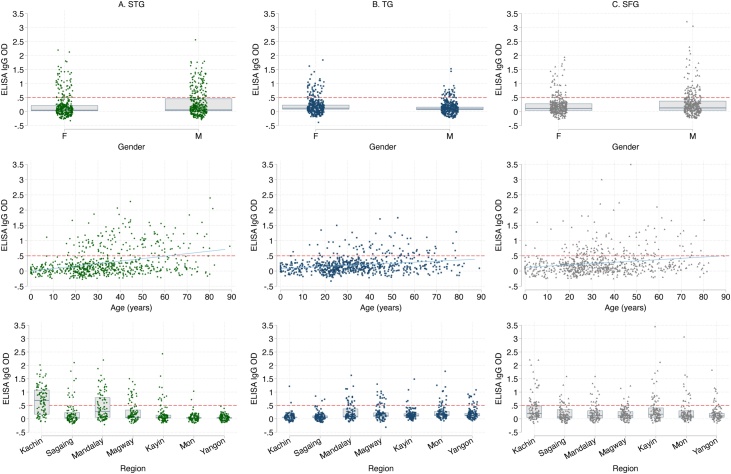
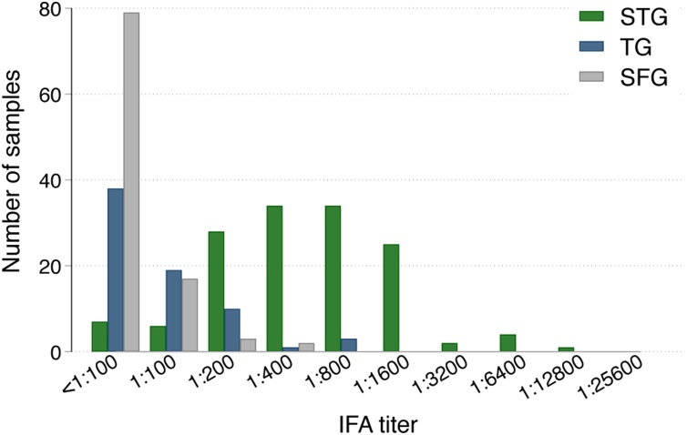
Methods: Seven hundred leftover blood samples from patients of all ages in primary care clinics and hospitals in seven regions of Myanmar were collected. Samples were screened for scrub typhus group (STG), typhus group (TG) and spotted fever group (SFG) IgG antibodies using enzyme-linked immunosorbent assays (ELISA). Immunofluorescence assays were performed for the same rickettsial groups to confirm seropositivity if ELISA optical density ≥0.5.

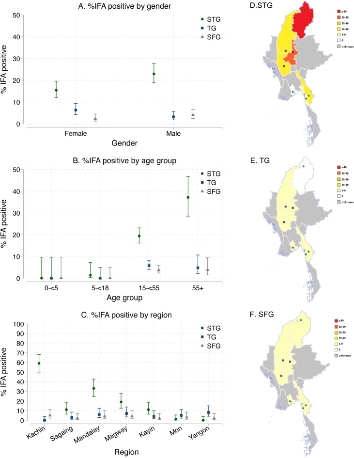
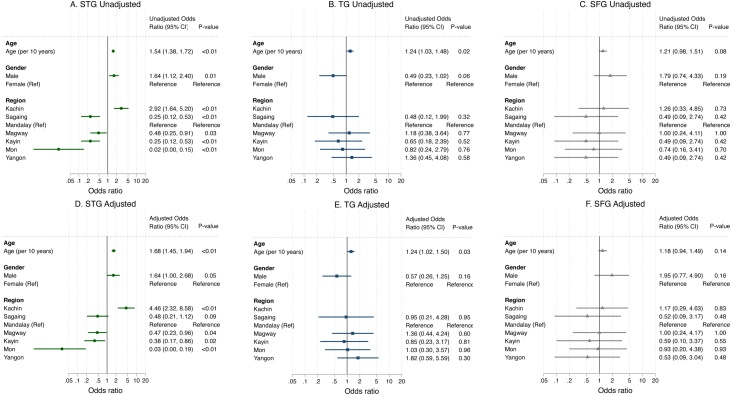
Results: Overall IgG seroprevalence was 19% [95% confidence interval (CI) 16-22%] for STG, 5% (95% CI 3-7%) for TG and 3% (95% CI: 2-5%) for SFG. The seroprevalence of STG was particularly high in northern and central Myanmar (59% and 19-33%, respectively). Increasing age was associated with higher odds of STG and TG seropositivity [per 10-year increase, adjusted odds ratio estimate 1.68 (p < 0.01) and 1.24 (p = 0.03), respectively].

Conclusion: Rickettsial infections are widespread in Myanmar, with particularly high seroprevalence of STG IgG antibodies in central and northern regions. Healthcare workers should consider rickettsial infections as common causes of fever in Myanmar.

Keywords: Murine typhus; Myanmar; Rickettsial infections; Scrub typhus; Seroprevalence; Spotted fever group.


Figures

Figure 1
Figure 1
Study sites. Blue squares, government hospitals; green circles, primary care clinics of Medical Action Myanmar, a medical non-governmental organization.
Figure 2
Figure 2
Optical densities (ODs) of enzyme-linked immunosorbent assays (ELISA) for each gender, age in years, and region for scrub typhus group (A), typhus group (B) and spotted fever group (C). Boxes denote quartiles (75th percentile, median, 25th percentile). The red dashed line indicates ELISA OD ≥ 0.5, which was used as the cut-off point for further testing with immunofluorescence assay. The blue line is a fitted linear regression line between age and ELISA IgG OD.
Figure 3
Figure 3
Immunofluorescence assay (IFA) titres of all samples with enzyme-linked immunosorbent assay optical density ≥0.5 for scrub typhus group (STG), typhus group (TG) and spotted fever group (SFG).
Figure 4
Figure 4
Percentages and 95 % confidence intervals of seropositive participants out of all participants displayed by gender (A), age group (B) and region (C) with maps (D, E, F) displaying the percentage in each region for scrub typhus group (STG), typhus group (TG) and spotted fever group (SFG), respectively. Seropositivity was defined as an immunofluorescence assay (IFA) titre ≥1:100 on samples with enzyme-linked immunosorbent assay (ELISA) optical density (OD) ≥ 0.5. All participants with ELISA OD < 0.5 were not tested with IFA, and were considered to be seronegative.
Figure 5
Figure 5
Unadjusted (A, B, C) and adjusted (D, E, F) odds ratios for seropositivity using logistic regression analysis. The variables that were entered were age per 10 years, gender (female as reference) and region (Mandalay as reference). Odds ratios with 95 % confidence intervals are displayed for scrub typhus group (STG), typhus group (TG) and spotted fever group (SFG). No participants were seropositive for STG in Yangon, and no participants were seropositive for TG in Kachin. Seropositivity was defined as immunofluorescence assay (IFA) titre ≥1:100 following enzyme-linked immunosorbent assay (ELISA) optical density (OD) ≥0.5. All participants with ELISA OD < 0.5 were not tested with IFA, and were considered to be seronegative.

References

    1. Abarca K., Martínez-Valdebenito C., Angulo J., Jiang J., Farris C.M., Richards A.L. Molecular description of a novel Orientia species causing scrub typhus in Chile. Emerg Infect Dis. 2020;26:2148–2156. - PMC - PubMed
    1. Acestor N., Cooksey R., Newton P.N., Ménard D., Guerin P.J., Nakagawa J. Mapping the aetiology of non-malarial febrile illness in Southeast Asia through a systematic review –terra incognita impairing treatment policies. PLoS One. 2012;7:e44269. - PMC - PubMed
    1. Althaus T., Thaipadungpanit J., Greer R.C., Swe M.M.M., Dittrich S., Peerawaranun P. Causes of fever in primary care in Southeast Asia and the performance of C-reactive protein in discriminating bacterial from viral pathogens. Int J Infect Dis. 2020;96:334–342. - PMC - PubMed
    1. Aung A.K., Spelman D.W., Murray R.J., Graves S. Rickettsial infections in Southeast Asia: implications for local populace and febrile returned travelers. Am J Trop Med Hyg. 2014;91:451–460. - PMC - PubMed
    1. Blacksell S.D., Bryant N.J., Paris D.H., Doust J.A., Sakoda Y., Day N.P.J. Scrub typhus serologic testing with the indirect immunofluorescence method as a diagnostic gold standard: a lack of consensus leads to a lot of confusion. Clin Infect Dis. 2007;44:391–401. - PubMed

LinkOut - more resources