

Detection of parvovirus B19 and human herpesvirus 6 in pediatric dilated cardiomyopathy: Impact after heart transplantation
- PMID: 33311918
- PMCID: PMC7727911
- DOI: 10.4103/apc.APC_124_19
Detection of parvovirus B19 and human herpesvirus 6 in pediatric dilated cardiomyopathy: Impact after heart transplantation
Abstract
Objectives: The aim of this study is to evaluate HHV-6 and PVB19 infection using polymerase chain reaction (PCR) and immunofluorescent assay (IFA) in the myocardium of pediatric patients with dilated cardiomyopathy (DCM) and the impact of viral persistence in the cardiac allograft after heart transplantation (HT).
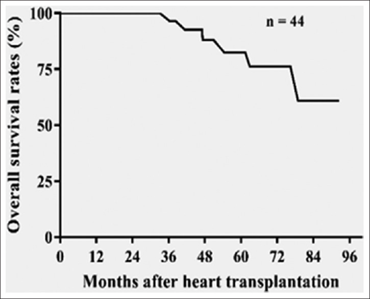
Methods: Multiplex droplet digital PCR was used to analyze the prevalence of viral sequences in myocardial samples from 48 pediatric DCM patients and 10 control subjects. Of the 48 DCM patients, 44 underwent HT. After HT, consecutive endomyocardial biopsy (EMB) samples were analyzed for the presence of PVB19 and HHV-6 antigens using IFA and the patients were evaluated for rejections, coronary vasculopathy, and graft loss.
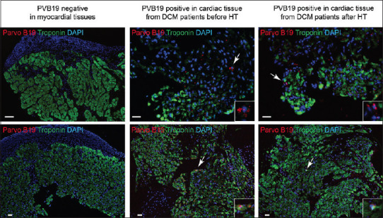
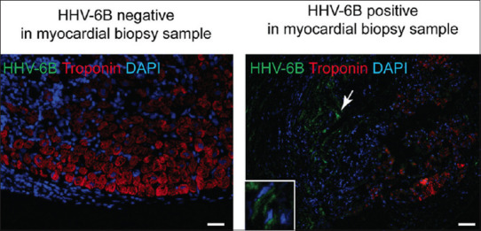
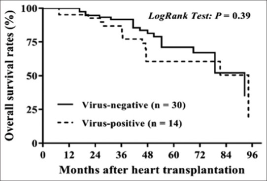
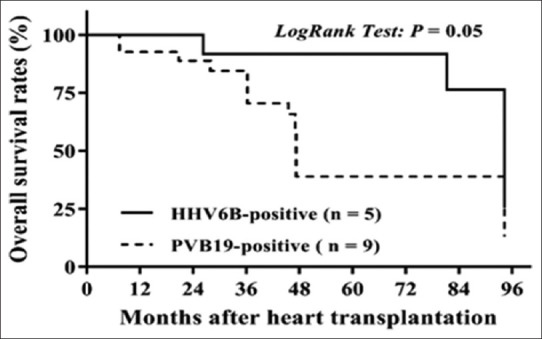
Results: Of the 48 DCM patients, 14 had positive viral PCR results in explanted/autopsy hearts. Among them, PVB19 was found in 8/48, HHV6 in 4/48, both PVB19 and HHV6 in 1/48, and enterovirus in one, but no adenovirus was found. The EMB samples obtained after HT were positive for PVB19 and HHV-6 in 7/44 and 3/44 cases, respectively. Viral presence in both the explanted heart and the cardiac allograft was demonstrated in 4 patients, 3 of whom were positive for PVB19, and one of whom was positive for HHV-6 pretransplant. Coronary vasculopathy and graft loss were more common in patients with PVB19-positive myocardial tissues versus those who were PVB19-negative.
Conclusions: There is an association between PVB19 and HHV-6 infection and DCM in children. The study suggests the persistence of PVB19 and HHV-6 in the host can lead to subsequent viral reactivation in the transplanted heart, even in those recipients who do not have active myocarditis. PVB19 in the cardiac allograft tended toward higher adverse post-HT events.
Keywords: Cardiotropic viruses; coronary vasculopathy; dilated cardiomyopathy; immunofluorescent assay; pediatric heart transplantation; polymerase chain reaction.
Copyright: © 2020 Annals of Pediatric Cardiology.
Conflict of interest statement
There are no conflict of interest.
Figures
Similar articles
-
Viral persistence in the myocardium is associated with progressive cardiac dysfunction.Circulation. 2005 Sep 27;112(13):1965-70. doi: 10.1161/CIRCULATIONAHA.105.548156. Epub 2005 Sep 19. Circulation. 2005. PMID: 16172268
-
Frequent detection of parvovirus B19 genome in the myocardium of adult patients with idiopathic dilated cardiomyopathy.Med Microbiol Immunol. 2004 May;193(2-3):75-82. doi: 10.1007/s00430-003-0211-0. Epub 2003 Dec 20. Med Microbiol Immunol. 2004. PMID: 14689308
-
Viral epidemiologic shift in inflammatory heart disease: the increasing involvement of parvovirus B19 in the myocardium of pediatric cardiac transplant patients.J Heart Lung Transplant. 2010 Jul;29(7):739-46. doi: 10.1016/j.healun.2010.03.003. Epub 2010 Apr 24. J Heart Lung Transplant. 2010. PMID: 20456978 Free PMC article.
-
Human herpesvirus 6-induced inflammatory cardiomyopathy in immunocompetent children.Ann Pediatr Cardiol. 2017 Sep-Dec;10(3):259-268. doi: 10.4103/apc.APC_54_17. Ann Pediatr Cardiol. 2017. PMID: 28928612 Free PMC article. Review.
-
Pathophysiology and aetiological diagnosis of inflammatory myocardial diseases with a special focus on parvovirus B19.J Vet Med B Infect Dis Vet Public Health. 2005 Sep-Oct;52(7-8):344-7. doi: 10.1111/j.1439-0450.2005.00873.x. J Vet Med B Infect Dis Vet Public Health. 2005. PMID: 16316398 Review.
Cited by
-
Molecular Mechanisms behind Persistent Presence of Parvovirus B19 in Human Dilated Myocardium.Adv Exp Med Biol. 2022;1376:181-202. doi: 10.1007/5584_2021_702. Adv Exp Med Biol. 2022. PMID: 35025080
-
Advancing Precision Medicine in Myocarditis: Current Status and Future Perspectives in Endomyocardial Biopsy-Based Diagnostics and Therapeutic Approaches.J Clin Med. 2023 Jul 31;12(15):5050. doi: 10.3390/jcm12155050. J Clin Med. 2023. PMID: 37568452 Free PMC article. Review.
-
Viral Myocarditis as a Factor Leading to the Development of Heart Failure Symptoms, Including the Role of Parvovirus B19 Infection-Systematic Review.Int J Mol Sci. 2024 Jul 25;25(15):8127. doi: 10.3390/ijms25158127. Int J Mol Sci. 2024. PMID: 39125696 Free PMC article.
-
Cardiovascular consequences of viral infections: from COVID to other viral diseases.Cardiovasc Res. 2021 Nov 22;117(13):2610-2623. doi: 10.1093/cvr/cvab315. Cardiovasc Res. 2021. PMID: 34609508 Free PMC article. Review.
-
Immune mechanisms of group B coxsackievirus induced viral myocarditis.Virulence. 2023 Dec;14(1):2180951. doi: 10.1080/21505594.2023.2180951. Virulence. 2023. PMID: 36827455 Free PMC article. Review.
References
-
- Kühl U, Pauschinger M, Seeberg B, Lassner D, Noutsias M, Poller W, et al. Viral persistence in the myocardium is associated with progressive cardiac dysfunction. Circulation. 2005;112:1965–70. - PubMed
-
- Bock CT, Klingel K, Kandolf R. Human parvovirus B19-associated myocarditis. N Engl J Med. 2010;362:1248–9. - PubMed
-
- Schowengerdt KO, Ni J, Denfield SW, Gajarski RJ, Bowles NE, Rosenthal G, et al. Association of parvovirus B19 genome in children with myocarditis and cardiac allograft rejection: Diagnosis using the polymerase chain reaction. Circulation. 1997;96:3549–54. - PubMed
LinkOut - more resources
Full Text Sources