. 2020 Dec 4:15:3213-3225.
doi: 10.2147/COPD.S276429. eCollection 2020.

LINC00987 Ameliorates COPD by Regulating LPS-Induced Cell Apoptosis, Oxidative Stress, Inflammation and Autophagy Through Let-7b-5p/SIRT1 Axis

Affiliations

LINC00987 Ameliorates COPD by Regulating LPS-Induced Cell Apoptosis, Oxidative Stress, Inflammation and Autophagy Through Let-7b-5p/SIRT1 Axis

Yuanyuan Wang et al. Int J Chron Obstruct Pulmon Dis. .

Abstract

Background: Chronic obstructive pulmonary disease (COPD) is the third cause of disease-related death and brings a heavy burden to human health. Long non-coding RNA (lncRNA) was revealed to participate in COPD pathogenesis. This study aims to establish the effects and regulatory mechanism of lncRNA long intergenic non-coding 00987 (LINC00987) in lipopolysaccharide (LPS)-induced apoptosis, oxidative stress, inflammation and autophagy in BEAS-2B cells.

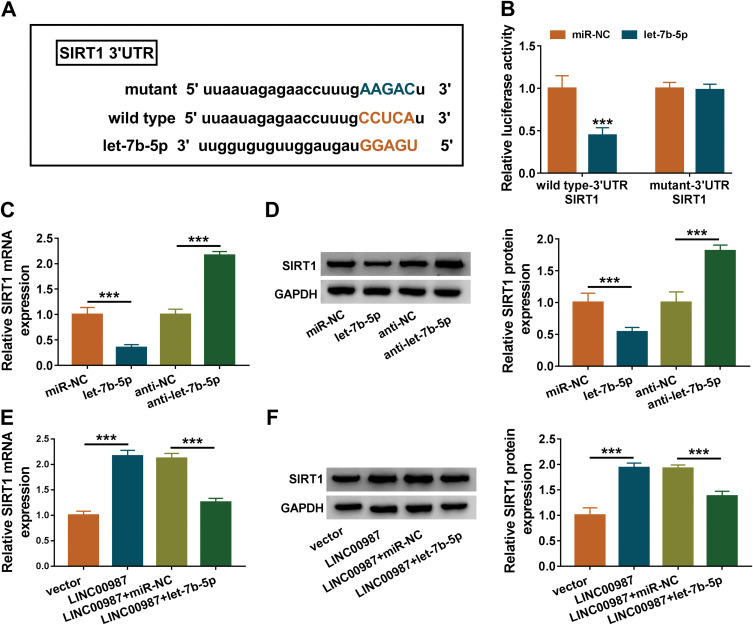
Methods: The expression levels of LINC00987 and let-7b-5p were detected by real-time quantitativepolymerase chain reaction (RT-qPCR). The expression of apoptosis-associated proteins, oxidative stress (ROS)-related proteins, autophagy-related proteins and sirtuin1 (SIRT1) protein was determined by Western blot. Cell viability was illustrated by cell counting kit-8 (CCK-8) assay. Cell apoptosis was investigated by caspase3 activity and apoptosis analysis assays. ROS, inflammation and autophagy were demonstrated by detecting reactive ROS level and superoxide dismutase (SOD) activity, enzyme-linked immunosorbent assay (ELISA) and Western blot analysis, respectively. The binding sites between let-7b-5p and LINC00987 or SIRT1 were predicted by lncBase or miRWalk online database, and identified by dual-luciferase reporter assay.

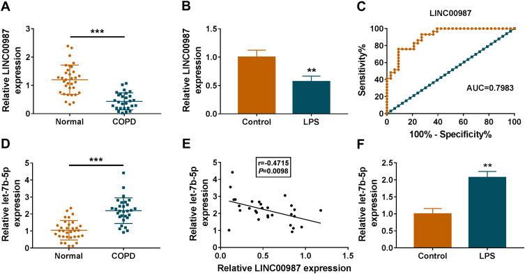
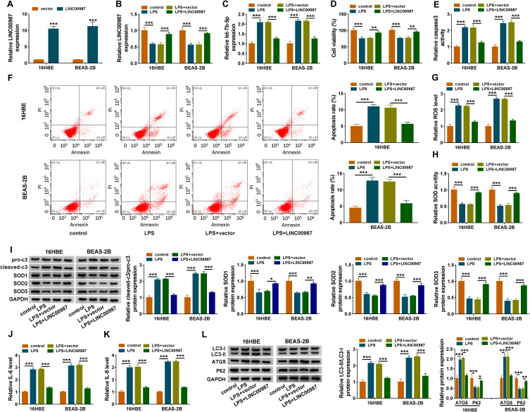
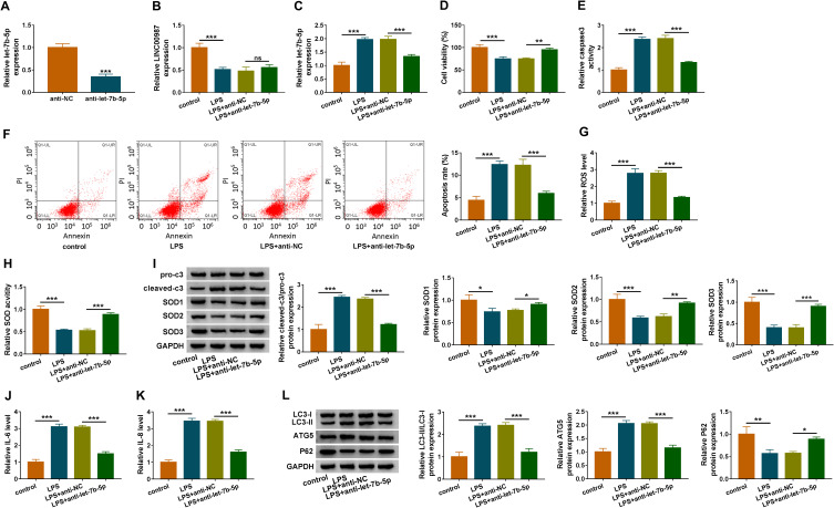
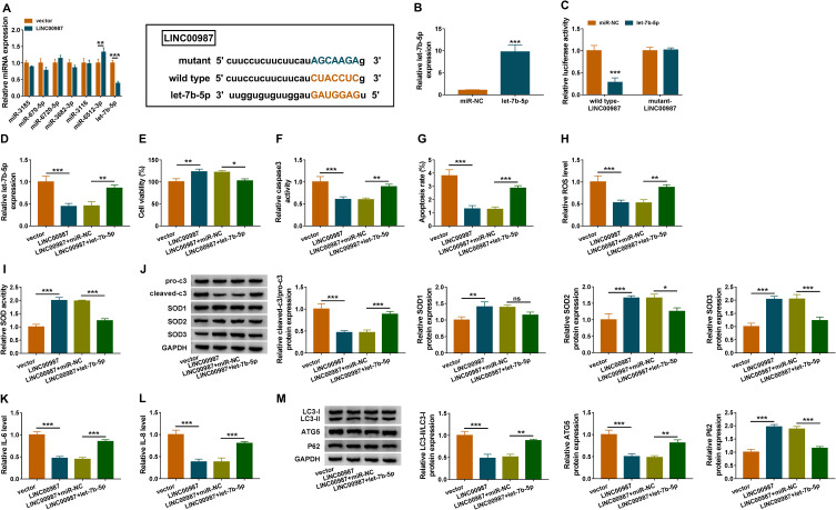
Results: LINC00987 expression was strikingly downregulated and let-7b-5p expression was obviously upregulated in COPD tissues and LPS-induced BEAS-2B cells compared with control groups. LINC00987 overexpression promoted BEAS-2B cells against LPS-mediated viability, apoptosis, oxidative stress, inflammation and autophagy, whereas these effects were attenuated by let-7b-5p mimic or SIRT1 knockdown. Furthermore, LINC00987 sponged let-7b-5p and let-7b-5p bound to SIRT1.

Conclusion: LINC00987 ameliorated COPD through modulating LPS-induced cell apoptosis, oxidative stress, inflammation and autophagy via sponging let-7b-5p to associate with SIRT1. This finding will provide a theoretical basis for the research of LncRNA-mediated treatment in COPD.

Keywords: COPD; LINC00987; LPS; SIRT1; let-7b-5p.


Conflict of interest statement

The authors declare that they have no financial or non-financial conflicts of interest.

Figures

Figure 1
Figure 1
LINC00987 had low expression and let-7b-5p had high expression in LPS-induced BEAS-2B cells. (A and D) RT-qPCR was performed to detect LINC00987 and let-7b-5p expression in normal (N=33) and COPD tissues (N=29). (B and F) The expression levels of LINC00987 and let-7b-5p were determined by RT-qPCR in LPS-induced BEAS-2B cells. (C) ROC curve was used to investigate the value of LINC00987 in COPD diagnosis. (E) Spearman correlation analysis was employed to reveal the relationship between LINC00987 expression and let-7b-5p expression. **P<0.01 and ***P<0.001.
Figure 2
Figure 2
LINC00987 inhibited cell apoptosis, oxidative stress, inflammation and autophagy in LPS-mediated 16HBE and BEAS-2B cells. (A) The transfection efficiency of LINC00987 was detected by RT-qPCR in LPS-induced 16HBE and BEAS-2B cells. (B and C) The effects of LPS treatment and LINC00987 overexpression on LINC00987 and let-7b-5p expression were demonstrated by RT-qPCR. (D) CCK-8 assay was performed to illustrate the impacts between LPS and LINC00987 on cell viability in 16HBE and BEAS-2B cells. (E and F) Caspase3 activity and apoptosis analysis assays were conducted to present the influences between LPS and LINC00987 overexpression on caspase3 activity and cell apoptosis, respectively, in 16HBE and BEAS-2B cells. (G and H) ROS detection kit and SOD activity assays were carried out to uncover the impacts between LPS and enforced LINC00987 expression on oxidative stress in 16HBE and BEAS-2B cells. (I) Western blot assay was employed to determine the impacts of LINC00987 on the protein levels of pro-c3, cleaved-c3, SOD1, SOD2 and SOD3 in LPS-induced 16HBE and BEAS-2B cells. (J and K) ELISA kit assays were employed to exhibit the effects between LPS exposure and LINC00987 on the production of IL-6 and IL-8 in 16HBE and BEAS-2B cells. (L) The effects of LPS treatment and LINC00987 overexpression on the level of IC3-II/IC3-I and the protein expression of ATG5 and P62 were demonstrated by Western blot in 16HBE and BEAS-2B cells. *P<0.05, **P<0.01 and ***P<0.001.
Figure 3
Figure 3
Let-7b-5p inhibitor attenuated LPS-induced apoptosis, oxidative stress, inflammation and autophagy in BEAS-2B cells. (A) RT-qPCR was performed to detect the transfection efficiency of anti-let-7b-5p in LPS-induced BEAS-2B cells. (B and C) The impact of anti-let-7b-5p on LINC00987 and let-7b-5p expression after LPS treatment was determined by RT-qPCR in BEAS-2B cells. (D) CCK-8 assay was carried out to illustrate the impacts between LPS and let-7b-5p absence on cell viability in BEAS-2B cells. (E and F) Caspase3 activity and apoptosis analysis assays were employed to reveal the influences between LPS exposure and let-7b-5p deletion on caspase3 activity and cell apoptosis, respectively, in BEAS-2B cells. (G and H) ROS detection kit and SOD activity assays were used to reveal the impacts between LPS treatment and let-7b-5p repression on oxidative stress in BEAS-2B cells. (I) Western blot assay was employed to determine the influences of let-7b-5p inhibitor on the protein levels of pro-c3, cleaved-c3, SOD1, SOD2 and SOD3 in LPS-induced BEAS-2B cells. (J and K) ELISA kits were purchased to disclose the influences of anti-let-7b-5p on the levels of IL-6 and IL-8 under LPS treatment in BEAS-2B cells. (L) Western blot was performed to detect the impacts between LPS and let-7b-5p inhibitor on the levels of IC3-II/IC3-I, ATG5 and P62 in BEAS-2B cells. *P<0.05, **P<0.01 and ***P<0.001.
Figure 4
Figure 4
LINC00987 overexpression inhibited cell apoptosis, oxidative stress, inflammation and autophagy by binding to let-7b-5p in LPS-mediated BEAS-2B cells. (A) The expression levels of miRNAs possessing the binding sites of LINC00987 were determined by RT-qPCR in BEAS-2B cells transfected with LINC00987 or vector, and the binding sites between LINC00987 and let-7b-5p were predicted by lncBase online database. (B) The transfection efficiency of let-7b-5p mimic was determined by RT-qPCR. (C) Dual-luciferase reporter assay was performed to detect luciferase activity in LPS-induced BEAS-2B cells. (D) The effects between LINC00987 and let-7b-5p on let-7b-5p expression were revealed by RT-qPCR. (E) CCK-8 assay was employed to illustrate the impacts between LINC00987 and let-7b-5p on cell viability in LPS-induced BEAS-2B cells. (F and G) Caspase3 activity and apoptosis analysis assays were carried out to demonstrate the impacts between LINC00987 and let-7b-5p mimic on the apoptosis of LPS-induced BEAS-2B cells. (H and I) ROS detection kit and SOD activity assays were conducted to present the influences between LINC00987 overexpression and let-7b-5p mimic on oxidative stress in LPS-induced BEAS-2B cells. (J) The influences between LINC00987 and let-7b-5p on the expression of apoptosis-related proteins (pro-caspase3 and cleaved-caspase3) and ROS-associated proteins (SOD1, SOD2 and SOD3) were revealed by Western blot. (K and L) ELISA assays were used to disclose the impacts between LINC00987 overexpression and let-7b-5p mimic on inflammation response in LPS-induced BEAS-2B cells. (M) Western blot was performed to detect the influences between LINC00987 and let-7b-5p on the levels of LC3-II/IC3-I, ATG5 and P62 in LPS-mediated BEAS-2B cells. *P<0.05, **P<0.01 and ***P<0.001.
Figure 5
Figure 5
LINC00987 upregulated SIRT1 expression via binding to let-7b-5p. (A) MiRWalk online database was used to predict the binding sites between let-7b-5p and SIRT1 3ʹUTR. (B) Luciferase activities were detected by dual-luciferase reporter assay. (C) The effects of let-7b-5p mimic and inhibitor on SIRT1 mRNA level were revealed by RT-qPCR. (D) The effects of let-7b-5p mimic and inhibitor on SIRT1 protein level were investigated by Western blot. (E and F) The effects between LINC00987 and let-7b-5p on SIRT1 expression at mRNA and protein levels were illustrated via RT-qPCR and Western blot, respectively. ***P<0.001.
Figure 6
Figure 6
LINC00987 ameliorated LPS-induced disorders by regulating SITR1. (A and B) The knockdown efficiency of si-SIRT1#1, si-SIRT1#2 and si-SIRT1#3 was determined by detecting the mRNA and protein levels of SIRT1 via RT-qPCR and Western blot, respectively. (C) The effects of LINC00987 and SIRT1 knockdown on the mRNA level of SIRT1 were illustrated by RT-qPCR. (D) The effects of LINC00987 and SIRT1 knockdown on the protein expression of SIRT1 were illustrated by Western blot. (E) CCK-8 assay was performed to illustrate the effects between LINC00987 and SIRT1 silencing on cell viability in LPS-induced BEAS-2B cells. (F and G) Caspase3 activity and apoptosis analysis assays were employed to demonstrate the impacts between LINC00987 and SIRT1 deletion on cell apoptosis in LPS-induced BEAS-2B cells. (H and I) ROS detection kit and SOD activity assays were carried out to display the influences between LINC00987 overexpression and SIRT1 absence on oxidative stress in LPS-induced BEAS-2B cells. (J) The impacts between ectopic LINC00987 expression and SIRT1 downregulation on the protein expression of pro-c3, cleaved-c3, SOD1, SOD2 and SOD3 were presented by Western blot assay. (K and L) ELISA kit assays were conducted to investigate the impacts between LINC00987 overexpression and SIRT1 deletion on IL-6 and IL-8 production in LPS-induced BEAS-2B cells. (M) The effects between LINC00987 overexpression and SIRT1 knockdown on the levels of LC3-II/IC3-I, ATG5 and P62 were presented by Western blot in LPS-induced BEAS-2B cells. *P<0.05, **P<0.01 and ***P<0.001.

Similar articles

Cited by

References

    1. Barnes PJ. Targeting cytokines to treat asthma and chronic obstructive pulmonary disease. Nat Rev Immunol. 2018;18(7):454–466. doi:10.1038/s41577-018-0006-6 - DOI - PubMed
    1. Osei ET, Florez-Sampedro L, Timens W, Postma DS, Heijink IH, Brandsma CA. Unravelling the complexity of COPD by microRNAs: it’s a small world after all. Eur Respir J. 2015;46(3):807–818. doi:10.1183/13993003.02139-2014 - DOI - PubMed
    1. Roffel MP, Bracke KR, Heijink IH, Maes T. miR-223: a key regulator in the innate immune response in asthma and COPD. Front Med (Lausanne). 2020;7:196. doi:10.3389/fmed.2020.00196 - DOI - PMC - PubMed
    1. Jemal A, Ward E, Hao Y, Thun M. Trends in the leading causes of death in the United States, 1970-2002. JAMA. 2005;294(10):1255–1259. doi:10.1001/jama.294.10.1255 - DOI - PubMed
    1. Djebali S, Davis CA, Merkel A, et al. Landscape of transcription in human cells. Nature. 2012;489(7414):101–108. doi:10.1038/nature11233 - DOI - PMC - PubMed