

Morphological and Molecular Analysis of Osteoblasts Differentiated from Mesenchymal Stem Cells in Polycaprolactone/Magnesium Oxide/Graphene Oxide Scaffold
- PMID: 33312462
- PMCID: PMC7722513
Morphological and Molecular Analysis of Osteoblasts Differentiated from Mesenchymal Stem Cells in Polycaprolactone/Magnesium Oxide/Graphene Oxide Scaffold
Abstract
Background: The loss or dysfunction of bone tissue that observed after bone tumor resections and severe nonunion fractures afflicts 200 million people worldwide. Bone tissue engineering is a promising approach to repair osteoporotic fractures.
Objective: In this paper, polycaprolactone (PCL)/magnesium oxide (MgO)/graphene oxide (GO) nanofibrous scaffold was fabricated by electrospining method, and its biocompatibility and osteogenic differentiation of adipose-derived mesenchymal stem cells (MSCs) on this scaffold were evaluated and compared with pure PCL nanofibrous scaffold.
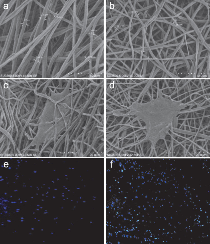
Methods: SEM analysis, DAPI staining and MTT assay were used to evaluation biocompatibility of PCL/MgO/GO composite scaffold. In addition by ALP assay and proteomic approach, osteostimulatory effect of electrospun composite scaffold was investigated and the expression level of osteogenic markers including Runt-related transcription factor cbfa1/runx2 (runx2), collagen type I (Col1a1) and osteopontin (OPN) in MSCs seeded on PCL/MgO/GO composite scaffold was determined and compared with pure PCL scaffold. Then, RT-PCR technique was used to validate the level expression of these genes.
Results: The obtained results showed that adhesion, viability and ALP activity of MSCs on PCL/MgO/GO scaffold considerably enhanced compared with pure PCL. As well as proteomic and real-time analysis illustrated the expression of osteogenic markers including runx2, Col1a1 and OPN increased (>2-fold) in cells seeded on PCL/MgO/GO composite scaffold.
Conclusion: It was concluded that MgO and GO nanoparticles could improve the biocompatibility of PCL scaffold and enhance the osteogenic differentiation of MSCs.
Keywords: Bone tissue engineering; Mesenchymal stem cells; Osteoblast; Proteomics; Scaffold.
Figures


Similar articles
-
Osteogenic Differentiation Potential of Adipose-Derived Mesenchymal Stem Cells Cultured on Magnesium Oxide/Polycaprolactone Nanofibrous Scaffolds for Improving Bone Tissue Reconstruction.Adv Pharm Bull. 2022 Jan;12(1):142-154. doi: 10.34172/apb.2022.015. Epub 2020 Sep 22. Adv Pharm Bull. 2022. PMID: 35517875 Free PMC article.
-
Evaluation of osteoconductive effect of polycaprolactone (PCL) scaffold treated with Aloe vera on adipose-derived mesenchymal stem cells (ADSCs).Am J Stem Cells. 2023 Oct 20;12(4):83-91. eCollection 2023. Am J Stem Cells. 2023. PMID: 38021455 Free PMC article.
-
PCL/Col I-based magnetic nanocomposite scaffold provides an osteoinductive environment for ADSCs in osteogenic cues-free media conditions.Stem Cell Res Ther. 2022 Apr 4;13(1):143. doi: 10.1186/s13287-022-02816-0. Stem Cell Res Ther. 2022. PMID: 35379318 Free PMC article.
-
MiR-221-inhibited adipose tissue-derived mesenchymal stem cells bioengineered in a nano-hydroxy apatite scaffold.In Vitro Cell Dev Biol Anim. 2016 Apr;52(4):479-87. doi: 10.1007/s11626-015-9992-x. Epub 2016 Jan 28. In Vitro Cell Dev Biol Anim. 2016. PMID: 26822432
-
Polycaprolactone in Bone Tissue Engineering: A Comprehensive Review of Innovations in Scaffold Fabrication and Surface Modifications.J Funct Biomater. 2024 Aug 24;15(9):243. doi: 10.3390/jfb15090243. J Funct Biomater. 2024. PMID: 39330219 Free PMC article. Review.
Cited by
-
The beneficial effects of simultaneous supplementation of Lactobacillus reuteri and calcium fluoride nanoparticles on ovariectomy-induced osteoporosis.BMC Complement Med Ther. 2023 Sep 26;23(1):340. doi: 10.1186/s12906-023-04167-6. BMC Complement Med Ther. 2023. PMID: 37752485 Free PMC article.
-
Injectable and Assembled Calcium Sulfate/Magnesium Silicate 3D Scaffold Promotes Bone Repair by In Situ Osteoinduction.Bioengineering (Basel). 2025 May 31;12(6):599. doi: 10.3390/bioengineering12060599. Bioengineering (Basel). 2025. PMID: 40564415 Free PMC article.
-
Osteogenic Differentiation Potential of Adipose-Derived Mesenchymal Stem Cells Cultured on Magnesium Oxide/Polycaprolactone Nanofibrous Scaffolds for Improving Bone Tissue Reconstruction.Adv Pharm Bull. 2022 Jan;12(1):142-154. doi: 10.34172/apb.2022.015. Epub 2020 Sep 22. Adv Pharm Bull. 2022. PMID: 35517875 Free PMC article.
-
Fabrication of Polymer/Graphene Biocomposites for Tissue Engineering.Polymers (Basel). 2022 Mar 4;14(5):1038. doi: 10.3390/polym14051038. Polymers (Basel). 2022. PMID: 35267861 Free PMC article. Review.
References
-
- Stevens MM. Biomaterials for bone tissue engineering. Materials today. 2008;11:18–25.
-
- Roseti L, Parisi V, Petretta M, et al. Scaffolds for Bone Tissue Engineering: State of the art and new perspectives. Materials Science and Engineering Mater Sci Eng C Mater Biol Appl. 2017;78:1246–62. - PubMed
-
- Xu W, Liao X, Li B, Li T. Biomaterials and bone tissue engineering. Bioelectronics and Bioinformatics (ISBB), 2011. International Symposium on bioelectronics and bioinformatics 2011. :224–7.
-
- Bouët G, Marchat D, Cruel M, et al. In vitro three-dimensional bone tissue models: from cells to controlled and dynamic environment. Tissue Engineering Part B: Reviews. 2014;21:133–56. - PubMed
LinkOut - more resources
Full Text Sources
Research Materials
Miscellaneous