

Clinical and radiological examination of bony-mediated shoulder instability
- PMID: 33312708
- PMCID: PMC7722946
- DOI: 10.1302/2058-5241.5.200049
Clinical and radiological examination of bony-mediated shoulder instability
Abstract
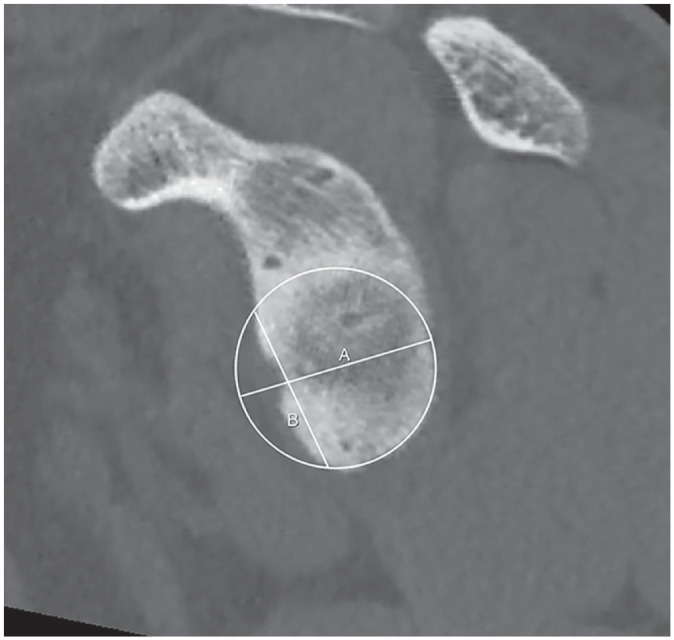
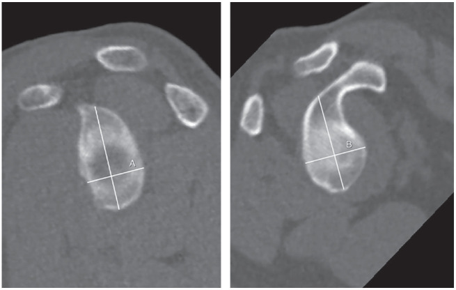
The coexistence of glenoid and humeral head bone defects may increase the risk of recurrence of instability after soft tissue repair.Revealed factors in medical history such as male gender, younger age of dislocation, an increasing number of dislocations, contact sports, and manual work or epilepsy may increase the recurrence rate of instability.In physical examination, positive bony apprehension test, catching and crepitations in shoulder movement may suggest osseous deficiency.Anteroposterior and axial views allow for the detection of particular bony lesions in patients with recurrent anterior shoulder instability.Computed Tomography (CT) with multiplanar reconstruction (MPR) and various types of 3D rendering in 2D (quasi-3D-CT) and 3D (true-3D-CT) space allows not only detection of glenoid and humeral bone defects but most of all their quantification and relations (engaging/not-engaging and on-track/off-track) in the context of bipolar lesion.Magnetic resonance imaging (MRI) is increasingly developing and can provide an equally accurate measurement tool for bone assessment, avoiding radiation exposure for the patient. Cite this article: EFORT Open Rev 2020;5:815-827. DOI: 10.1302/2058-5241.5.200049.
Keywords: Hill–Sachs lesion; glenoid bone loss; glenoid track; shoulder instability.
© 2020 The author(s).
Conflict of interest statement
ICMJE Conflict of interest statement: PL reports board membership of the Rehasport Clinic, employment by Rehasport Clinic, University of Medical Sciences, payment for lectures including service on speakers’ bureaus from Smith&Nephew and Arthrex, payment for development of educational presentations from Smith&Nephew and is an Associate Editor of EFORT Open Reviews, all outside the submitted work. The other authors declare no conflict of interest relevant to this work.
Figures


References
-
- Burkhart SS, De Beer JF. Traumatic glenohumeral bone defects and their relationship to failure of arthroscopic Bankart repairs: significance of the inverted-pear glenoid and the humeral engaging Hill-Sachs lesion. Arthroscopy 2000;16:677-694. - PubMed
-
- Tauber M, Resch H, Forstner R, Raffl M, Schauer J. Reasons for failure after surgical repair of anterior shoulder instability. J Shoulder Elbow Surg 2004;13:279-285. - PubMed
-
- Boileau P, Villalba M, Héry Jy, Balg F, Ahrens P. Risk factors for recurrence of shoulder instability after arthroscopic Bankart repair. J Bone Joint Surg Am 2006. August;88:1755-1763. - PubMed
-
- Balg F, Boileau P. The instability severity index score: a simple pre-operative score to select patients for arthroscopic or open shoulder stabilisation. J Bone Jt Surg – Ser B 2007;89:1470–1477. - PubMed
-
- Sugaya H., Moriishi J, Dohi M, Kon Y, Tsuchiya A. Glenoid rim morphology in recurrent anterior glenohumeral instability. J Bone Joint Surg Am 2003. May;85:878-884. - PubMed
Publication types
LinkOut - more resources
Full Text Sources
