Review
. 2020 Nov 13;5(11):835-844.
doi: 10.1302/2058-5241.5.190077. eCollection 2020 Nov.

Treatment options for aseptic tibial diaphyseal nonunion: A review of selected studies

Affiliations
Review

Treatment options for aseptic tibial diaphyseal nonunion: A review of selected studies

Elena Gálvez-Sirvent et al. EFORT Open Rev. .

Abstract

In aseptic tibial diaphyseal nonunions after failed conservative treatment, the recommended treatment is a reamed intramedullary (IM) nail.Typically, when an aseptic tibial nonunion previously treated with an IM nail is found, it is advisable to change the previous IM nail for a larger diameter reamed and locked IM nail (the rate of success of renailing is around 90%).A second change after an IM nail failure is also a good option, especially if bone healing has progressed after the first change.Fibular osteotomy is not routinely advised; it is only recommended when it interferes with the nonunion site.In delayed unions before 24 weeks, IM nail dynamization can be performed as a less invasive option before deciding on a nail change.If there is a bone defect, a bone graft must be recommended, with the gold standard being the autologous iliac crest bone graft (AICBG).A reamer-irrigator-aspirator (RIA) system might also obtain a bone autograft that is comparable to AICBG.Although the size of the bone defect suitable to perform bone transport techniques is a controversial issue, we believe that such techniques can be considered in bone defects > 3 cm.Non-invasive therapies and biologic therapies could be applied in isolation for patients with high surgical risk, or could be used as adjuvants to the aforementioned surgical treatments. Cite this article: EFORT Open Rev 2020;5:835-844. DOI: 10.1302/2058-5241.5.190077.

Keywords: aseptic nonunion; tibial diaphysis; treatment options.


Conflict of interest statement

ICMJE Conflict of interest statement: The authors declare no conflict of interest relevant to this work.

Figures
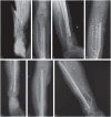
Fig. 1
Fig. 1
A 28-year-old male suffered a comminuted diaphyseal fracture of the tibia and fibula Gustilo IIIB (a). Anteroposterior (b) and lateral (c) radiographs after open reduction and bone fixation using a large fragment LCP (low compression plate), with simultaneous coverage by a latissimus dorsi flap. In (d) a plate rupture due to aseptic nonunion is observed. Anteroposterior (e) (f) and lateral (g) radiographs at 18 months after the plate was replaced by a locked reamed IM nail, in which bone healing is observed. In this case, plate fixation was chosen so as not to create a large bone defect, since the tibial fracture had multiple fragments. The external fixator was discarded because it did not stabilize the intermediate fragments, and, in association with the Plastic Surgery Department, it was possible to perform immediate coverage (fix and flap) with a latissimus dorsi free flap at the same surgical time.
Fig. 2
Fig. 2
A 41-year-old male suffered a comminuted diaphyseal fracture of the tibia Gustilo II (a). Anteroposterior (b) and lateral (c) radiographs at 13 months after treatment with a 10-mm diameter non-reamed intramedullary (IM) nail; note oligotrophic nonunion at the level of the tibial shaft. Anteroposterior (d) and lateral (e) radiographs at 12 months after changing the nail for a larger reamed IM nail (13 mm) with associated fibular osteotomy; note satisfactory bone healing. In the preoperative planning of this case, the clinical examination ruled out malrotation (correct thigh–foot angle). A preoperative measurement of the tibial canal at the level of the isthmus was performed, and it was found that the maximum thickness of the nail would be 13 mm, so a reaming of up to 14 mm was performed. Since the fibula was completely consolidated at the distal level, the plate was removed and a fibular osteotomy was added at the diaphyseal level.
Fig. 3
Fig. 3
A 51-year-old woman with an open Gustilo IIIB fracture in the proximal third of the tibial shaft was initially treated with an external fixator for damage control (a). Anteroposterior (b) and lateral (c) radiographs at 10 months after nailing with a non-reamed intramedullary (IM) nail 9 mm in diameter, in which nonunion is observed and a bone defect of the 50% anterior circumference of the tibia. Anteroposterior (d) and lateral (e) postsurgical radiographs after the change to a reamed nail of greater diameter (12 mm) and iliac crest autograft. In (f), an intraoperative image of the autograft implanted in the bone defect (arrow) is observed. Anteroposterior (g) and lateral (h) radiographs at nine months, in which bone healing is observed. In this case, damage control with external fixator plus initial debridement was initially performed, since this was a polytraumatized patient. In a second stage, after nine days, definitive osteosynthesis was performed with an IM nail and coverage with an anterolateral free thigh (ALT) flap. We did not add bone graft to the defect because of the possible increased risk of infection. Ten months later, lack of consolidation was observed with mobility at the fracture site, without analytical and clinical data of infection. For this reason, we decided to change the nail with the addition of bone graft, since the bone defect affected 50% of the tibial circumference. In this case the bone defect of more than 50% was given more importance than the risk of malreduction in recurvatum.

References

    1. Browner B, Jupiter J, Levine A, Trafton P, Krettek C. Skeletal trauma: basic science, management and reconstruction. Fourth ed. Philadelphia, Saunders, 2009.
    1. Rupp M, Biehl C, Budak M, Thormann U, Heiss C, Alt V. Diaphyseal long bone nonunions: types, aetiology, economics, and treatment recommendations. Int Orthop 2018;42:247–258. - PubMed
    1. Rodriguez-Merchan EC, Forriol F. Nonunion: general principles and experimental data. Clin Orthop Relat Res 2004;419:4–12. - PubMed
    1. Aldemir C, Duygun F. Outcome of locked compressive nailing in aseptic tibial diaphyseal nonunions without bone defect. Indian J Orthop 2019;53:251–256. - PMC - PubMed
    1. Kohlprath R, Assal M, Uçkay I, Holzer N, Hoffmeyer P, Suva D. Open fractures of the tibia in the adult: surgical treatment and complications. Rev Med Suisse 2011;7:2482, 2484–2488. - PubMed

LinkOut - more resources