Findings on intraprocedural non-contrast computed tomographic imaging following hepatic artery embolization are associated with development of contrast-induced nephropathy
- PMID: 33312900
- PMCID: PMC7701934
- DOI: 10.5527/wjn.v9.i2.33
Findings on intraprocedural non-contrast computed tomographic imaging following hepatic artery embolization are associated with development of contrast-induced nephropathy
Abstract
Background: Contrast-induced nephropathy (CIN) is a reversible form of acute kidney injury that occurs within 48-72 h of exposure to intravascular contrast material. CIN is the third leading cause of hospital-acquired acute kidney injury and accounts for 12% of such cases. Risk factors for CIN development can be divided into patient- and procedure-related. The former includes pre-existing chronic renal insufficiency and diabetes mellitus. The latter includes high contrast volume and repeated exposure over 72 h. The incidence of CIN is relatively low (up to 5%) in patients with intact renal function. However, in patients with known chronic renal insufficiency, the incidence can reach up to 27%.
Aim: To examine the association between renal enhancement pattern on non-contrast enhanced computed tomographic (CT) images obtained immediately following hepatic artery embolization with development of CIN.
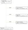
Methods: Retrospective review of all patients who underwent hepatic artery embolization between 01/2010 and 01/2011 (n = 162) was performed. Patients without intraprocedural CT imaging (n = 51), combined embolization/ablation (n = 6) and those with chronic kidney disease (n = 21) were excluded. The study group comprised of 84 patients with 106 procedures. CIN was defined as 25% increase above baseline serum creatinine or absolute increase ≥ 0.5 mg/dL within 72 h post-embolization. Post-embolization CT was reviewed for renal enhancement patterns and presence of renal artery calcifications. The association between non-contrast CT findings and CIN development was examined by Fisher's Exact Test.
Results: CIN occurred in 11/106 (10.3%) procedures (Group A, n = 10). The renal enhancement pattern in patients who did not experience CIN (Group B, n = 74 with 95/106 procedures) was late excretory in 93/95 (98%) and early excretory (EE) in 2/95 (2%). However, in Group A, there was a significantly higher rate of EE pattern (6/11, 55%) compared to late excretory pattern (5/11) (P < 0.001). A significantly higher percentage of patients that developed CIN had renal artery calcifications (6/11 vs 20/95, 55% vs 21%, P = 0.02).
Conclusion: A hyperdense renal parenchyma relative to surrounding skeletal muscle (EE pattern) and presence of renal artery calcifications on immediate post-HAE non-contrast CT images in patients with low risk for CIN are independently associated with CIN development.
Keywords: Contrast-induced nephropathy; Hepatic artery embolization; Intra-arterial; Non-contrast computed tomographic; Renal artery calcification; Renal enhancement pattern.
©The Author(s) 2020. Published by Baishideng Publishing Group Inc. All rights reserved.
Conflict of interest statement
Conflict-of-interest statement: All authors declare no conflicts of interest related to this article.
Figures


Similar articles
-
Contrast-induced nephropathy after peripheral vascular intervention: Long-term renal outcome and risk factors for progressive renal dysfunction.J Vasc Surg. 2019 Mar;69(3):913-920. doi: 10.1016/j.jvs.2018.06.196. Epub 2018 Oct 3. J Vasc Surg. 2019. PMID: 30292616
-
Incidence and risk factors of contrast-induced nephropathy after bronchial arteriography or bronchial artery embolization.Tuberc Respir Dis (Seoul). 2013 Apr;74(4):163-8. doi: 10.4046/trd.2013.74.4.163. Epub 2013 Apr 30. Tuberc Respir Dis (Seoul). 2013. PMID: 23678357 Free PMC article.
-
Use of NaCl saline hydration and N-Acetyl Cysteine to prevent contrast induced nephropathy in different populations of patients at high and low risk undergoing coronary artery angiography.Minerva Cardioangiol. 2010 Feb;58(1):35-40. Minerva Cardioangiol. 2010. PMID: 20145594
-
Is the risk of contrast-induced nephropathy a real contraindication to perform intravenous contrast enhanced Computed Tomography for non-traumatic acute abdomen in Emergency Surgery Department?Acta Biomed. 2018 Dec 17;89(9-S):158-172. doi: 10.23750/abm.v89i9-S.7891. Acta Biomed. 2018. PMID: 30561410 Free PMC article.
-
The clinical and renal consequences of contrast-induced nephropathy.Nephrol Dial Transplant. 2006 Jun;21(6):i2-10. doi: 10.1093/ndt/gfl213. Nephrol Dial Transplant. 2006. PMID: 16723349 Review.
Cited by
-
Artificial Intelligence Algorithm-Based Computed Tomography Image in Assessment of Acute Renal Insufficiency of Patients Undergoing Percutaneous Coronary Intervention.Contrast Media Mol Imaging. 2022 Feb 28;2022:2214583. doi: 10.1155/2022/2214583. eCollection 2022. Contrast Media Mol Imaging. 2022. PMID: 35291424 Free PMC article.
-
Exploring the relationship between post-contrast acute kidney injury and different baseline creatinine standards: A retrospective cohort study.Front Endocrinol (Lausanne). 2023 Jan 12;13:1042312. doi: 10.3389/fendo.2022.1042312. eCollection 2022. Front Endocrinol (Lausanne). 2023. PMID: 36714583 Free PMC article.
References
-
- Parfrey PS, Griffiths SM, Barrett BJ, Paul MD, Genge M, Withers J, Farid N, McManamon PJ. Contrast material-induced renal failure in patients with diabetes mellitus, renal insufficiency, or both. A prospective controlled study. N Engl J Med. 1989;320:143–149. - PubMed
-
- Gleeson TG, Bulugahapitiya S. Contrast-induced nephropathy. AJR Am J Roentgenol. 2004;183:1673–1689. - PubMed
-
- Hou SH, Bushinsky DA, Wish JB, Cohen JJ, Harrington JT. Hospital-acquired renal insufficiency: a prospective study. Am J Med. 1983;74:243–248. - PubMed
-
- Mehran R, Nikolsky E. Contrast-induced nephropathy: definition, epidemiology, and patients at risk. Kidney Int Suppl. 2006:S11–S15. - PubMed
LinkOut - more resources
Full Text Sources

