. 2020 Nov;8(21):1346.
doi: 10.21037/atm-20-3191.

Stromal cells promote chemoresistance of acute myeloid leukemia cells via activation of the IL-6/STAT3/OXPHOS axis

Affiliations

Stromal cells promote chemoresistance of acute myeloid leukemia cells via activation of the IL-6/STAT3/OXPHOS axis

Diyu Hou et al. Ann Transl Med. 2020 Nov.

Abstract

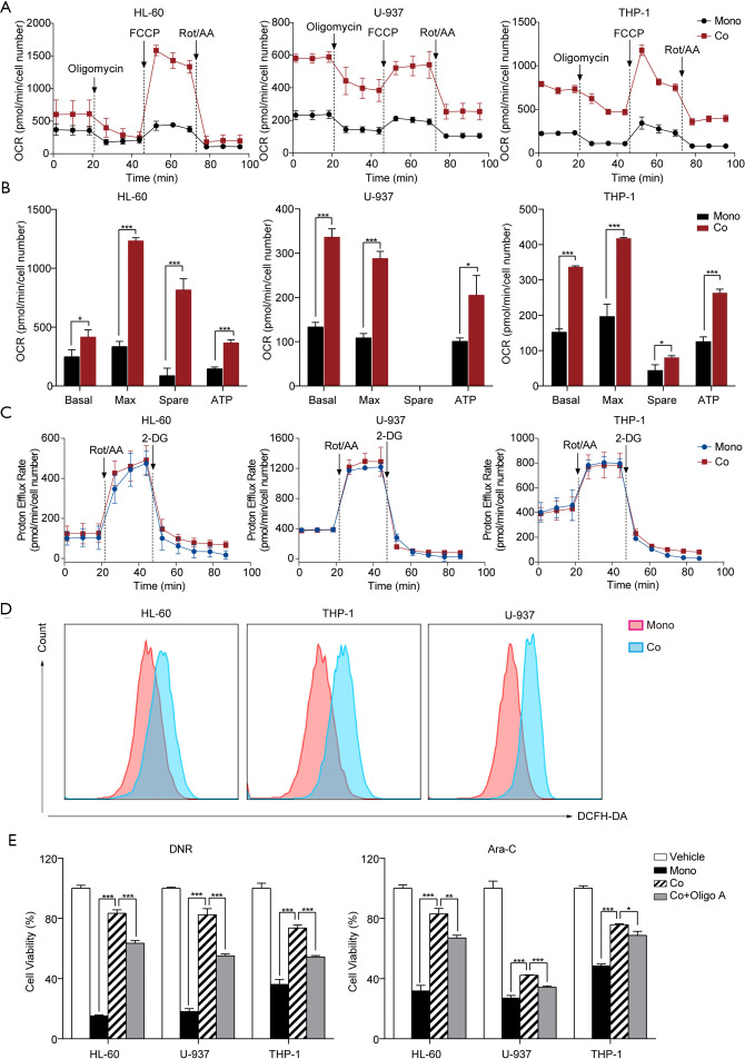
Background: Bone marrow stromal cells (BMSCs) are known to promote chemoresistance in acute myeloid leukemia (AML) cells. However, the molecular basis for BMSC-associated AML chemoresistance remains largely unexplored.

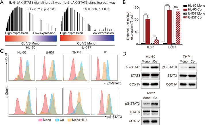
Methods: The mitochondrial oxidative phosphorylation (OXPHOS) levels of AML cells were measured by a Seahorse XFe24 cell metabolic analyzer. The activity of total or mitochondrial signal transducer and transcription activator 3 (STAT3) in AML cells was explored by flow cytometry and Western blotting. Real-time quantitative PCR, Western blotting and enzyme-linked immunosorbent assay (ELISA) were used to analyze expression of interleukin 6 (IL-6) in the human BMSC line HS-5, and IL-6 was knocked out in HS-5 cells by CRISPR/Cas9 system.

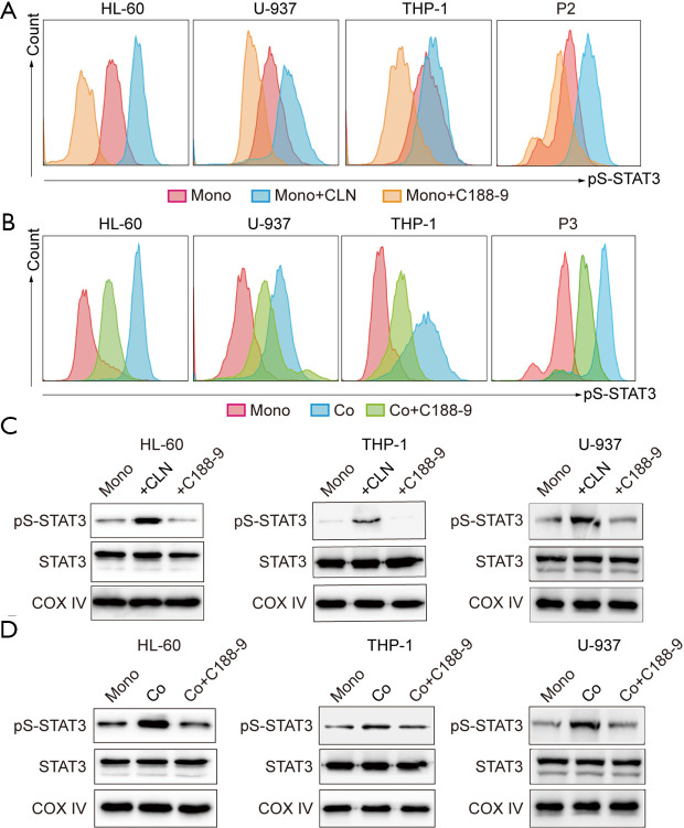
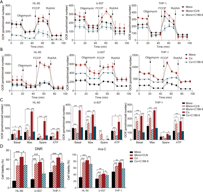
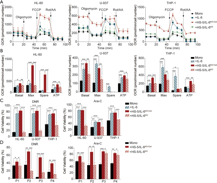
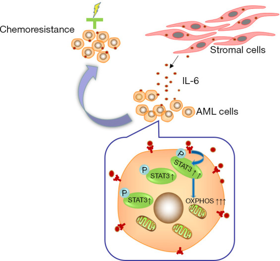
Results: In this study, we observed that co-culturing with BMSCs heightened OXPHOS levels in AML cells, thus promoting chemoresistance in these cells. HS-5 cell-induced upregulation of OXPHOS is dependent on the activation of STAT3, especially on that of mitochondrial serine phosphorylated STAT3 (pS-STAT3) in AML cells. The relationship among pS-STAT3, OXPHOS, and chemosensitivity of AML cells induced by BMSCs was demonstrated by the STAT3 activator and inhibitor, which upregulated and downregulated the levels of mitochondrial pS-STAT3 and OXPHOS, respectively. Intriguingly, AML cells remodeled HS-5 cells to secrete more IL-6, which augmented mitochondrial OXPHOS in AML cells and stimulated their chemoresistance. IL-6 knockout in HS-5 cells impaired the ability of these cells to activate STAT3, to increase OXPHOS, or to promote chemoresistance in AML cells.

Conclusions: BMSCs promoted chemoresistance in AML cells via the activation of the IL-6/STAT3/OXPHOS pathway. These findings exhibit a novel mechanism of chemoresistance in AML cells in the bone marrow microenvironment from a metabolic perspective.

Keywords: IL-6/STAT3/OXPHOS axis; acute myeloid leukemia (AML); chemosensitivity; stromal cells.


Conflict of interest statement

Conflicts of Interest: All authors have completed the ICMJE uniform disclosure form (available at http://dx.doi.org/10.21037/atm-20-3191). The authors have no conflicts of interest to declare.

Figures

Figure 1
Figure 1
Upregulation of OXPHOS in acute myeloid leukemia (AML) cells induced by HS-5 cells was associated with AML chemoresistance. (A) AML cell lines HL-60, U-937, and THP-1 were co-cultured with HS-5 cells (red) for 24 h, were exposed to oligomycin, FCCP, and Rot/AA, and were analyzed for the oxygen consumption rate (OCR); AML cells in mono-cultures (black) were used as control. The data were normalized to cell numbers. (B) Basal respiration (Basal), maximal respiratory capacity (Max), spare respiratory capacity (Spare), and ATP production (ATP) of AML cells co-cultured with HS-5 cells (red); AML cells in monocultures (black) were used as control. (C) The glycolytic proton efflux rate of AML cells co-cultured with HS-5 cells (red) was measured after exposure to Rot/AA and 2-DG; AML cells in monocultures (blue) were used as control. (D) Intracellular ROS levels in the monocultured or co-cultured AML cells. (E) Oligomycin A increased chemosensitivity of AML cells co-cultured with HS-5 cells. AML cells were co-cultured with HS-5 cells, exposed to oligomycin A for 24 h, and treated with either DNR (200 ng/mL) or Ara-C (10 µM); cell viability was analyzed using the CCK-8 assay. The data were normalized to vehicle-treated control cells. *, P<0.05; **, P<0.01; ***, P<0.001.
Figure 2
Figure 2
IL-6/STAT3 signaling and mitochondrial STAT3 were activated in acute myeloid leukemia (AML) cells after co-culturing with HS-5 cells. (A) Enrichment of genes involved in IL-6/JAK/STAT3 signaling in HL-60 and U-937 cells co-cultured with HS-5 cells. The color bar shows gene ranking according to expression levels after co-culturing with HS-5 cells (blue and red colors indicate low and high gene expression, respectively). Vertical bars represent genes; their position corresponds to the position of the gene in the ranked gene list, and the height corresponds to the running GSEA enrichment score. (B) Expression of IL-6R and IL-6ST genes in HL-60 and U-937 cells co-cultured with HS-5 cells was measured by RT-qPCR and normalized to β-actin. (C) STAT3 phosphorylation (pY-STAT3 and pS-STAT3) levels in HL-60, U-937, THP-1 cells, and primary AML cells (P1) co-cultured with HS-5 cells for 24 h were measured by flow cytometry. (D) Mitochondrial STAT3 and pS-STAT3 protein levels of the monocultured and co-cultured HL-60, U-937, and THP-1 cells were measured using immunoblotting with COX IV as a mitochondrial loading control. β-actin and Histone H3 were used as controls and indicators of mitochondrial purity (not shown). ***, P<0.001.
Figure 3
Figure 3
Effects of STAT3 phosphorylation activator or inhibitor on mitochondrial pS-STAT3 levels in acute myeloid leukemia (AML) cells. (A) Monocultured AML cell lines and primary AML cells (P2) were incubated with 100 nM colivelin (CLN) or 10 µM C188-9 for 24 h and analyzed for total pS-STAT3 levels by flow cytometry. (B) Co-cultured AML cell lines and primary AML cells (P3) were incubated with 10 µM C188-9 for 24 h and analyzed for total pS-STAT3 levels by flow cytometry. (C) Mitochondrial STAT3 and pS-STAT3 protein levels of CLN or C188-9 treated HL-60, THP-1 and U-937 cells were measured using immunoblotting with COX IV as a mitochondrial loading control. β-actin and histone H3 were used as controls and indicators of mitochondrial purity (not shown). (D) Co-cultured HL-60, THP-1 and U-937 cells were treated with C188-9 and analyzed for the mitochondrial STAT3 and pS-STAT3 protein levels using immunoblotting with COX IV as a mitochondrial loading control. β-actin and Histone H3 were used as controls and indicators of mitochondrial purity (not shown).
Figure 4
Figure 4
Upregulation of OXPHOS in acute myeloid leukemia (AML) cells by HS-5 cells was associated with STAT3 activation. (A,B,C) HL-60, U-937, and THP-1 cells co-cultured with HS-5 cells or monocultured for 24 h were incubated with colivelin (CLN) or C188-9 for 24 h and then analyzed for OCR (A,B), basal and maximal respiration, spare respiratory capacity, and ATP production (C). (D) Monocultured AML cells were treated with CLN for 24 h and co-cultured AML cells were incubated with C188-9 for 24 h. After the separate treatments, AML cells were treated with DNR (200 ng/mL) or Ara-C (10 µM), and cell viability was analyzed by using the CCK-8 assay. *, P<0.05; **, P<0.01; ***, P<0.001.
Figure 5
Figure 5
IL-6 secreted by bone marrow stromal cells (BMSCs) activated mitochondrial STAT3 in acute myeloid leukemia (AML) cells. (A) IL-6 protein and mRNA expression in HS-5 cells mono-cultured or co-cultured with AML cells respectively were measured by Western blotting and RT-qPCR and normalized to GAPDH levels. (B) The knockout efficiency of IL-6 in HS-5 cells was validated by Western blotting. (C) HL-60, U-937, THP-1 cells, and primary AML cells (P4) were incubated with IL-6 (50 ng/mL) or co-cultured with HS-5/IL-6KO or HS-5/IL-6KO-Con cells for 24 h and analyzed for total pS-STAT3 levels by flow cytometry. (D) Mitochondrial STAT3 and pS-STAT3 protein levels of HL-60, U-937, and THP-1 cells treated with IL-6 or co-cultured with HS-5/IL-6KO or HS-5/IL-6KO-Con cells were measured using immunoblotting with COX IV as a mitochondrial loading control. β-actin and histone H3 were used as controls for mitochondrial purity (not shown). **, P<0.01; ***, P<0.001.
Figure 6
Figure 6
IL-6 secreted by bone marrow stromal cells (BMSCs) enhanced mitochondrial OXPHOS in acute myeloid leukemia (AML) cells to confer chemoresistance. (A,B) HL-60, U-937, and THP-1 cells were incubated with IL-6 (50 ng/mL) or co-cultured with HS-5/IL-6KO or HS-5/IL-6KO-Con cells for 24 h respectively, the levels of the OCR (A), basal and maximal respiration, spare respiratory capacity, and ATP production (B) were measured by using a Seahorse XF Cell Mito Stress Test Kit. (C) AML cells treated with DNR (200 ng/mL) or Ara-C (10 μM) for 24 h were then analyzed for cell viability by the CCK-8 assay. (D) Primary AML cells were isolated from bone marrow aspirates of four patients (P1, P2, P3, and P4), co-cultured with HS-5/IL-6KO or HS-5/IL-6KO-Con cells for 24 h, treated with DNR (200 ng/mL) or Ara-C (10 μM), and analyzed for cell viability by the CCK-8 assay. *, P<0.05; **, P<0.01; ***, P<0.001.
Figure 7
Figure 7
Schematic models for the mechanism by which the stromal cells promote chemoresistance of acute myeloid leukemia (AML) cells via activation of the IL-6/STAT3/OXPHOS axis.

References

    1. Burnett A, Wetzler M, Lowenberg B. Therapeutic advances in acute myeloid leukemia. J Clin Oncol 2011;29:487-94. 10.1200/JCO.2010.30.1820 - DOI - PubMed
    1. Duarte D, Hawkins ED, Lo Celso C. The interplay of leukemia cells and the bone marrow microenvironment. Blood 2018;131:1507-11. 10.1182/blood-2017-12-784132 - DOI - PubMed
    1. Chen P, Jin Q, Fu Q, et al. Induction of Multidrug Resistance of Acute Myeloid Leukemia Cells by Cocultured Stromal Cells via Upregulation of the PI3K/Akt Signaling Pathway. Oncol Res 2016;24:215-23. 10.3727/096504016X14634208143021 - DOI - PMC - PubMed
    1. Kyurkchiev D, Bochev I, Ivanova-Todorova E, et al. Secretion of immunoregulatory cytokines by mesenchymal stem cells. World J Stem Cells 2014;6:552-70. 10.4252/wjsc.v6.i5.552 - DOI - PMC - PubMed
    1. Kumar R, Godavarthy PS, Krause DS. The bone marrow microenvironment in health and disease at a glance. J Cell Sci 2018;131:jcs201707. 10.1242/jcs.201707 - DOI - PubMed