

Human retinal pigment epithelial cells are protected against hypoxia by BNIP3
- PMID: 33313247
- PMCID: PMC7729317
- DOI: 10.21037/atm-20-7145
Human retinal pigment epithelial cells are protected against hypoxia by BNIP3
Abstract
Background: Hypoxia has been implicated in the process of retinal pigment epithelium (RPE) dysfunction. However, recent studies suggest that hypoxia contributes to survival rather than cell death through induction of Bcl-2/adenovirus E1B 19-kDa interacting protein 3 (BNIP3)-dependent autophagy. In contrast, persistent oxidative stress was found to result in autophagy dysregulation in RPE cells. These seemingly contradictory findings led us to investigate the potential role of BNIP3, a crucial mediator of hypoxia-induced autophagy, in the context of hypoxic RPE cells.
Methods: Human RPE D407 cells were treated with low-oxygen conditions, and cell growth, apoptosis, and autophagy was assessed by Cell Counting Kit-8 assay, flow cytometry analysis and immunofluorescence staining, respectively.
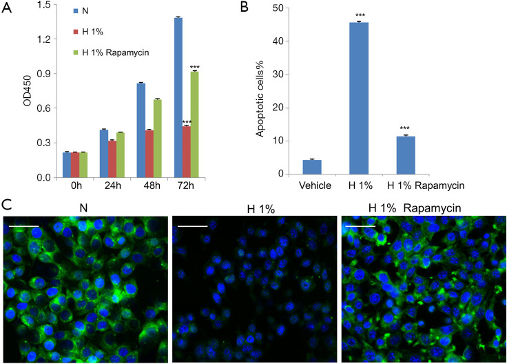
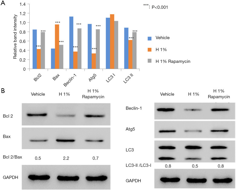
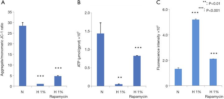
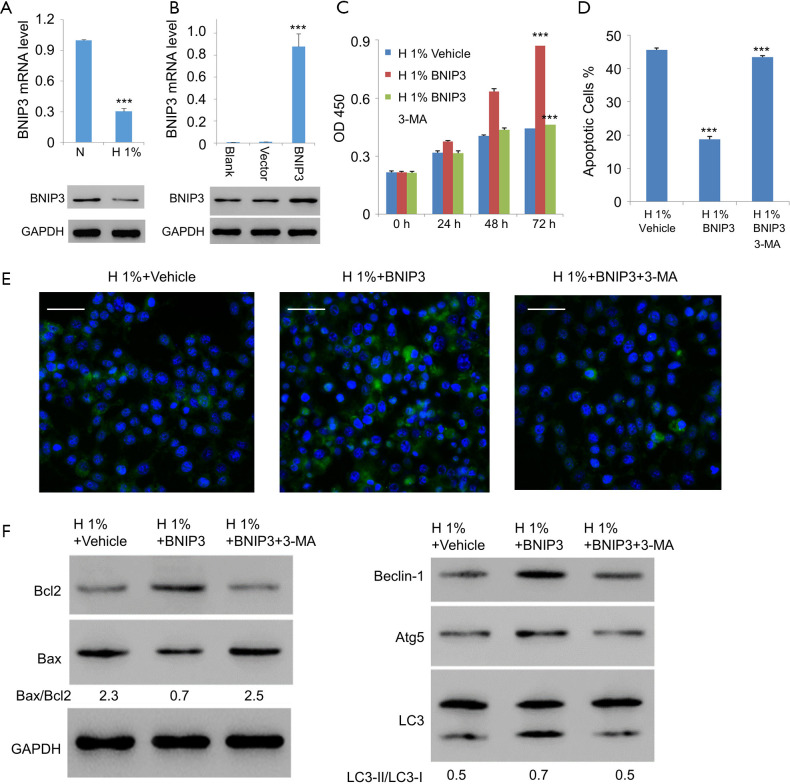
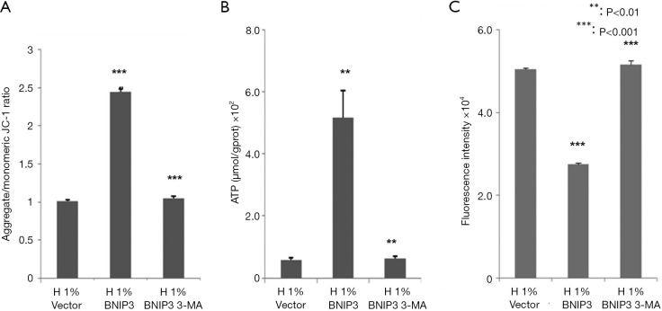
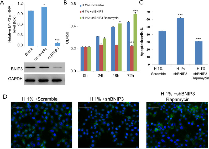
Results: Hypoxic conditions simultaneously triggered a large amount of apoptosis and inhibited autophagy. Moreover, hypoxia led to severe impairments, including the stimulation of reactive oxygen species, and reduction of mitochondrial membrane potential, and adenosine triphosphate production. The stimulation of autophagy by rapamycin inhibited hypoxia-induced severe impairments to a great extent. Interestingly, similar results were observed for BNIP3 overexpression, which can be largely blocked by 3-MA, a well-defined inhibitor of autophagy. Moreover, BNIP3 knockdown further aggravated hypoxia-induced impairments in D407 cells, which can be reversed by rapamycin.
Conclusions: Collectively, these results indicated that BNIP3 can protect human retinal pigmented epithelial cells under hypoxic conditions by inducing autophagy.
Keywords: Bcl-2/adenovirus E1B 19-kDa interacting protein 3 (BNIP3); Retinal pigment epithelium (RPE); apoptosis; autophagy; hypoxia.
2020 Annals of Translational Medicine. All rights reserved.
Conflict of interest statement
Conflicts of Interest: All authors have completed the ICMJE uniform disclosure form (available at http://dx.doi.org/10.21037/atm-20-7145). The authors have no conflicts of interest to declare.
Figures
Similar articles
-
BNIP3-mediated Autophagy Induced Inflammatory Response and Inhibited VEGF Expression in Cultured Retinal Pigment Epithelium Cells Under Hypoxia.Curr Mol Med. 2019;19(6):395-404. doi: 10.2174/1566524019666190509105502. Curr Mol Med. 2019. PMID: 31072291
-
BNIP3 induces apoptosis and protective autophagy under hypoxia in esophageal squamous cell carcinoma cell lines: BNIP3 regulates cell death.Dis Esophagus. 2017 Sep 1;30(9):1-8. doi: 10.1093/dote/dox059. Dis Esophagus. 2017. PMID: 28859361
-
Bcl-2 interacting protein 3 (BNIP3) promotes tumor growth in breast cancer under hypoxic conditions through an autophagy-dependent pathway.Bioengineered. 2022 Mar;13(3):6280-6292. doi: 10.1080/21655979.2022.2036399. Bioengineered. 2022. PMID: 35200106 Free PMC article.
-
Mechanisms and biology of B-cell leukemia/lymphoma 2/adenovirus E1B interacting protein 3 and Nip-like protein X.Antioxid Redox Signal. 2011 May 15;14(10):1959-69. doi: 10.1089/ars.2010.3772. Epub 2011 Mar 4. Antioxid Redox Signal. 2011. PMID: 21126215 Free PMC article. Review.
-
Bnip3 as a dual regulator of mitochondrial turnover and cell death in the myocardium.Pediatr Cardiol. 2011 Mar;32(3):267-74. doi: 10.1007/s00246-010-9876-5. Epub 2011 Jan 6. Pediatr Cardiol. 2011. PMID: 21210091 Free PMC article. Review.
Cited by
-
A simplified protocol to induce hypoxia in a standard incubator: A focus on retinal cells.Exp Eye Res. 2023 Nov;236:109653. doi: 10.1016/j.exer.2023.109653. Epub 2023 Oct 2. Exp Eye Res. 2023. PMID: 37793495 Free PMC article.
-
LXA4 protects against blue-light induced retinal degeneration in human A2E-laden RPE cells and Balb-c mice.Ann Transl Med. 2021 Aug;9(15):1249. doi: 10.21037/atm-21-3390. Ann Transl Med. 2021. PMID: 34532386 Free PMC article.
-
YTHDF1-regulated ALOX5 in retinal pigment epithelial cells under hypoxia enhances VEGF expression and promotes viability, migration, and angiogenesis of vascular endothelial cells.Sci Rep. 2024 Oct 5;14(1):23226. doi: 10.1038/s41598-024-72388-x. Sci Rep. 2024. PMID: 39369033 Free PMC article.
-
Real-Time Monitoring the Effect of Cytopathic Hypoxia on Retinal Pigment Epithelial Barrier Functionality Using Electric Cell-Substrate Impedance Sensing (ECIS) Biosensor Technology.Int J Mol Sci. 2021 Apr 27;22(9):4568. doi: 10.3390/ijms22094568. Int J Mol Sci. 2021. PMID: 33925448 Free PMC article.
-
Exploring the therapeutic potential of MOTS-c in age-related macular degeneration: from cellular responses to patient-derived cybrids.Hum Cell. 2025 Feb 17;38(2):57. doi: 10.1007/s13577-025-01188-w. Hum Cell. 2025. PMID: 39961901
References
LinkOut - more resources
Full Text Sources