. 2021 Feb 12;144(1):162-185.
doi: 10.1093/brain/awaa385.

Neuregulin-1 beta 1 is implicated in pathogenesis of multiple sclerosis

Affiliations

Neuregulin-1 beta 1 is implicated in pathogenesis of multiple sclerosis

Hardeep Kataria et al. Brain. .

Abstract

Multiple sclerosis is characterized by immune mediated neurodegeneration that results in progressive, life-long neurological and cognitive impairments. Yet, the endogenous mechanisms underlying multiple sclerosis pathophysiology are not fully understood. Here, we provide compelling evidence that associates dysregulation of neuregulin-1 beta 1 (Nrg-1β1) with multiple sclerosis pathogenesis and progression. In the experimental autoimmune encephalomyelitis model of multiple sclerosis, we demonstrate that Nrg-1β1 levels are abated within spinal cord lesions and peripherally in the plasma and spleen during presymptomatic, onset and progressive course of the disease. We demonstrate that plasma levels of Nrg-1β1 are also significantly reduced in individuals with early multiple sclerosis and is positively associated with progression to relapsing-remitting multiple sclerosis. The functional impact of Nrg-1β1 downregulation preceded disease onset and progression, and its systemic restoration was sufficient to delay experimental autoimmune encephalomyelitis symptoms and alleviate disease burden. Intriguingly, Nrg-1β1 therapy exhibited a desirable and extended therapeutic time window of efficacy when administered prophylactically, symptomatically, acutely or chronically. Using in vivo and in vitro assessments, we identified that Nrg-1β1 treatment mediates its beneficial effects in EAE by providing a more balanced immune response. Mechanistically, Nrg-1β1 moderated monocyte infiltration at the blood-CNS interface by attenuating chondroitin sulphate proteoglycans and MMP9. Moreover, Nrg-1β1 fostered a regulatory and reparative phenotype in macrophages, T helper type 1 (Th1) cells and microglia in the spinal cord lesions of EAE mice. Taken together, our new findings in multiple sclerosis and experimental autoimmune encephalomyelitis have uncovered a novel regulatory role for Nrg-1β1 early in the disease course and suggest its potential as a specific therapeutic target to ameliorate disease progression and severity.

Keywords: disease pathogenesis; experimental autoimmune encephalomyelitis; immune regulation; multiple sclerosis; neuregulin-1.


Figures

Figure 1
Figure 1
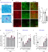
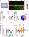
Nrg-1β1 expression levels are declined in CNS and peripherally in EAE mice. (A) Representative Luxol fast blue-haematoxylin and eosin (LFB-HE) stained spinal cord tissue from naïve and EAE mice at the peak of the disease indicating demyelinating lesions. (B) Immunohistological examination for myelin (MBP) and Nrg-1β1 revealed that Nrg-1β1 expression is depleted in demyelinated regions whereas adjacent myelinated normal appearing white matter area (normal-appearing white matter) as well as naive mice tissue indicated a strong expression of MBP and Nrg-1β1. (C) Quantitative immunofluorescence intensity in EAE spinal cord lesions showed 48% reduction in Nrg-1β1 as compared to the adjacent normal-appearing white matter and naïve mice tissue. Values are represented as fold change in intensity normalized to naïve. (DF) Longitudinal assessment of Nrg-1β1 levels was performed on spinal cord (D), plasma (E) and spleen (F) of EAE mice at 7 dpi, onset (10 dpi), peak (14–16 dpi), 7 dpp, 14 dpp and 28 dpp. Nrg-1β1 was significantly depleted in plasma, spleen and spinal cord of EAE mice at 7 dpi, onset and peak of the disease. It was restored in plasma and spleen during the recovery phase (7 dpp, 14 dpp and 28 dpp). However, in the spinal cord there was another reduction Nrg-1β1 levels at 14 dpp in which persisted until 28 dpp. Values represent mean ± SEM. *P <0.05; One-way ANOVA followed by Holm-Sidak post hoc test. n = 3–5. Naïve mean values were compared to each time point for post hoc test in DF.
Figure 2
Figure 2
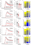
Nrg-1β1 treatment ameliorates neurological disability in EAE mice. (A) Mice were assessed daily for EAE symptoms on the basis of tail and hind limb functional deficits. Treatment with recombinant human Nrg-1β1 peptide (300 ng/day, 600 ng/day and 1200 ng/day) was administered subcutaneously (s.c.) starting at the peak of the disease (Day 16 post induction) for 4 weeks. Nrg-1β1 treatment improved functional deficits in a dose dependent manner in EAE mice. Daily clinical scores were expressed as mean ± SEM, *P <0.05. Two-way-ANOVA followed by Holm-Sidak post hoc test. (B) Cumulative disease burden for each animal was calculated as area under the curve. The 600 ng/day and 1200 ng/day Nrg-1β1-treated groups showed significant reduction in their mean cumulative disease burden as compared to the vehicle-treated group. *P <0.05. n = 10 for vehicle, 300 ng/day and 600 ng/day Nrg-1β1 groups. n = 5 for 1200 ng/day Nrg-1β1 group. (C) Clinical score of each mouse in vehicle and NRG1 treatment group (600 ng/day) are plotted as a heat map. Sustained daily Nrg-1β1 treatment (600 ng/day) significantly improved clinical score and reduced the cumulative burden of disease when administered at different paradigms including symptomatically at the onset of EAE (DF), prophylactically at the time of EAE induction (GI), and delayed at 4 days after reaching the peak of the disease (JL). (MO) Transient Nrg-1β1 therapy for 7 days starting at peak of EAE did not confer beneficial effects when assessed. Clinical scores are expressed as average (mean ± SEM). *P <0.05. Two-way-ANOVA followed by Holm-Sidak post hoc test. n = 7–10. *P <0.05; Mann-Whitney non-parametric test for area under curve graph statistics.
Figure 3
Figure 3
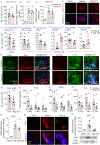
Nrg-1β1 treatment attenuates leucocyte infiltration and inflammation foci in the spinal cord of EAE mice. (A) Representative images of haematoxylin and eosin-Luxol fast blue stained spinal cord tissue show active inflammatory and demyelinating lesions (black arrows) in the white matter (WM) from vehicle and Nrg-1β1 treated animals 2 weeks after peak treatment. (B and C) Nrg-1β1 treatment significantly reduced the area and number of EAE lesions as compared to the vehicle group. *P <0.05; Student’s t-test. n = 5. (D) Representative images of perivascular and spinal cord tissue stained with the leucocyte marker CD45 and laminin in naïve, vehicle and Nrg-1β1 treated group. (E) Higher magnified images of CD45+/DAPI+ in the spinal cord. (F) Infiltrating CD45+ cells were significantly reduced after Nrg-1β1 treatment as compared to vehicle group. *P <0.05; Student’s t-test. n = 5. (GI) Multiplex Mesoscale ELISA at 2, 7 and 14 dpp revealed that Nrg-1β1 treatment significantly reduces the expression levels of chemokines involved in (G) recruitment of neutrophils, CXCL1/2 (KC-GRO), (H) MCP1 and (I) chemokine for T cells, CXCL10. Data represent mean ± SEM, *P <0.05; Student’s t-test. n = 4–8. (J) Gelatin zymography for MMP2 and MMP9 activity was performed on the spinal cord lysate samples of EAE mice treated with vehicle or Nrg-1β1. MMP9 activity was significantly elevated as the result of EAE, which was significantly reduced by Nrg-1β1 treatment to a level close to the basal level detected in naïve non-EAE spinal cord. (K) No alteration in activity of MMP2 was observed in vehicle or Nrg-1β1 treated group as compared to the naïve group. (L) Representative gelatin zymogram of MMP2 and MMP9 activity in the spinal cord lysate samples of EAE mice treated with vehicle or Nrg-1β1. Data represent mean fold change in expression ± SEM normalized to the naïve group. *P <0.05. One-way ANOVA followed by Holm-Sidak post hoc test. n = 4–6.
Figure 4
Figure 4
Nrg-1β1 treatment attenuates the expression of CSPGs in EAE lesions. (A) Representative images of immunohistochemical staining of CSPGs, microglia/macrophage (Iba-1) and astrocytes (GFAP) in naïve, vehicle and Nrg-1β1 treated groups. Treatments were administered for 2 weeks starting at peak of the disease. (B) CSPGs were highly upregulated in association with both astrocytes and microglia/macrophages as demonstrated by co-labelling with Iba-1 and GFAP in the high-magnification images of marked area. Yellow arrows show co-labelling with Iba-1 and GFAP, respectively. (C and D) Quantitative immunofluorescence intensity and slot blot analysis showed Nrg-1β1 treatment significantly abated the EAE-induced expression of CSPGs. Data represent mean fold change in expression ± SEM normalized to the naïve group. *P <0.05. One-way ANOVA followed by Holm-Sidak post hoc test. n = 4–5.
Figure 5
Figure 5
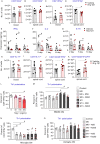
Nrg-1β1 suppresses monocyte expansion and infiltration and attenuates pro-inflammatory phenotype of microglia and macrophages in EAE mice. (A) Flow cytometric analysis of spinal cord from vehicle and Nrg-1β1 treated animals at 7 dpp showed that Nrg-1β1 did not change the overall population of CD3/CD11b+ microglia and macrophages. (B and C) Immunohistochemical cell density analysis of microglia/macrophage common marker, Iba-1 and microglia specific marker TMEM119 also confirmed that Nrg-1β1 did not alter the recruitment/activation of macrophages and resident microglia in the spinal cord as compared to the vehicle EAE groups. *P <0.05. One-way ANOVA, n = 3–4. (D) Representative images of TMEM119 immunostaining from spinal cord lesions of naïve, vehicle and NRG1 treated groups are shown. (E and F) Nrg-1β1 treatment significantly reduced circulating monocytes (CD11clo/CD11bhi/Ly6g/NK1.1) in the blood and infiltrating macrophages (CD3/CD49e+/CD11c/Ly6c+) in the spinal cord of Nrg-1β1 treated animals as compared to vehicle group. (G and H) Nrg-1β1 treatment also significantly reduced pro-inflammatory M1 (CD3/CD11b+/CD80+) microglia/macrophages, while promoted an anti-inflammatory M2 (CD3/CD11b+/CD206+) phenotype. (I and J) Monocyte derived M1 macrophages (CD3/CD49e+/CD80+) were also decreased in the spinal cord Nrg-1β1 treated mice, while ‘M2’ macrophages were increased (CD3/CD49e+/CD206+). (K and L) Representative images of EAE spinal cord immunostained with M1 marker (CD80) or ‘M2’ marker (CD206) co-labelled with microglia/macrophage markers Iba-1 or OX-42, respectively, show the M1 to M2 phenotype shift in microglia/macrophage population in Nrg-1β1 treated group as compared to vehicle group at the 7 dpp time point. (M) Flow cytometric assessment showed a significant reduction in total antigen presenting cells (CD3IA/IE+) in the EAE spinal cord under Nrg-1β1 treatment as compared to vehicle group. (NP) Cytokine analysis by ELISA in spinal cord tissues also revealed Nrg-1β1 treatment significantly reduced pro-inflammatory cytokines IL-1β, TNF-α, and IL-6. (Q and R) Reactive oxygen species (ROS) was detected in EAE mice by conversion of DHE to ethidium in the spinal cord tissue. EAE resulted in substantial increase in reactive oxygen species levels in the white matter of the spinal cord in which was significantly reduced by Nrg-1β1 treatment. (S) Slot blot analysis of oxidized lipids (E06) was performed on spinal cord lysates at 14 days after Nrg-1β1 treatment. EAE induced E06 levels, which was reduced significantly after Nrg-1β1 treatment as compared to the vehicle treated group. Data represent mean ± SEM. *P <0.05; Student’s t-test. n = 6–8.
Figure 6
Figure 6
Nrg-1β1 treatment promotes a T regulatory response directly and indirectly by modulating macrophages. (A and B) Flow-cytometry of vehicle and Nrg-1β1 treated mice after 7 days of treatment revealed no change in the total number of CD4+ T cells in the blood and spinal cord. (C and D) While the total number of effector T cells (CD4+IFNγ+) remained unchanged in the blood, there was a significant reduction in CD4+IFNγ+ cells in the spinal cord of Nrg-1β1 treated animals as compared to vehicle group. (EG) Cytokine assessment in spinal cord tissue with ELISA showed that Nrg-1β1 treatment significantly reduced key drivers of Th1 cell differentiation IFNγ, IL-2 and IL-16. *P <0.05; Student’s t-test. n = 6–8. (H and I) Flow-cytometry for CD4+IL-17+ effector T cell population after 7 days of Nrg-1β1 treatment did not show any change in this population in the blood or spinal cord. (J and K) A significant increase in anti-inflammatory T regulatory (CD4+/CD25+/FR4+) and CD4+/CD25+/FoxP3+ cells was observed with Nrg-1β1 treatment as compared to vehicle group. *P <0.05; Student’s t-test. n = 6–8. (LO) Naïve CD4+ T cells were polarized in vitro under Th1 conditions and were cultured with different concentration of Nrg-1β1, conditioned medium (CM) from normal (M0) and M1 polarized (IFNγ+LPS treated) microglia and BMDMs treated with Nrg-1β1 50 ng/ml or 200 ng/ml for 72 h. (L) Flow cytometric assessment revealed higher dose of Nrg-1β1 (200 ng/ml) significantly reduced CD4+IFNγ+ T cells. (M) While there was a significant increase in CD4+IFNγ+ cell population under BMDM M1 conditioned media, Nrg-1β1 200 ng/ml treated M1 BMDM conditioned media was able to diminish this increase significantly. (N) M0 Microglial conditioned media reduced the total number of Th1 polarized CD4+IFNγ+ T cells, (O) while astrocytes conditioned media did not alter Th1 cell population. *P <0.05. One-way ANOVA followed by Holm-Sidak post hoc test. n = 4–5.
Figure 7
Figure 7
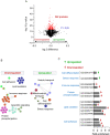
Proteomic analysis asserts that lipid oxidation and immune modulation are key Nrg-1β1 mediated mechanisms in EAE recovery. (A) Volcano plot illustrates differentially abundant proteins in the spinal cord of vehicle and Nrg-1β1 treated mice at 7 days post-peak of EAE. The −log10 is plotted against the log2 (fold change). The horizontal line denotes P =0.05, which was set as significance threshold (prior to logarithmic transformation). (B) Functional analysis of differentially expressed proteins using ClueGo plug-in in cytoscape software shows the interactions among the significantly different biological functions associated with upregulated and downregulated proteins in this study. Based on the κ score level, biological functions are depicted as coloured nodes linked to related groups. (C) Differentially expressed proteins were further analysed using DAVID software. The x-axis represents the fold enrichment of each biological function GO term. Only statistically significant GO terms are shown. Key for the GO terms are: GO:0098609, cell, cell adhesion; GO:0098641, cadherin binding involved in cell, cell adhesion; GO:0005913, cell, cell adherens junction; GO:0034440, lipid oxidation; GO:0005777, peroxisome; GO:0019395, fatty acid oxidation; GO:0009062, fatty acid catabolic process; GO:0006635, fatty acid beta, oxidation; GO:0004721, phosphoprotein phosphatase activity; GO:0006470, protein dephosphorylation; GO:0043123, positive regulation of I, kappaB kinase/NF, kappaB signaling; GO:0045087, innate immune response; GO:0033555, multicellular organismal response to stress; GO:0019771, negative regulation of cell morphogenesis involved in differentiation.
Figure 8
Figure 8
Nrg-1β1 is depleted in plasma and brain lesions of multiple sclerosis patients. (A) Post-mortem brain samples from multiple sclerosis patients were stained for Luxol fast blue-haematoxylin and eosin (HE-LFB) to identify demyelinating lesion in the white matter (B) Representative images of normal-appearing white matter (NAWM) or lesion area from multiple sclerosis (MS) brain sections stained with antibodies against Nrg-1β1 and MBP. (C) Quantification for Nrg-1β1 immunofluorescence intensity was performed from six different multiple sclerosis brain samples comparing the Nrg-1β1 intensity in lesion to the normal-appearing white matter from same section. Values are represented as fold change in intensity normalized to normal-appearing white matter for each sample which is shown as dotted baseline. There was 39% reduction in Nrg-1β1 expression within multiple sclerosis lesions as compared to the adjacent normal-appearing white matter. (DG) ELISA was performed for Nrg-1β1 on human plasma samples from normal individuals and multiple sclerosis patients. (D) Box plots show range of Nrg-1β1 levels (minimum to maximum) in normal controls (HC, n = 30) and multiple sclerosis patients (n = 136). (E) multiple sclerosis patients were categorized into disease modifying therapy (DMT, n = 88) or no DMT (n = 48) users at the time of sample collection. Nrg-1β1 levels of DMT users are indicated with dotted box plots. (F) CIS individuals showed a significant reduction in Nrg-1β1 levels in plasma as compared to normal individual samples. *P <0.05; Mann-Whitney U-test. (G) Nrg-1β1 plasma levels of CIS individuals were further categorized based on their subsequent diagnosis of multiple sclerosis (RRMS) in the follow-up years and compared to normal patient samples. Six of 11 CIS patients converted to RRMS and represented lower levels of Nrg-1β1 as compared to those who did not progress to multiple sclerosis (non-converter). (H) Multiple sclerosis patients were further categorized based on clinical diagnosis into different multiple sclerosis type/stage and whether they received any DMT or not (NDMT). Nrg-1β1 levels in plasma samples from CIS (n = 11; NDMT = 7, DMT = 4), RRMS (n = 113; NDMT = 31, DMT = 82) and SPMS (n = 12; NDMT = 10, DMT = 2) were analysed. Nrg-1β1 levels in CIS and SPMS patients who did not receive DMT were significantly reduced as compared to normal individuals. *P <0.05; Mann-Whitney U-test. (I) Nrg-1β1 plasma levels of normal individuals and multiple sclerosis patients (with or without DMT) were analysed against their EDSS. Nrg-1β1 levels were stable in DMT receiving patients irrespective of EDSS score, while multiple sclerosis patients without any DMT demonstrated lower levels of Nrg-1β1 as compared to normal individuals. The plus symbol indicates the mean value of Nrg-1β1 levels among analysed samples.

Comment in

References

    1. Agrawal SM, Silva C, Tourtellotte WW, Yong VW.. EMMPRIN: a novel regulator of leukocyte transmigration into the CNS in multiple sclerosis and experimental autoimmune encephalomyelitis. J Neurosci 2011; 31: 669–77. - PMC - PubMed
    1. Ajami B, Bennett JL, Krieger C, McNagny KM, Rossi FM.. Infiltrating monocytes trigger EAE progression, but do not contribute to the resident microglia pool. Nat Neurosci 2011; 14: 1142–9. - PubMed
    1. Alizadeh A, Dyck SM, Kataria H, Shahriary GM, Nguyen DH, Santhosh KT, et al.Neuregulin-1 positively modulates glial response and improves neurological recovery following traumatic spinal cord injury. Glia 2017; 65: 1152–75. - PubMed
    1. Alizadeh A, Santhosh KT, Kataria H, Gounni AS, Karimi-Abdolrezaee S.. Neuregulin-1 elicits a regulatory immune response following traumatic spinal cord injury. J Neuroinflammation 2018; 15: 53. - PMC - PubMed
    1. Asher RA, Morgenstern DA, Fidler PS, Adcock KH, Oohira A, Braistead JE, et al.Neurocan is upregulated in injured brain and in cytokine-treated astrocytes. J Neurosci 2000; 20: 2427–38. - PMC - PubMed

Publication types

MeSH terms