Review
. 2021 Apr;595(8):1025-1061.
doi: 10.1002/1873-3468.14024. Epub 2020 Dec 22.

The Diseased Mitoribosome

Affiliations
Review

The Diseased Mitoribosome

Alberto Ferrari et al. FEBS Lett. 2021 Apr.

Abstract

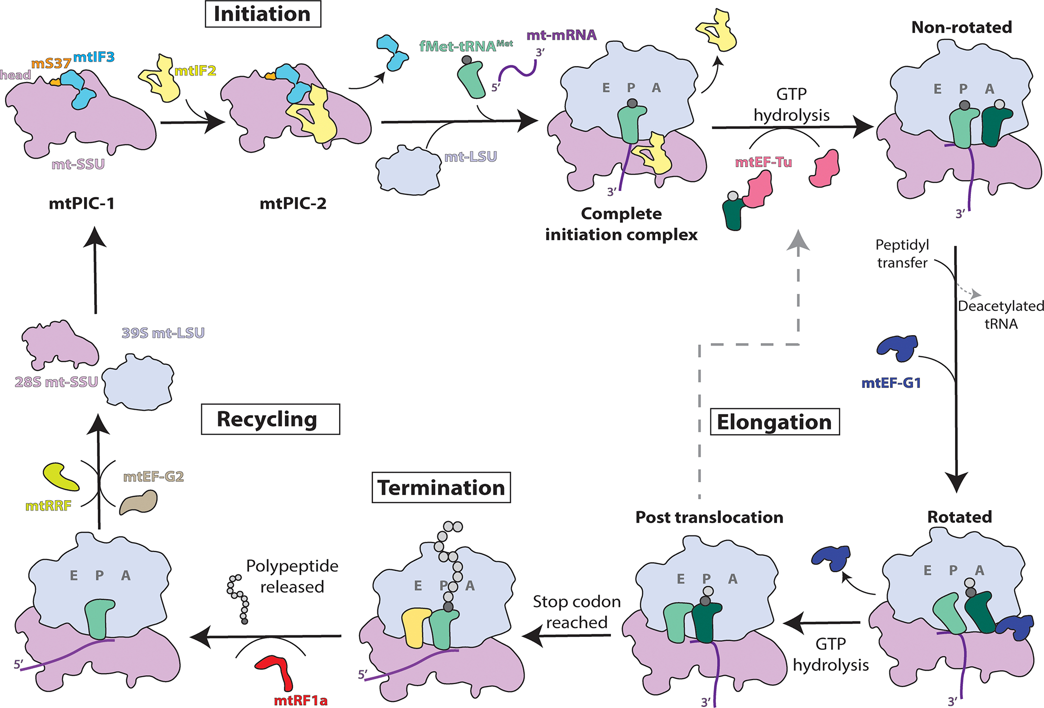
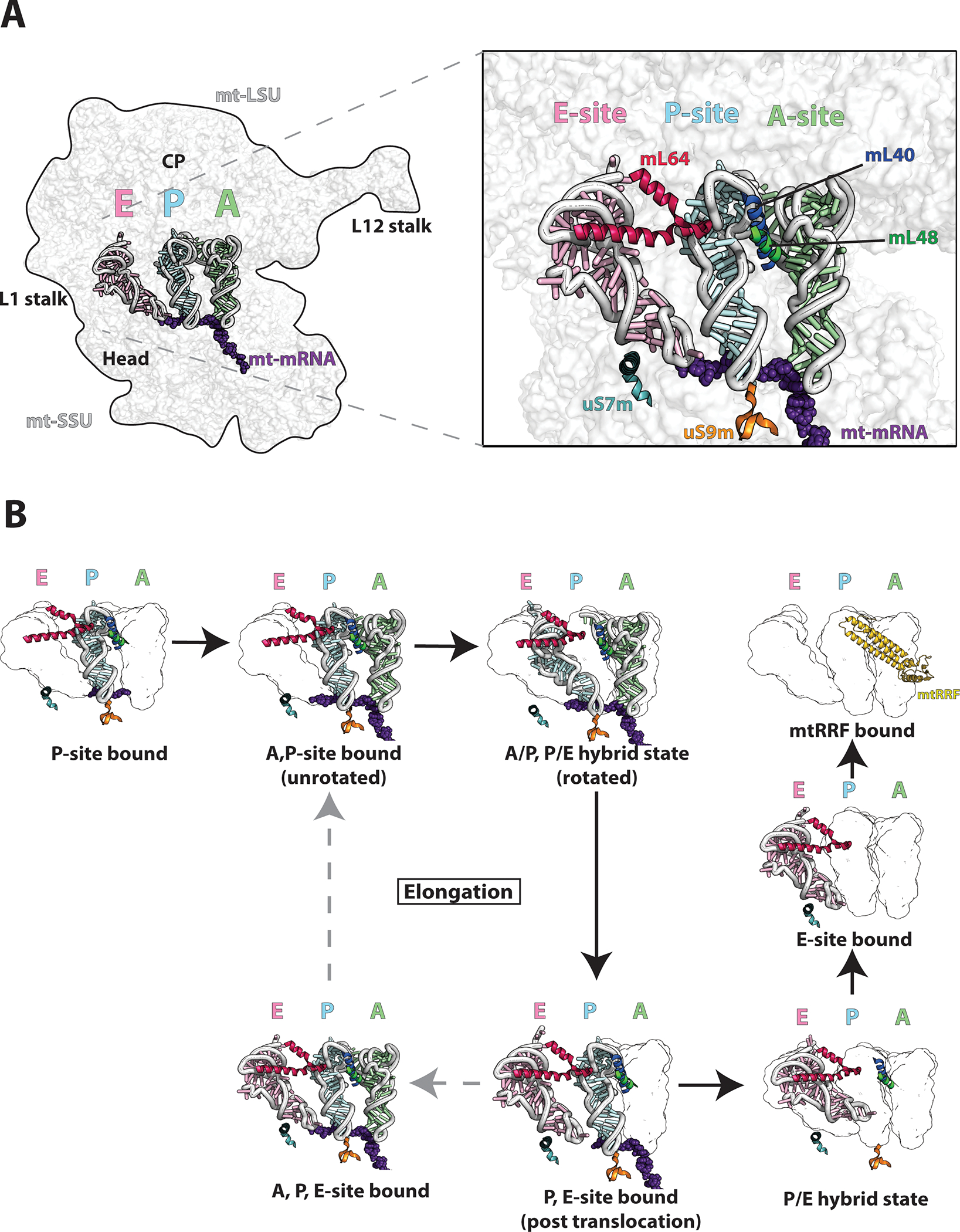
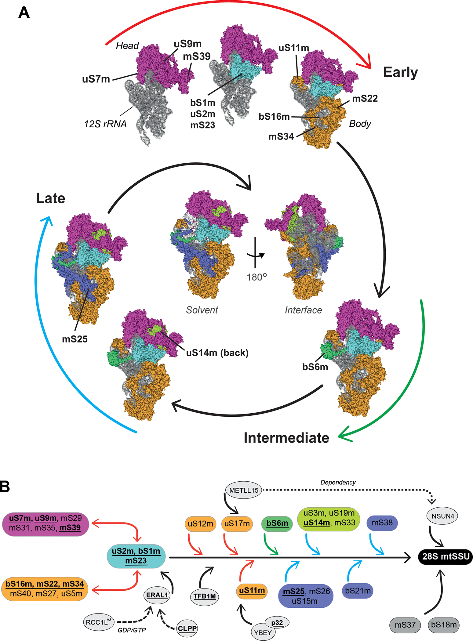
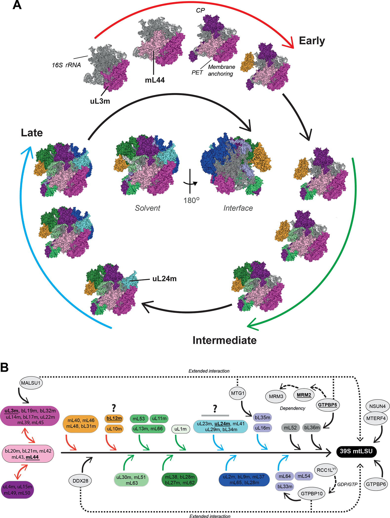
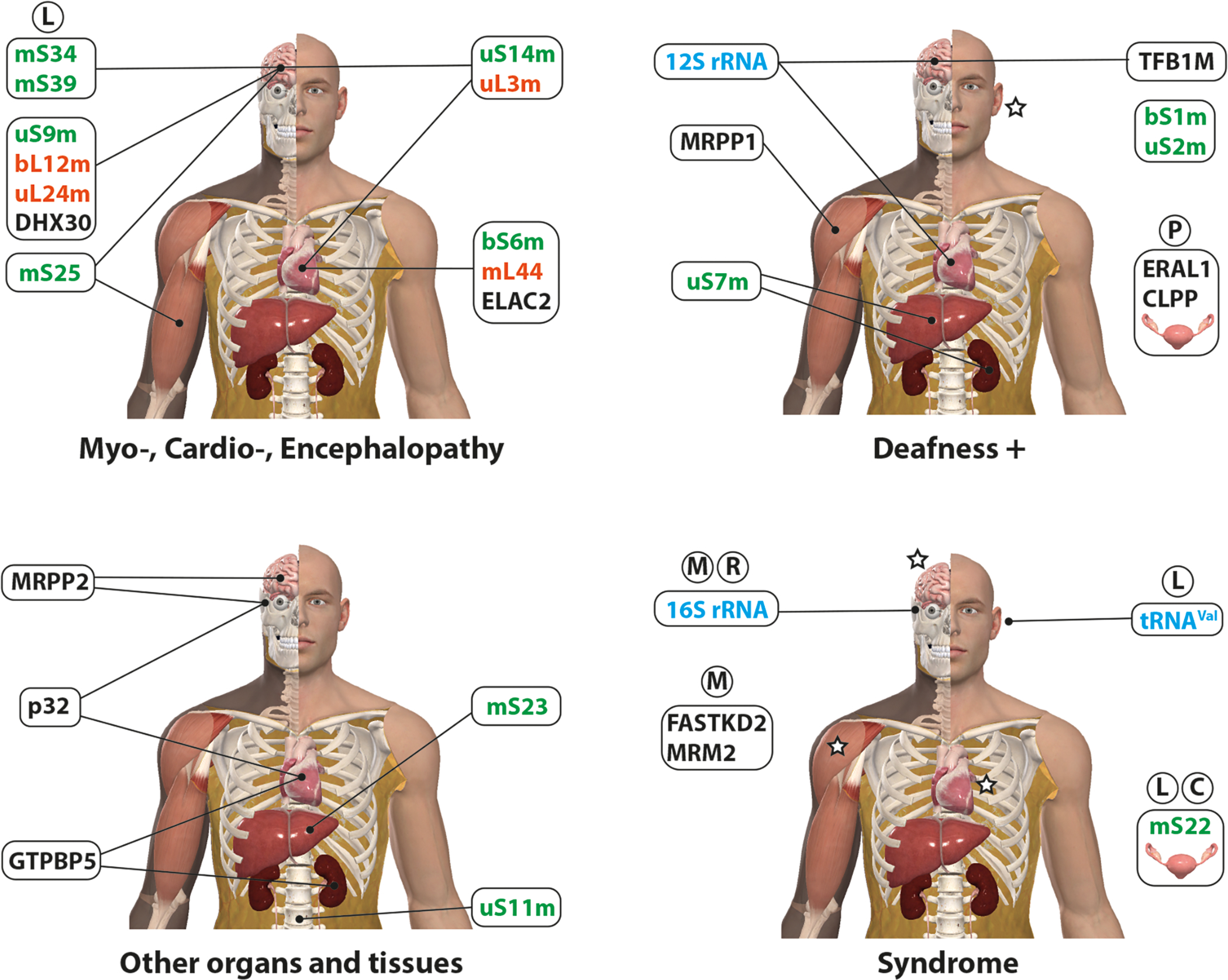
Mitochondria control life and death in eukaryotic cells. Harboring a unique circular genome, a by-product of an ancient endosymbiotic event, mitochondria maintains a specialized and evolutionary divergent protein synthesis machinery, the mitoribosome. Mitoribosome biogenesis depends on elements encoded in both the mitochondrial genome (the RNA components) and the nuclear genome (all ribosomal proteins and assembly factors). Recent cryo-EM structures of mammalian mitoribosomes have illuminated their composition and provided hints regarding their assembly and elusive mitochondrial translation mechanisms. A growing body of literature involves the mitoribosome in inherited primary mitochondrial disorders. Mutations in genes encoding mitoribosomal RNAs, proteins, and assembly factors impede mitoribosome biogenesis, causing protein synthesis defects that lead to respiratory chain failure and mitochondrial disorders such as encephalo- and cardiomyopathy, deafness, neuropathy, and developmental delays. In this article, we review the current fundamental understanding of mitoribosome assembly and function, and the clinical landscape of mitochondrial disorders driven by mutations in mitoribosome components and assembly factors, to portray how basic and clinical studies combined help us better understand both mitochondrial biology and medicine.

Keywords: OXPHOS deficiency; mitochondrial disease; mitochondrial ribosome; mitochondrial translation; mitoribosome assembly.


Conflict of interest statement

Conflict of interest statement.

None declared.

Figures

Figure 1.
Figure 1.. The mitochondrial translation process.
Schematic overview of mitochondrial translation indicating the major steps of initiation, elongation, termination, and recycling. Mitochondrial pre-initiation complexes are denoted mtPIC-1 and mtPIC-2. See full explanation in the text.
Figure 2.
Figure 2.. Structure of mt-tRNAs in the translating mitoribosome.
A) The cryo-EM structure (PDB-6ZS9) [15] is used to depict the mitoribosome bound to A,P, and E-site tRNAs highlighting key structural features (left). Detailed view of A,P,E-site tRNAs with bound mt-mRNA. mL40 N-terminal helix (blue) interacts with both A and P-site tRNA elbows with supporting hydrophobic interactions from the mL48 C-terminal helix (green). uS9m C-terminal tail (orange) binds the P-site tRNA anticodon stem loop. C-terminal helix of mL64 (red) interacts with the E-site tRNA elbow. uS7m C-terminal helix (teal) stabilizes the E-site tRNA through alignment with the major groove of the E-site tRNA anticodon stem. B) Eight representative cryo-EM structures [15] indicating tRNA translocation during elongation and termination/recycling. The figures were prepared using PYMOL and Adobe Illustrator software.
Figure 3.
Figure 3.. Mitoribosome SSU assembly.
(A) Model of human 28S mt-SSU biogenesis depicting a hierarchical and module-based protein assembly pathway as reported [21]. All modules are color-coded. The 12S rRNA is shown in grey. The assembly process is divided in three stages: early, intermediate, and late (see explanation in the text). Solvent-facing views of the cryo-EM structure (PDB-3J9M) [13] are used to depict the assembly pathway. The localization of mt-SSU protein components that have been found mutated in patients suffering from primary mitochondrial disorders is indicated. In the center of the spiral scheme, complete solvent-facing and interface-facing views of the 28S mt-SSU are presented. The figures were prepared using PYMOL and Adobe Illustrator software. (B) Human 28S mt-SSU assembly pathway as determined by SILAC-proteomics [21], including known assembly factors at their approximate stage of incorporation. Boxes, highlighted with the same color used in the spiral assembly, represent different protein clusters at different assembly stages: early (red arrows), intermediate (green arrows) late (blue arrows). Proteins in gray boxes were not assigned to any assembly stage [21]. Disease-driven mitoribosome proteins and assembly factors are in bold and underlined. Continuous arrows highlight the activity of assembly factors during the assembly process while dotted arrows indicate dependency or extended interaction until the mt-SSU formation.
Figure 4.
Figure 4.. Mitoribosome LSU assembly.
(A) Model of human 39S mt-LSU biogenesis depicting a hierarchical and module-based protein assembly pathway as reported [21]. All modules are color-coded. The 16S rRNA is shown in grey. The assembly process is divided in three stages: early, intermediate, and late (see explanation in the text). Solvent-facing views of the cryo-EM structure (PDB-3J9M) [13] are used to depict the assembly pathway. The localization of mt-SSU protein components that have been found mutated in patients suffering from primary mitochondrial disorders is indicated. In the center of the spiral scheme, complete solvent-facing and interface-facing views of the 28S mt-SSU are presented. The figures were prepared using PYMOL and Adobe Illustrator software. CP, central protuberance; PET, polypeptide exit tunnel. (B) Human 39S mt-LSU assembly pathway as determined by SILAC-proteomics [21], incorporating known assembly factors at their approximate stage of incorporation. Boxes, highlighted with the same color used in the spiral assembly, represent different protein clusters at different assembly stages: early (red arrows), intermediate (green arrows) late (blue arrows). Proteins in gray boxes were not assigned to any assembly stage [21]. Disease-driven mitoribosome proteins and assembly factors are in bold and underlined. Continuous arrows highlight the activity of assembly factors during the assembly process while dotted arrows their dependency or extended interaction until the mt-LSU formation. Question marks highlight proteins whose proposed assembly kinetics [21] differ from bacterial and yeast mitoribosome systems (see explanation in the text).
Figure 5.
Figure 5.. Defective mitochondrial ribosome assembly and human disease.
Schematic representation of organs and tissues affected by mutations in mitoribosome components: assembly factors (black), tRNA or rRNA (light blue), mt-SSU proteins (green) and mt-LSU proteins (orange). For clarity, four human body models, generated by using the Complete Anatomy software, are presented.= indicate organs or tissues commonly affected by all the mitoribosome components in the same group, C = Cornelia de Lange syndrome, L = Leigh syndrome, M = MELAS, P= Perrault Syndrome, R = Rett Syndrome.

References

    1. McLean JR, Cohn GL, Brandt IK and Simpson MV (1958). Incorporation of labeled amino acids into the protein of muscle and liver mitochondria. J. Biol. Chem. 233, 657–63. - PubMed
    1. Rendi R (1959). On the occurrence of intramitochondrial ribonucleoprotein particles. Exp. Cell Res. 17, 585–7. - PubMed
    1. Kalf GF (1963). The incorporation of leucine-1-C-14 into the protein of rat heart sarcosomes: an investigation of optimal conditions. Arch. Biochem. Biophys. 101, 350–59. - PubMed
    1. Swift H (1965). Nucleic Acids of Mitochondria and Chloroplasts. The American Naturalist 99, 201–214.
    1. Küntzel H and Noll H (1967). Mitochondrial and cytoplasmic polysomes from Neurospora crassa. Nature 215, 1340–5. - PubMed

Publication types

MeSH terms