. 2021 May;238(5):1106-1115.
doi: 10.1111/joa.13367. Epub 2020 Dec 11.

Id(entifying) the inhibitor of DNA binding 3 in the brain of Nothobranchius furzeri upon aging

Affiliations

Id(entifying) the inhibitor of DNA binding 3 in the brain of Nothobranchius furzeri upon aging

Adele Leggieri et al. J Anat. 2021 May.

Abstract

Inhibitors of DNA (Id) are key transcription factors (TFs) regulating neurogenic processes. They belong to the helix-loop-helix (HLH) TF family and are dominant negative regulators of basic HLH proteins (bHLHs). Specifically, they inhibit cell differentiation and enhance cell proliferation and motility. The Id family includes four members, Id1, Id2, Id3, and Id4, which have been identified in nearly all vertebrates. The transcript catalog of the African turquoise killifish, Nothobranchius furzeri, contains all four TFs and has evolved showing positive selection for Id3. N. furzeri, a teleost, is the short-lived vertebrate and is gaining increasing scientific interest as a new model organism in aging research. It is characterized by embryonic diapause, explosive sexual maturation, and rapid aging. In this study, we investigated both the expression and the role of Id3 in the brain of this model organism. Interestingly, Id3 was upregulated age-dependently along with a distribution pattern resembling that of other vertebrates. Additionally, the gene has undergone positive selection during evolution and shows a high degree of conservation relative to that of other vertebrates. These features make N. furzeri a valid tool for aging studies and a potential model in translational research.

Keywords: Id3; aging; central nervous system; fish.


Conflict of interest statement

None declared.

Figures

FIGURE 1
FIGURE 1
(a) Id3 evolutionary history and (b) protein alignment. The neighbor‐joining method was employed. The optimal tree with the sum of branch length = 79,78044216 is shown. The tree is drawn to scale with branch lengths in the same units as those of the evolutionary distances used to infer the phylogenetic tree. The evolutionary distances were computed using the number of differences method and are in the units of the number of base differences per sequence. The analysis involved 22 nucleotide sequences. Codon positions included were 1st+2nd+3rd+Noncoding. All positions containing gaps and missing data were eliminated. There were a total of 1856 positions in the final dataset. Protein sequence alignment of human, mouse, zebrafish, and Nothobranchius furzeri Id3 was created with the Clustal W program (Thompson et al., 1994). “*” indicates fully conserved residues, and “:” and “.” indicate conserved residues of strong or weaker groups as defined by (Thompson et al., 1997)
FIGURE 2
FIGURE 2
Id3 mRNA levels upon aging. qPCR was assessed to evaluate brain expression of Nothobranchius furzeri Id3 mRNA in young (5 wph) and old (27 wph) animals. The analysis has shown that Id3 mRNA is slightly upregulated during aging (p = 0.0006), for p ≤ 0.05. Expression levels were analyzed by the ΔΔCt method and normalized to TBP. The relative ΔΔ curve threshold was built on fold change values
FIGURE 3
FIGURE 3
Id3 mRNA distribution in transverse sections of the brain of young Nothobranchius furzeri. (a) Positive staining in the dorsal telencephalon; (b, b′) High labeling in diencephalic regions, optic tectum, and longitudinal tori. In schematic drawings of N. furzeri brain, red dots indicate Id3 mRNA distribution over the different brain areas. Scale bars: a ‐, b′ = 100 µm; b = 200 µm. Abbreviations: Dlv, ventrolateral zone of dorsal telencephalon; Hd, dorsal hypothalamus; Hl, lateral hypothalamus; NG, glomerular nucleus; PGl, lateral preglomerular nucleus; PGZ, periventricular gray zone; Tl, longitudinal tori
FIGURE 4
FIGURE 4
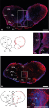
Id3 mRNA distribution in transverse sections of the brain of old Nothobranchius furzeri. Positive staining in: (a, a′) olfactory bulbs and dorsal telencephalon, (b, b′) diencephalon. In schematic drawings of N. furzeri brain, red dots indicateId3 mRNA distribution over the different brain areas. Scale bars: a, b, =100 µm; a’, =200 µm; b’ =300 µm. Abbreviations: A, anterior thalamic nucleus; CPN, central pretectal nucleus; Dc, central zone of dorsal telencephalon; Dd, dorsal zone of dorsal telencephalon; Dld, dorsolateral zone of dorsal telencephalon; Dlv, ventrolateral zone of dorsal telencephalon; Dm, medial zone of dorsal telencephalon; ECL, external cellular layer; Ha, habenulae; Hd, dorsal hypothalamic; Hv, central hypothalamus; I, intermediate thalamic nucleus; ICL, internal cellular layer; lfb, lateral forebrain bundle; OT, optic tectum; PGa, anterior preglomerular nucleus; VAO, ventral accessory nucleus; VM, ventromedial thalamic nucleus; VL, ventrolateral thalamic nucleus
FIGURE 5
FIGURE 5
Id3 mRNA distribution in transverse sections of the brain of old Nothobranchius furzeri. Positive staining in: (a–c) mesencephalon, and (d–f) rhombencephalon. In schematic drawings of N. furzeri brain, red dots indicate Id3 mRNA distribution over the different brain areas. Scale bars: a, c = 100 µm; a′= 200 µm; b, c, d = 300 µm; b, f: 500 µm. Abbreviations: CC, cerebellar cristae; CCe, corpus of cerebellum; EG, granular eminentiae; Hd, dorsal hypothalamus; Hl, hypothalamus subdivision; NSF, nucleus of solitary fascicle; OT, optic tectum; PGZ, periventricular gray zone; Tl, longitudinal tori

References

    1. Afouda, A.B. , Reyonaud‐Deonauth, S. , Mohun, T. & Spohr, G. (1999) Localized XId3 mRNA activation in Xenopus embryos by cytoplasmatic polyadenylation. Mechanisms of Development, 88(1), 15–31. - PubMed
    1. Andres‐Barquin, P. , Hernandez, M.C. & Israel, M.A. (2000) Id genes in nervous system development. Histology and Histopathology, 15(2), 603–618. - PubMed
    1. Armant, O. , Marz, M. , Schmidt, R. , Ferg, M. , Diotel, N. , Ertzer, R. et al. (2013) Genome‐wide, whole mount in situ analysis of transcriptional regulators in zebrafish embryos. Developmental Biology, 380(2), 351–362. - PMC - PubMed
    1. Baumgart, M. , Groth, M. , Priebe, S. et al. (2014) RNA‐seq of the aging brain in the short‐lived fish N. furzeri ‐ conserved pathways and novel genes associated with neurogenesis. Aging Cell, 13(6), 965–974. - PMC - PubMed
    1. Benezra, R. (1993) Id as a general negative regulator of the helix‐loop‐helix family of DNA‐binding proteins. Pharmacology of Cell Differentiation, 1016, 171–180.

Publication types

Substances