Changes in left ventricular electromechanical relations during targeted hypothermia
- PMID: 33315166
- PMCID: PMC7736464
- DOI: 10.1186/s40635-020-00363-7
Changes in left ventricular electromechanical relations during targeted hypothermia
Abstract
Background: Targeted hypothermia, as used after cardiac arrest, increases electrical and mechanical systolic duration. Differences in duration of electrical and mechanical systole are correlated to ventricular arrhythmias. The electromechanical window (EMW) becomes negative when the electrical systole outlasts the mechanical systole. Prolonged electrical systole corresponds to prolonged QT interval, and is associated with increased dispersion of repolarization and mechanical dispersion. These three factors predispose for arrhythmias. The electromechanical relations during targeted hypothermia are unknown. We wanted to explore the electromechanical relations during hypothermia at 33 °C. We hypothesized that targeted hypothermia would increase electrical and mechanical systolic duration without more profound EMW negativity, nor an increase in dispersion of repolarization and mechanical dispersion.
Methods: In a porcine model (n = 14), we registered electrocardiogram (ECG) and echocardiographic recordings during 38 °C and 33 °C, at spontaneous and atrial paced heart rate 100 beats/min. EMW was calculated by subtracting electrical systole; QT interval, from the corresponding mechanical systole; QRS onset to aortic valve closure. Dispersion of repolarization was measured as time from peak to end of the ECG T wave. Mechanical dispersion was calculated by strain echocardiography as standard deviation of time to peak strain.
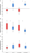
Results: Electrical systole increased during hypothermia at spontaneous heart rate (p < 0.001) and heart rate 100 beats/min (p = 0.005). Mechanical systolic duration was prolonged and outlasted electrical systole independently of heart rate (p < 0.001). EMW changed from negative to positive value (- 20 ± 19 to 27 ± 34 ms, p = 0.001). The positivity was even more pronounced at heart rate 100 beats/min (- 25 ± 26 to 41 ± 18 ms, p < 0.001). Dispersion of repolarization decreased (p = 0.027 and p = 0.003), while mechanical dispersion did not differ (p = 0.078 and p = 0.297).
Conclusion: Targeted hypothermia increased electrical and mechanical systolic duration, the electromechanical window became positive, dispersion of repolarization was slightly reduced and mechanical dispersion was unchanged. These alterations may have clinical importance. Further clinical studies are required to clarify whether corresponding electromechanical alterations are accommodating in humans.
Keywords: Echocardiography; Electromechanical relations; Electromechanical window; Myocardial function; Targeted hypothermia; Ventricular arrhythmia.
Conflict of interest statement
The authors declare that they have no competing interests.
Figures


References
-
- Nolan JP, Soar J, Cariou A, Cronberg T, Moulaert VR, Deakin CD, Bottiger BW, Friberg H, Sunde K, Sandroni C. European Resuscitation Council and European Society of Intensive Care Medicine Guidelines for Post-resuscitation care 2015: Section 5 of the European Resuscitation Council Guidelines for resuscitation 2015. Resuscitation. 2015;95:202–222. doi: 10.1016/j.resuscitation.2015.07.018. - DOI - PubMed
LinkOut - more resources
Full Text Sources

