. 2020 Dec 14;16(12):e1009242.
doi: 10.1371/journal.pgen.1009242. eCollection 2020 Dec.

Accurate mapping of mitochondrial DNA deletions and duplications using deep sequencing

Affiliations

Accurate mapping of mitochondrial DNA deletions and duplications using deep sequencing

Swaraj Basu et al. PLoS Genet. .

Abstract

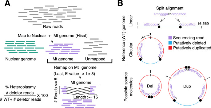
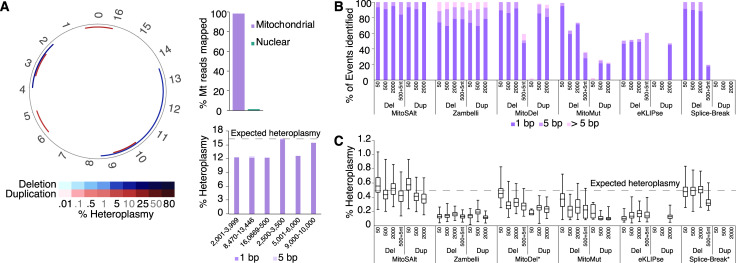
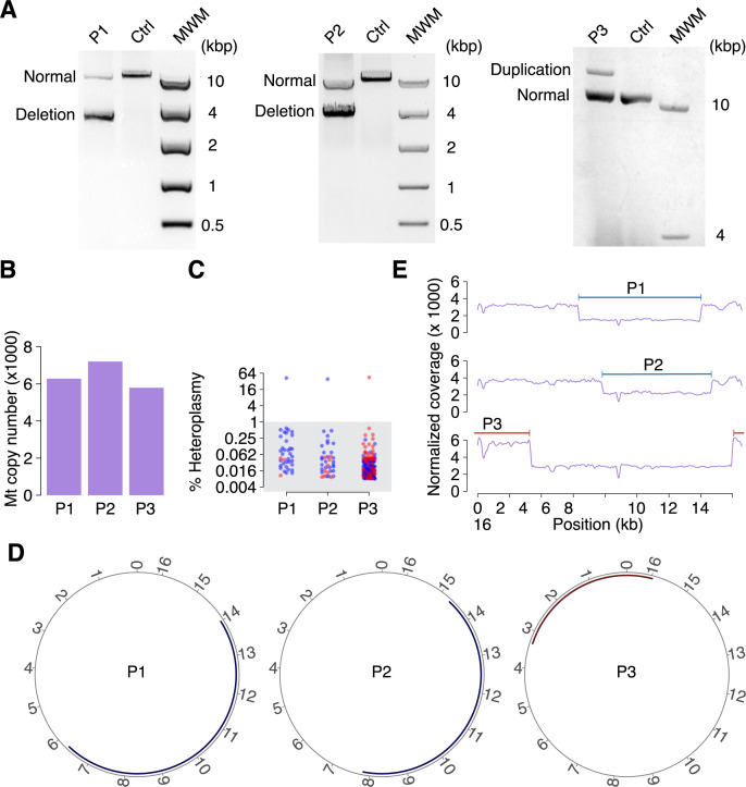
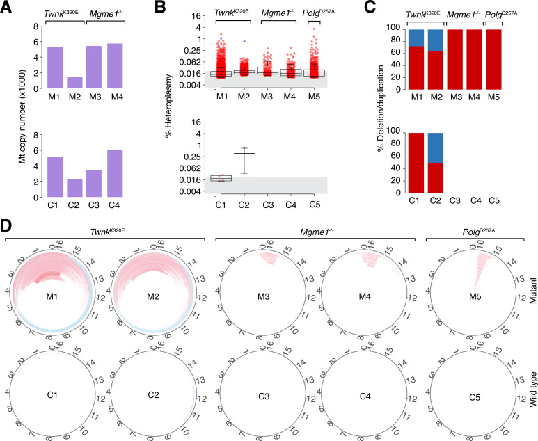
Deletions and duplications in mitochondrial DNA (mtDNA) cause mitochondrial disease and accumulate in conditions such as cancer and age-related disorders, but validated high-throughput methodology that can readily detect and discriminate between these two types of events is lacking. Here we establish a computational method, MitoSAlt, for accurate identification, quantification and visualization of mtDNA deletions and duplications from genomic sequencing data. Our method was tested on simulated sequencing reads and human patient samples with single deletions and duplications to verify its accuracy. Application to mouse models of mtDNA maintenance disease demonstrated the ability to detect deletions and duplications even at low levels of heteroplasmy.


Conflict of interest statement

The authors have declared that no competing interests exist.

Figures

Fig 1
Fig 1. MitoSAlt pipeline overview.
(A) Raw sequencing reads are mapped first to the nuclear and mitochondrial (Mt) genomes using a fast aligner, followed by precision alignment of unmapped and Mt mapped reads to the Mt genome to identify “split” reads informative of structural breakpoints. (B) Dual interpretation of split alignments: a split read can represent either a deletion or a complementary arc duplication, and these scenarios are indistinguishable using short-read sequencing.
Fig 2
Fig 2. MitoSAlt pipeline performance on simulated data.
(A) Evaluation on simulated sequencing data harboring two synthetic deletions and 4 duplications, each at 16.7% heteroplasmy (2 × 126 bp, 10,000,000 reads, resulting in ~2,000× mtDNA coverage). The circular plot shows deleted (blue) or duplicated (red) segments. The upper bar graph indicates the fraction of Mt reads mapped to the mitochondrial genome, while the lower shows heteroplasmy levels estimated by MitoSAlt for each event (events with 1 bp or 5 bp of the expected breakpoints are quantified separately). (B) Evaluation of sensitivity on simulated sequencing datasets containing large numbers of low heteroplasmy deletions and duplications of various sizes. Each data set contained 200 minor or major arc events, each at 0.5% heteroplasmy (2 × 126 bp, 50,000,000 reads, resulting in ~6,000× mtDNA coverage. “500+5nt” refers to 500 bp deletions with 5 bp non-template random insertions at the breakpoint. (C) Box and whisker plot of heteroplasmy levels estimated by different pipelines. The boxes show 25th to 75th percentiles, and whiskers show the minimum and maximum value. *, These tools do not directly report heteroplasmy levels, and estimates were instead made based on the reported number of reads supporting each event and the average mitochondrial read-depth.
Fig 3
Fig 3. Assessment of MitoSAlt on patient samples with a single deletion or duplication.
(A) Total DNA from patients (P1, P2 and P3) and controls were analyzed by LX-PCR using two different primer sets. A single deletion was detected in patients P1 and P2 using primers LX1 and LX2, while a single duplication was detected in P3 with primers LX3 and LX4. Amplicons from wild type mtDNA (denoted “normal”) were also detected in all patients. (B) Predicted mtDNA copy number in the patients. (C) Heteroplasmy levels for the identified deletions/duplications (marked in blue and red, respectively) in the patient samples. All cases have single events at heteroplasmy levels (> 35%), in addition to multiple low-heteroplasmy alterations (<1%, grey area). (D) Circular plots showing deletions/duplications at heteroplasmy >1%, all being consistent with the LX-PCR results. (E) Read coverage depth across the Mt genome for the human samples shows drastic changes in the regions identified as being deleted or duplicated (marked in blue and red respectively). MWM, molecular weight marker.
Fig 4
Fig 4. Identification of mtDNA structural alterations in wild-type and Mgme1, Twnk or Polg mutant mice using the MitoSAlt pipeline.
(A) Predicted copy number for the given mutant and wild-type samples (denoted M and C, respectively). Copy number could not be estimated for the Polg samples due to use of an mtDNA enrichment protocol. (B) Heteroplasmy levels for the deletions (blue) and duplications (red) identified in the mutant and wild-type samples. The grey area delineates low-heteroplasmy events (<0.02%). (C) Fraction deletions (blue) and duplications (red) in each sample. (D) Circular plots showing the deletions (blue) and duplications (red) identified in the mutant and wild-type samples. For visual clarity, a heteroplasmy cut-off of 0.02% was used for all samples.

References

    1. Anderson S, Bankier AT, Barrell BG, de Bruijn MH, Coulson AR, Drouin J, et al. Sequence and organization of the human mitochondrial genome. Nature. 1981;290(5806):457–65. Epub 1981/04/09. 10.1038/290457a0 . - DOI - PubMed
    1. Andrews RM, Kubacka I, Chinnery PF, Lightowlers RN, Turnbull DM, Howell N. Reanalysis and revision of the Cambridge reference sequence for human mitochondrial DNA. Nat Genet. 1999;23(2):147 Epub 1999/10/03. 10.1038/13779 . - DOI - PubMed
    1. Van Goethem G, Dermaut B, Lofgren A, Martin JJ, Van Broeckhoven C. Mutation of POLG is associated with progressive external ophthalmoplegia characterized by mtDNA deletions. Nat Genet. 2001;28(3):211–2. Epub 2001/06/30. 10.1038/90034 . - DOI - PubMed
    1. Spelbrink JN, Li FY, Tiranti V, Nikali K, Yuan QP, Tariq M, et al. Human mitochondrial DNA deletions associated with mutations in the gene encoding Twinkle, a phage T7 gene 4-like protein localized in mitochondria. Nat Genet. 2001;28(3):223–31. Epub 2001/06/30. 10.1038/90058 . - DOI - PubMed
    1. Goffart S, Cooper HM, Tyynismaa H, Wanrooij S, Suomalainen A, Spelbrink JN. Twinkle mutations associated with autosomal dominant progressive external ophthalmoplegia lead to impaired helicase function and in vivo mtDNA replication stalling. Hum Mol Genet. 2009;18(2):328–40. Epub 2008/10/31. 10.1093/hmg/ddn359 - DOI - PMC - PubMed

Publication types

MeSH terms

Substances

LinkOut - more resources