. 2021 Jan:148:105223.
doi: 10.1016/j.nbd.2020.105223. Epub 2020 Dec 11.

Neural endophenotypes and predictors of laryngeal dystonia penetrance and manifestation

Affiliations

Neural endophenotypes and predictors of laryngeal dystonia penetrance and manifestation

Sanaz Khosravani et al. Neurobiol Dis. 2021 Jan.

Abstract

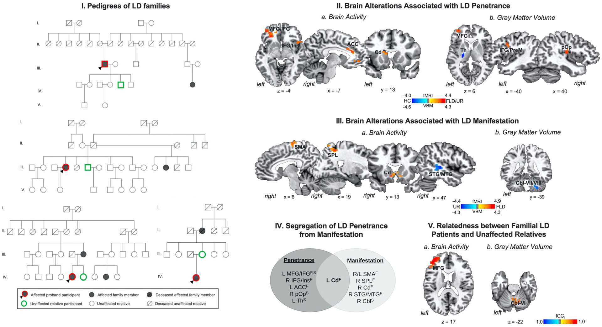
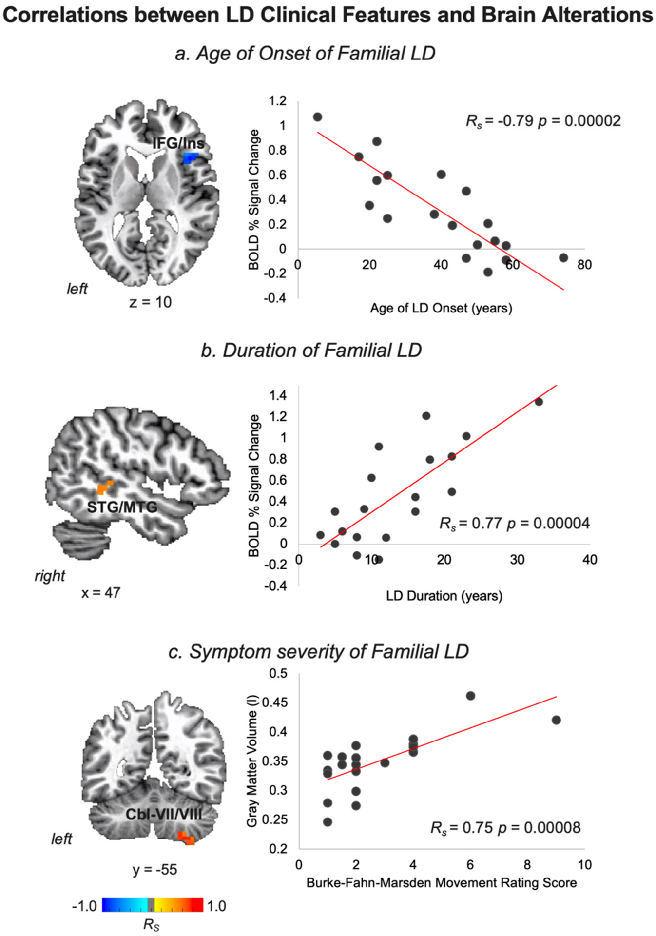
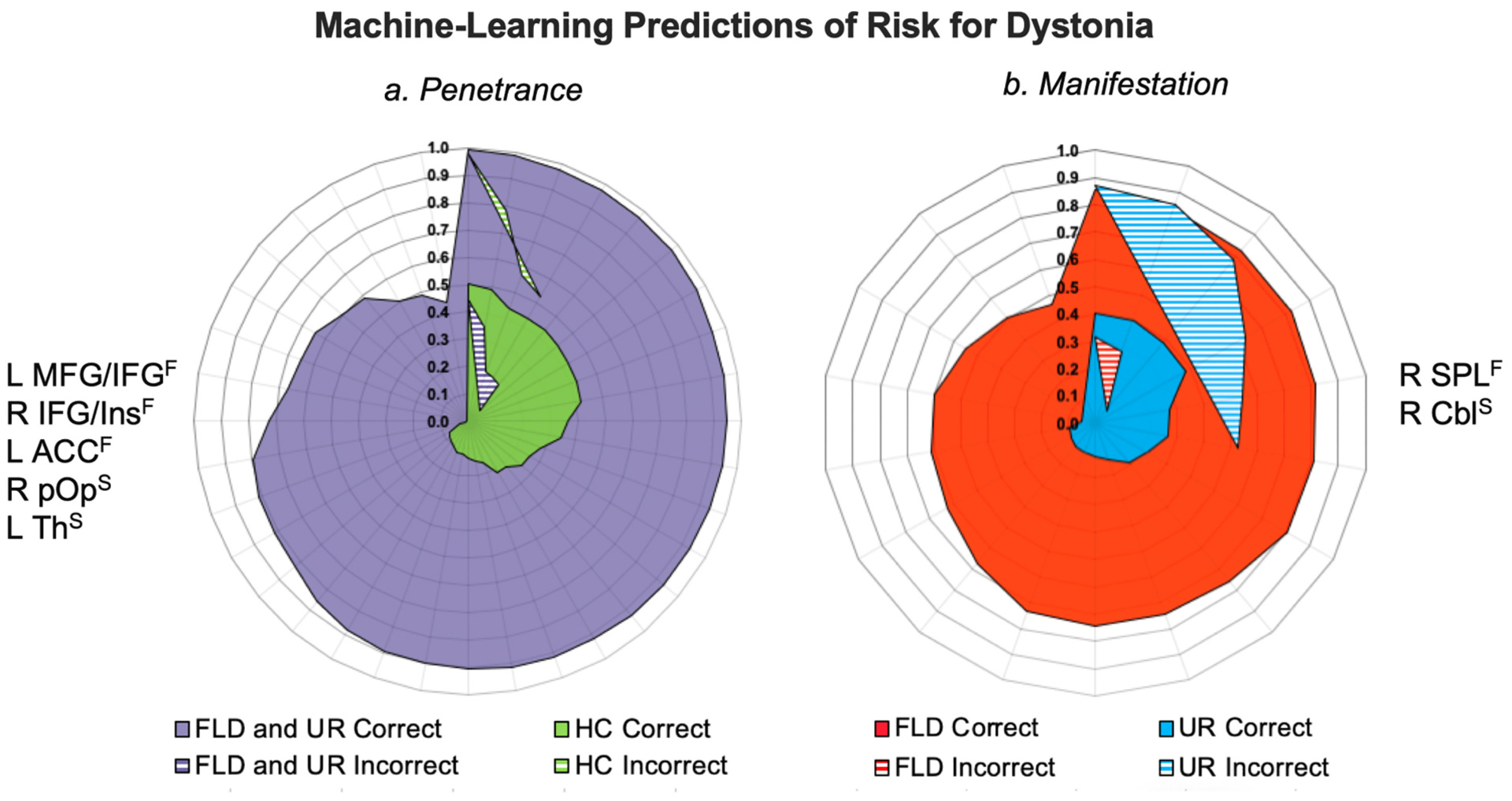
Focal dystonias are the most common forms of isolated dystonia; however, the etiopathophysiological signatures of disorder penetrance and clinical manifestation remain unclear. Using an imaging genetics approach, we investigated functional and structural representations of neural endophenotypes underlying the penetrance and manifestation of laryngeal dystonia in families, including 21 probands and 21 unaffected relatives, compared to 32 unrelated healthy controls. We further used a supervised machine-learning algorithm to predict the risk for dystonia development in susceptible individuals based on neural features of identified endophenotypes. We found that abnormalities in prefrontal-parietal cortex, thalamus, and caudate nucleus were commonly shared between patients and their unaffected relatives, representing an intermediate endophenotype of laryngeal dystonia. Machine learning classified 95.2% of unaffected relatives as patients rather than healthy controls, substantiating that these neural alterations represent the endophenotypic marker of dystonia penetrance, independent of its symptomatology. Additional abnormalities in premotor-parietal-temporal cortical regions, caudate nucleus, and cerebellum were present only in patients but not their unaffected relatives, likely representing a secondary endophenotype of dystonia manifestation. Based on alterations in the parietal cortex and caudate nucleus, the machine learning categorized 28.6% of unaffected relative as patients, indicating their increased lifetime risk for developing clinical manifestation of dystonia. The identified endophenotypic neural markers may be implemented for screening of at-risk individuals for dystonia development, selection of families for genetic studies of novel variants based on their risk for disease penetrance, or stratification of patients who would respond differently to a particular treatment in clinical trials.

Keywords: Brain imaging; Dystonia; Endophenotypes.


Conflict of interest statement

Declaration of Competing Interest

The authors report no conflict of interest.

Figures

Fig. 1.
Fig. 1.
(I) Example pedigrees of families of patients with laryngeal dystonia (LD). (II) Brain alterations associated with LD penetrance are based on differences in (a) brain activity and (b) gray matter volume in familial LD patients and their unaffected relatives vs. healthy controls. (III) Brain alterations associated with LD manifestation are based on differences in (a) brain activity and (b) gray matter volume in familial LD patients vs. their unaffected relatives. (IV) Schematic of functional and structural alterations segregating LD penetrance from manifestation. (V) Relatedness between patients with familial LD and their unaffected relatives as a function of (a) brain activity and (b) gray matter volume. Brain alterations are shown on a series of axial, sagittal or coronal brain slices in the AFNI standard Talairach-Tournoux space. The color bar indicates the z-statistics in (II, III) and the intraclass correlation index (ICCi) in (V). Abbreviations: ACC - anterior cingulate cortex, Cbl - cerebellum, Cd - caudate nucleus, F – functional alteration, FLD – familial laryngeal dystonia patients, HC – healthy controls, ICCi – intraclass correlation index, IFG - inferior frontal gyrus, Ins – insula, L – left, MFG - middle frontal gyrus, MTG - middle temporal gyrus, PrM - premotor cortex, pOp - parietal operculum, R – right, SMA - supplementary motor area, SPL - superior parietal lobule, STG - superior temporal gyrus, S – structural alteration, Th – thalamus, UR – unaffected relatives.
Fig. 2.
Fig. 2.
Correlations between LD clinical features and brain alterations are depicted in a series of axial, sagittal, or coronal brain slices in the AFNI standard Talairach-Tournoux space and the corresponding scatter plots. Abbreviations: Cbl - cerebellum, IFG - inferior frontal gyrus, Ins – insula, LD - laryngeal dystonia, MTG - middle temporal gyrus, STG - superior temporal gyrus. The color bar indicates Spearman rank correlation coefficients.
Fig. 3.
Fig. 3.
(I) Machine-learning predictions of risk for dystonia relevant to disorder (a) penetrance and (b) manifestation. Radial diagrams depict posterior probability of the subject-specific classification score [range 0.0–1.0]. Corresponding neural alterations used in each classification are shown on the left or right side of the diagrams. Solid areas indicate correctly classified subjects; dashed areas indicate incorrectly identified subjects. (II) Pedigrees of LD families where obligate carriers participated as unaffected relatives and were assigned to the same class as LD patients by the support vector machine. Abbreviations: ACC - anterior cingulate cortex, Cbl - cerebellum, F – functional alteration, FLD – familial laryngeal dystonia patients, HC – healthy controls, IFG - inferior frontal gyrus, Ins – insula, L – left, MFG - middle frontal gyrus, pOp - parietal operculum, R – right, SPL - superior parietal lobule, S – structural alteration, Th – thalamus, UR – unaffected relatives.

References

    1. Argyelan M, et al., 2009. Cerebellothalamocortical connectivity regulates penetrance in dystonia. J. Neurosci 29, 9740–9747. - PMC - PubMed
    1. Arrighi P, et al., 2016. EEG Theta dynamics within frontal and parietal cortices for error processing during reaching movements in a prism adaptation study altering visuo-motor predictive planning. PLoS One 11, e0150265. - PMC - PubMed
    1. Battistella G, et al., 2016. Cortical sensorimotor alterations classify clinical phenotype and putative genotype of spasmodic dysphonia. Eur. J. Neurol 23, 1517–1527. - PMC - PubMed
    1. Battistella G, et al., 2017. Isolated focal dystonia as a disorder of large-scale functional networks. Cereb. Cortex 27, 1203–1215. - PMC - PubMed
    1. Bianchi S, et al., 2017. Phenotype- and genotype-specific structural alterations in spasmodic dysphonia. Mov. Disord 32, 560–568. - PMC - PubMed

Publication types

MeSH terms

LinkOut - more resources