. 2020 Dec 9;21(24):9365.
doi: 10.3390/ijms21249365.

UBAC1/KPC2 Regulates TLR3 Signaling in Human Keratinocytes through Functional Interaction with the CARD14/CARMA2sh-TANK Complex

Affiliations

UBAC1/KPC2 Regulates TLR3 Signaling in Human Keratinocytes through Functional Interaction with the CARD14/CARMA2sh-TANK Complex

Pellegrino Mazzone et al. Int J Mol Sci. .

Abstract

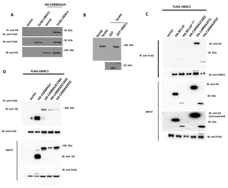
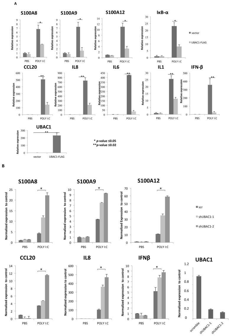
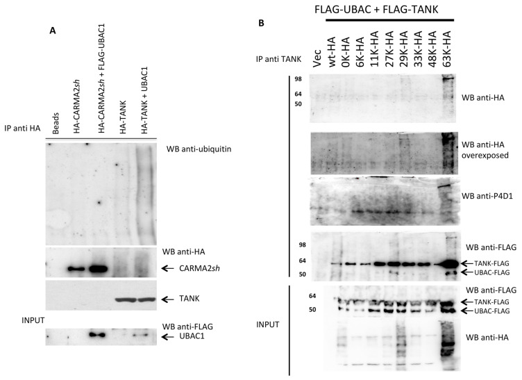
CARD14/CARMA2 is a scaffold molecule whose genetic alterations are linked to human inherited inflammatory skin disorders. However, the mechanisms through which CARD14/CARMA2 controls innate immune response and chronic inflammation are not well understood. By means of a yeast two-hybrid screening, we identified the UBA Domain Containing 1 (UBAC1), the non-catalytic subunit of the E3 ubiquitin-protein ligase KPC complex, as an interactor of CARMA2sh, the CARD14/CARMA2 isoform mainly expressed in human keratinocytes. UBAC1 participates in the CARMA2sh/TANK complex and promotes K63-linked ubiquitination of TANK. In human keratinocytes, UBAC1 negatively regulates the NF-κF-activating capacity of CARMA2sh following exposure to poly (I:C), an agonist of Toll-like Receptor 3. Overall, our data indicate that UBAC1 participates in the inflammatory signal transduction pathways involving CARMA2sh.

Keywords: BCL10; CARD14; CARMA2sh; NF-κB; TANK; UBAC1; Ubiquitin; psoriasis.


Conflict of interest statement

The authors declare no conflict of interest.

Figures

Figure 1
Figure 1
UBAC1 associates to CARMA2sh. (A) HEK293T cells were co-transfected with a plasmid encoding for FLAG-tagged UBAC1 together with an HA-tagged expression vector empty or encoding for CARMA2sh. A total of 24 h later, lysates were immunoprecipitated with anti-HA antibodies and analyzed for co-precipitating FLAG-UBAC1 by western blot assay. Data shown are representative of three independent experiments. (B) Affinity purified GST-UBAC1 was incubated with HaCaT cell lysates and western blot analysis was carried out with anti-CARMA2 and re-probing the membrane with anti-GST antibody. Data shown are representative of three independent experiments. (C,D) HEK293T cells were co-transfected with a plasmid encoding for FLAG-tagged UBAC1 together with expression vectors encoding for the indicated HA-tagged polypeptides. A total of 24 h later, lysates were immunoprecipitated with anti-FLAG antibodies and analyzed for coprecipitating proteins by western blot assay. Data shown are representative of three independent experiments.
Figure 2
Figure 2
UBAC1 represses TLR3 signaling. (A) NHEK cells were infected with a lentiviral vector expressing UBAC1 or control GFP. A total of 48 h later, cells were left in PBS or exposed poly (I:C) (20 μg/mL) for 24 h, and the expression level of selected NF-κB and IRF3 target genes was monitored by real time PCR. Graphs show the fold-change with respect to the cells left in PBS. Data shown are representative of five independent experiments done in triplicate. Data were analyzed by Student’s t-test (* p-value ≤ 0.05; ** p-value ≤ 0.02). (B) NHEK cells were infected with retroviral vectors encoding for two different shRNAs targeting UBAC1 or a control shRNA (scramble). After selection, cells were left in PBS or exposed to poly (I:C) (20 μg/mL) for 24 h, and the expression level of selected NF-κB target genes were quantified by real-time PCR.
Figure 3
Figure 3
UBAC1 promotes K63-linked ubiquitination of TANK. (A) HEK293 cells were transfected with the indicated expression vectors. A total of 24 h later, cell lysates were immunoprecipitated with anti-HA antibody, separated by SDS-PAGE and transferred onto membranes subsequently probed with anti-ubiquitin. Statistical analysis for the blot shown in provided as supplementary Figure S1. (B) HEK293 cells were co-transfected with HA-tagged ubiquitin mutants and FLAG-tagged UBAC1 and TANK. The number indicates the only lysine residue remaining in the ubiquitin molecule. Immunoprecipitates with anti-TANK antibody were resolved by SDS-PAGE and blotted onto a membrane subsequently probed with anti-HA or anti- ubiquitin (P4D1). Statistical analysis for the blot shown in provided as supplementary Figure S1.
Figure 4
Figure 4
UBAC1 affects viability and proliferation in primary keratinocytes. (A) NHEK cells were infected with a lentiviral vector empty or expressing UBAC1. A total of 48 h later, cells were trypsinized, counted in a Neubauer chamber, and 104 cells were seeded in each well of a 96-multi well plate. Cell viability was determined by MTT assay at seeding (right after cell attached to the bottom of the well) and 24, 48 and 72 h later. Representative phase contrast micrographs of transfected NHEK are also shown (magnification 4×). Graphs show the relative Optical Density at 570 nm at different time points. Data shown are representative of five independent experiments done in triplicate. Data were analyzed by Student’s t-test (* p-value ≤ 0.05). (B) NHEK cells were infected with a lentiviral vector expressing UBAC1-silencing shRNAs or scramble. A total of 48 h later, cells were trypsinized, counted with Neubauer chamber, and 104 (lower confluence) or 3 × 10⁴ (higher confluence) were seeded in each well of a 96-multi well plate. MTT data and micrographs and statistical analysis were performed as in Figure 4A. When seeded at higher confluence, differences between UBAC1-silenced NHEK and control cells are evident only 144 h after seeding (micrographs not shown).

References

    1. Scudiero I., Vito P., Stilo R.N. The three CARMA sisters: So different, so similar: A portrait of the three CARMA proteins and their involvement in human disorders. J. Cell Physiol. 2014;229:990–997. doi: 10.1002/jcp.24543. - DOI - PubMed
    1. Zotti T., Polvere I., Voccola S., Vito P., Stilo R. CARD14/CARMA2 Signaling and its Role in Inflammatory Skin Disorders. Front. Immunol. 2018;9:2167. doi: 10.3389/fimmu.2018.02167. - DOI - PMC - PubMed
    1. Mellett M. Regulation and dysregulation of CARD14 signalling and its physiological consequences in inflammatory skin disease. Cell Immunol. 2020;354:104147. doi: 10.1016/j.cellimm.2020.104147. - DOI - PubMed
    1. Van Nuffel E., Schmitt A., Afonina I.S., Schulze-Osthoff K., Beyaert R., Hailfinger S. CARD14-Mediated Activation of Paracaspase MALT1 in Keratinocytes: Implications for Psoriasis. J. Investig. Dermatol. 2017;137:569–575. doi: 10.1016/j.jid.2016.09.031. - DOI - PubMed
    1. Jordan C.T., Cao L., Roberson E.D., Pierson K.C., Yang C.F., Joyce C.E., Ryan C., Duan S., Helms C.A., Liu Y., et al. PSORS2 is due to mutations in CARD14. Am. J. Hum. Genet. 2012;90:784–795. doi: 10.1016/j.ajhg.2012.03.012. - DOI - PMC - PubMed

LinkOut - more resources