

Antibacterial Multi-Layered Nanocellulose-Based Patches Loaded with Dexpanthenol for Wound Healing Applications
- PMID: 33317206
- PMCID: PMC7764272
- DOI: 10.3390/nano10122469
Antibacterial Multi-Layered Nanocellulose-Based Patches Loaded with Dexpanthenol for Wound Healing Applications
Abstract
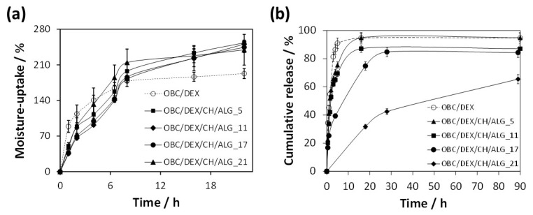
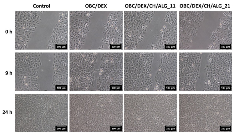
Antibacterial multi-layered patches composed of an oxidized bacterial cellulose (OBC) membrane loaded with dexpanthenol (DEX) and coated with several chitosan (CH) and alginate (ALG) layers were fabricated by spin-assisted layer-by-layer (LbL) assembly. Four patches with a distinct number of layers (5, 11, 17, and 21) were prepared. These nanostructured multi-layered patches reveal a thermal stability up to 200 °C, high mechanical performance (Young's modulus ≥ 4 GPa), and good moisture-uptake capacity (240-250%). Moreover, they inhibited the growth of the skin pathogen Staphylococcus aureus (3.2-log CFU mL-1 reduction) and were non-cytotoxic to human keratinocytes (HaCaT cells). The in vitro release profile of DEX was prolonged with the increasing number of layers, and the time-dependent data imply a diffusion/swelling-controlled drug release mechanism. In addition, the in vitro wound healing assay demonstrated a good cell migration capacity, headed to a complete gap closure after 24 h. These results certify the potential of these multi-layered polysaccharides-based patches toward their application in wound healing.
Keywords: alginate; chitosan; dexpanthenol; layer-by-layer assembly; multi-layered patches; oxidized bacterial cellulose; wound healing.
Conflict of interest statement
The authors declare no conflict of interest.
Figures


References
-
- Klemm D., Cranston E.D., Fischer D., Gama M., Kedzior S.A., Kralisch D., Kramer F., Kondo T., Lindström T., Nietzsche S., et al. Nanocellulose as a natural source for groundbreaking applications in materials science: Today’s state. Mater. Today. 2018;21:720–748. doi: 10.1016/j.mattod.2018.02.001. - DOI
Grants and funding
- UIDB/50011/2020 & UIDP/50011/2020/Fundação para a Ciência e a Tecnologia
- UIDB/50017/2020 & UIDP/50017/2020/Fundação para a Ciência e a Tecnologia
- PD/BD/115621/2016/Fundação para a Ciência e a Tecnologia
- CEECIND/04050/2017/Fundação para a Ciência e a Tecnologia
- CEECIND/00263/2018/Fundação para a Ciência e a Tecnologia
LinkOut - more resources
Full Text Sources
Miscellaneous
