. 2020 Jan-Dec;12(1):1854923.
doi: 10.1080/19420862.2020.1854923.

Identification and quantitation of hinge cysteinylation on endogenous IgG2 antibodies from human serum

Affiliations

Identification and quantitation of hinge cysteinylation on endogenous IgG2 antibodies from human serum

Luladey Ayalew et al. MAbs. 2020 Jan-Dec.

Abstract

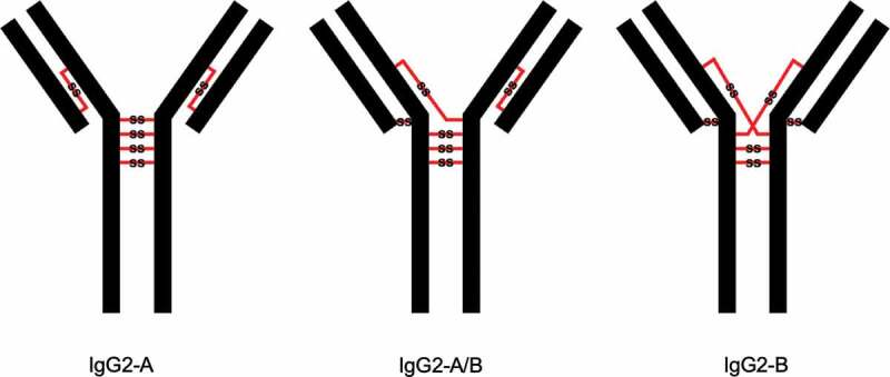
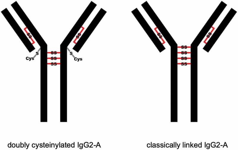
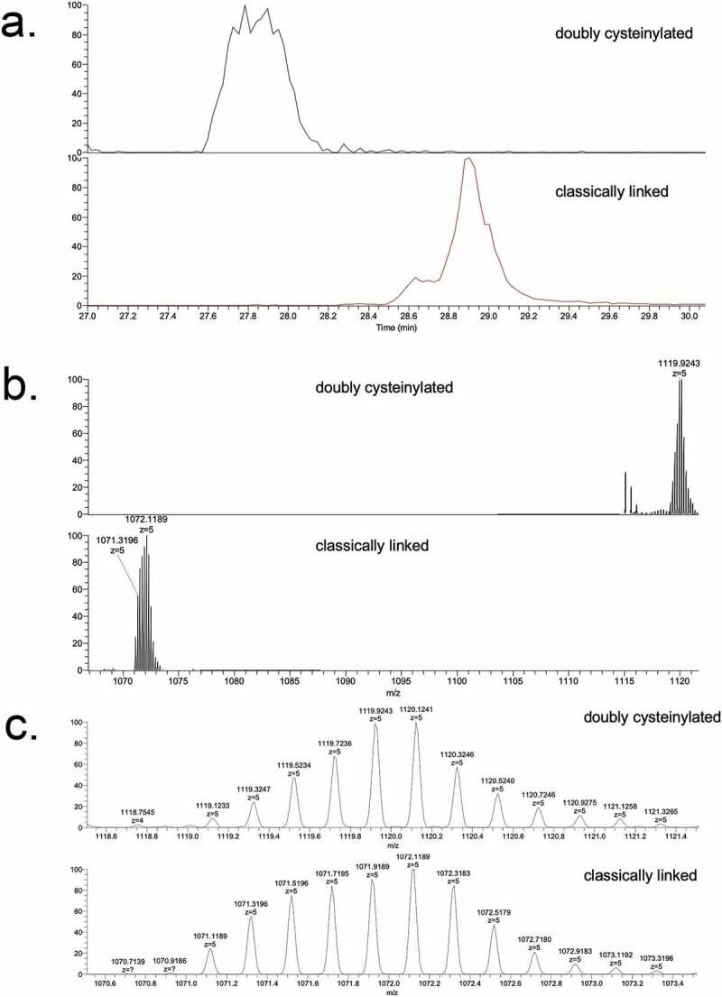
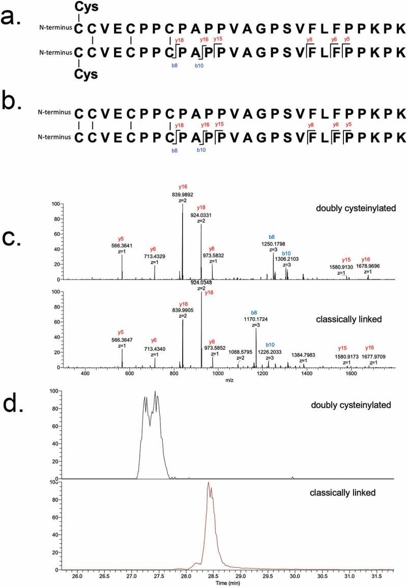
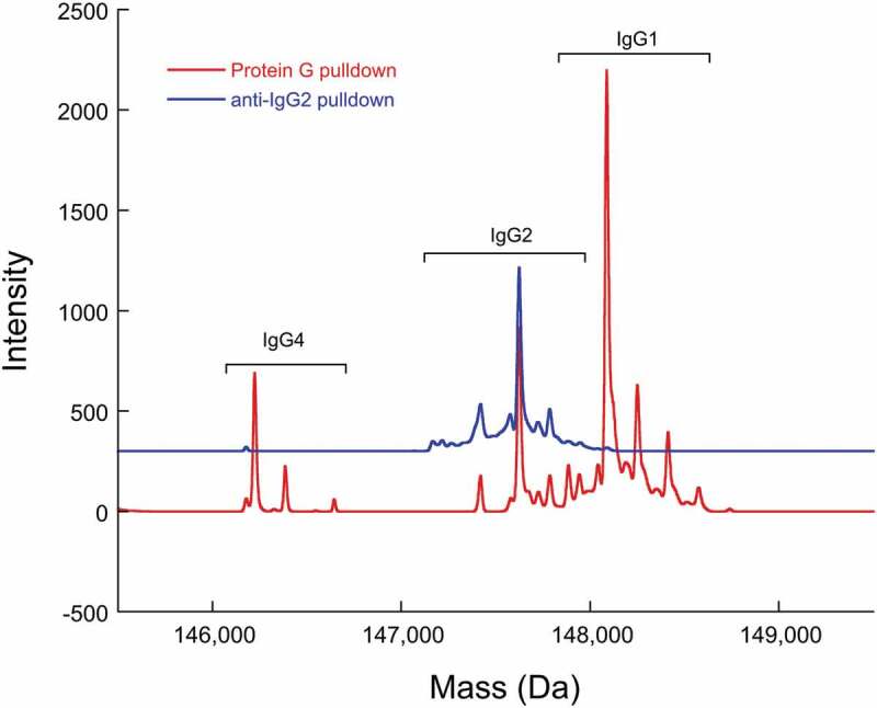
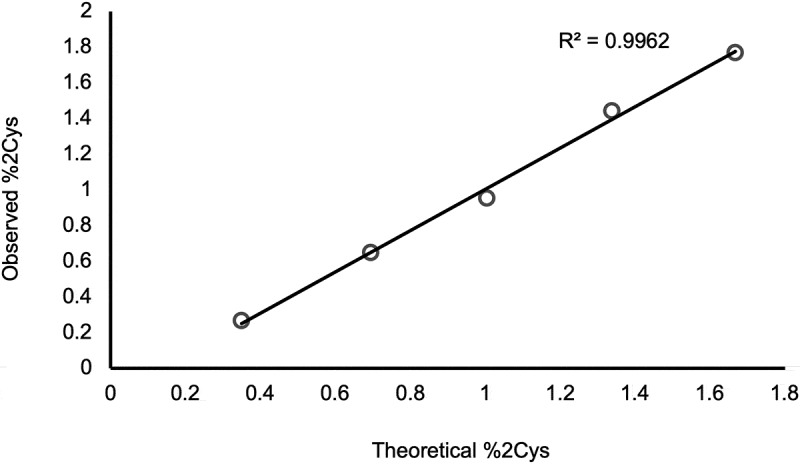
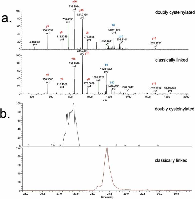
Cysteinylation is a post-translational modification (PTM) that occurs when a cysteine residue on a protein forms a disulfide bond with a terminal cysteine molecule. This PTM has been found in the hinge region of several recombinant therapeutic IgG2 antibodies, but the impact of cysteinylation on the safety and immunogenicity of therapeutics remains unclear. In this study, we characterized recombinant and endogenous IgG2 antibodies to quantify their levels of hinge cysteinylation, if present. To the best of our knowledge, this is the first study to identify and quantify hinge cysteinylation in endogenous IgG2 antibodies from healthy human serum. We used anti-IgG2 immunopurification of human serum to specifically enrich for endogenous IgG2 antibodies, and then subjected the resulting samples to Lys-C peptide mapping coupled with targeted mass spectrometry techniques. Using this analytical workflow, we found that all healthy human serum samples tested (N = 10) contained quantifiable levels of hinge cysteinylation (0.8 ± 0.3%) in their endogenous human IgG2s (IgG2-A isoform). These findings demonstrate that hinge cysteinylation in therapeutic IgG2s, at least up to a certain level, is well tolerated in humans and pose minimal safety or immunogenicity risks.

Keywords: IgG2; cysteinylation; disulfide; endogenous antibodies; hinge region; human serum.


Figures

Figure 1.
Figure 1.
Three structural isoforms of IgG2 (A, A/B, and B) and their associated disulfide connections depicted with red lines. Modified and used with permission of American Chemical Society, from ref.; permission conveyed through Copyright Clearance Center, Inc
Figure 2.
Figure 2.
Depictions of doubly cysteinylated and classically linked IgG2-A forms. “Cys” denotes a terminal cysteine molecule that is disulfide bonded to a cysteine on the antibody. Modified and used with permission of American Chemical Society, from ref.; permission conveyed through Copyright Clearance Center, Inc
Figure 3.
Figure 3.
SIM experiments of mAb1-Y Lys-C peptide maps. (a) Extracted ion chromatograms of doubly cysteinylated and classically linked IgG-A hinge Lys-C dipeptides (10 ppm mass tolerance for extraction). (b) Corresponding MS1 spectra using Orbitrap detection (60 000 resolution). (c) Corresponding MS1 spectra that are zoomed-in on the mass features of interest
Figure 4.
Figure 4.
PRM experiments of mAb1-Y Lys-C peptide maps. Sequences of (a) doubly cysteinylated and (b) classically linked IgG2-A hinge Lys-C dipeptides annotated with b- and y-ion fragmentation sites. (c) Corresponding annotated PRM spectra using CID fragmentation and Orbitrap detection (30 000 resolution, 10 ppm mass tolerance for assignments). (d) Corresponding extracted ion chromatograms of precursor/b8 product ion transitions (10 ppm mass tolerance for extraction)
Figure 5.
Figure 5.
Deconvoluted mass spectra of a recombinant IgG cocktail sample (including IgG1, IgG2, and IgG4 antibodies) that has been subjected to Protein G pulldown or anti-IgG2 pulldown. The heterogeneity within each recombinant IgG subclass is predominantly attributable to Fc glycosylation
Figure 6.
Figure 6.
PRM analysis of doubly cysteinylated IgG2 standards (prepared by co-mixing mAb1-Y and mAb1-Z) showing a linear response
Figure 7.
Figure 7.
PRM experiments of endogenous IgG2 Lys-C peptide maps. (a) PRM spectra using CID fragmentation and Orbitrap detection (30 000 resolution, 10 ppm mass tolerance for assignments). (b) Corresponding extracted ion chromatograms of precursor/b8 product ion transitions (10 ppm mass tolerance for extraction). Depicted here is data of endogenous IgG2s affinity purified from a single human subject, as an example (in total 10 human subject samples were analyzed)

References

    1. Smith SL. Ten years of orthoclone OKT3 (Muromonab-CD3): a review. J Transpl Coord. 1996;6(3):109–9. doi:10.1177/090591999600600304. - DOI - PubMed
    1. Reichert JM, Rosensweig CJ, Faden LB, Dewitz MC.. Monoclonal antibody successes in the clinic. Nat Biotechnol. 2005;23(9):1073–78. doi:10.1038/nbt0905-1073. - DOI - PubMed
    1. Newsome BW, Ernstoff MS. The clinical pharmacology of therapeutic monoclonal antibodies in the treatment of malignancy; have the magic bullets arrived? Br J Clin Pharmacol. 2008;66(1):6–19. doi:10.1111/j.1365-2125.2008.03187.x. - DOI - PMC - PubMed
    1. Brekke OH, Sandlie I. Therapeutic antibodies for human diseases at the dawn of the twenty-first century. Nat Rev Drug Discov. 2003;2(1):52–62. doi:10.1038/nrd984. - DOI - PubMed
    1. Vlasak J, Ionescu R. Fragmentation of monoclonal antibodies. mAbs. 2011;3(3):253–63. doi:10.4161/mabs.3.3.15608. - DOI - PMC - PubMed

Publication types

LinkOut - more resources