

Biological effects of the oxygen molecule in critically ill patients
- PMID: 33317639
- PMCID: PMC7734465
- DOI: 10.1186/s40560-020-00505-9
Biological effects of the oxygen molecule in critically ill patients
Abstract
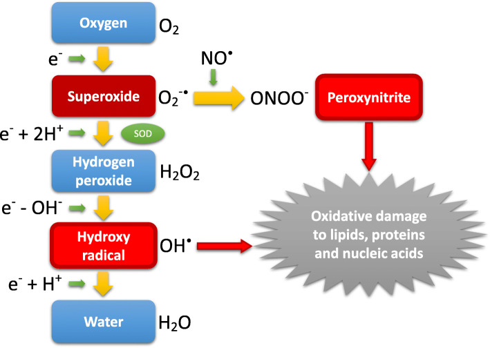
The medical use of oxygen has been widely and frequently proposed for patients, especially those under critical care; however, its benefit and drawbacks remain controversial for certain conditions. The induction of oxygen therapy is commonly considered for either treating or preventing hypoxia. Therefore, the concept of different types of hypoxia should be understood, particularly in terms of their mechanism, as the effect of oxygen therapy principally varies by the physiological characteristics of hypoxia. Oxygen molecules must be constantly delivered to all cells throughout the human body and utilized effectively in the process of mitochondrial oxidative phosphorylation, which is necessary for generating energy through the formation of adenosine triphosphate. If the oxygen availability at the cellular level is inadequate for sustaining the metabolism, the condition of hypoxia which is characterized as heterogeneity in tissue oxygen tension may develop, which is called dysoxia, a more physiological concept that is related to hypoxia. In such hypoxic patients, repetitive measurements of the lactate level in blood are generally recommended in order to select the adequate therapeutic strategy targeting a reduction in lactate production. Excessive oxygen, however, may actually induce a hyperoxic condition which thus can lead to harmful oxidative stress by increasing the production of reactive oxygen species, possibly resulting in cellular dysfunction or death. In contrast, the human body has several oxygen-sensing mechanisms for preventing both hypoxia and hyperoxia that are employed to ensure a proper balance between the oxygen supply and demand and prevent organs and cells from suffering hyperoxia-induced oxidative stress. Thus, while the concept of hyperoxia is known to have possible adverse effects on the lung, the heart, the brain, or other organs in various pathological conditions of critically ill patients, and no obvious evidence has yet been proposed to totally support liberal oxygen supplementation in any subset of critically ill patients, relatively conservative oxygen therapy with cautious monitoring appears to be safe and may improve the outcome by preventing harmful oxidative stress resulting from excessive oxygen administration. Given the biological effects of oxygen molecules, although the optimal target levels remain controversial, unnecessary oxygen administration should be avoided, and exposure to hyperoxemia should be minimized in critically ill patients.
Keywords: Critical care; Hyperoxia; Hypoxia; Lactate; Oxidative stress; Oxygen; Reactive oxygen species.
Conflict of interest statement
The author declares that he has no competing interests.
Figures

References
-
- West JB, Luks AB. West’s pulmonary pathophysiology: the essentials 9th ed. Philadelphia: Wolters Kluwer; 2017.
-
- Hall J. Guyton and Hall Textbook of Medical Physiology. 13. Philadelphia: Elsevier; 2016.
Publication types
LinkOut - more resources
Full Text Sources
