Case Reports
. 2020 Dec 14;11(1):6385.
doi: 10.1038/s41467-020-19761-2.

Treatment of COVID-19 with remdesivir in the absence of humoral immunity: a case report

Affiliations
Case Reports

Treatment of COVID-19 with remdesivir in the absence of humoral immunity: a case report

Matthew S Buckland et al. Nat Commun. .

Abstract

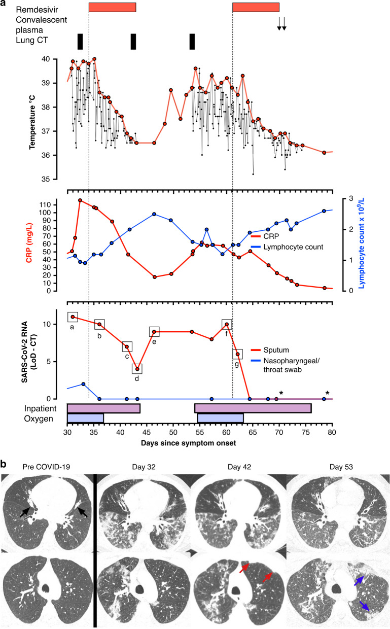
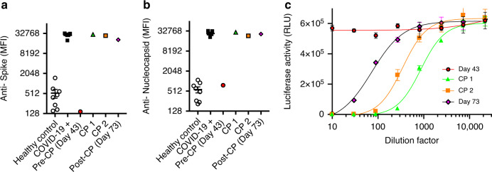
The response to the coronavirus disease 2019 (COVID-19) pandemic has been hampered by lack of an effective severe acute respiratory syndrome coronavirus 2 (SARS-CoV-2) antiviral therapy. Here we report the use of remdesivir in a patient with COVID-19 and the prototypic genetic antibody deficiency X-linked agammaglobulinaemia (XLA). Despite evidence of complement activation and a robust T cell response, the patient developed persistent SARS-CoV-2 pneumonitis, without progressing to multi-organ involvement. This unusual clinical course is consistent with a contribution of antibodies to both viral clearance and progression to severe disease. In the absence of these confounders, we take an experimental medicine approach to examine the in vivo utility of remdesivir. Over two independent courses of treatment, we observe a temporally correlated clinical and virological response, leading to clinical resolution and viral clearance, with no evidence of acquired drug resistance. We therefore provide evidence for the antiviral efficacy of remdesivir in vivo, and its potential benefit in selected patients.


Conflict of interest statement

E.J.M.T. is an employee of Hycult Biotechnology Ltd. All other authors declare no competing interests.

Figures

Fig. 1
Fig. 1. Clinical and virological assessment of the response to remdesivir.
a Temperature, CRP, total lymphocyte count, and cycle-threshold (CT) value for viral RNA amplification from patient samples, deducted from the CT value at the limit of detection (LoD) of the assay, plotted by day since symptom onset, aligned with clinical interventions. Samples tested in the clinical laboratory at the Royal London Hospital are highlighted (*). Indicated viral isolates ag were analyzed by Nanopore sequencing (Supplementary Fig. 2 and Table 5). n = 1 biologically independent samples. b Computerized tomography (CT) images at the level of the inferior pulmonary veins (top) and aortic arch (bottom) obtained at indicated time points. Prior to COVID-19 infection, there is moderate bronchiectasis, mucoid impaction and small airway obstruction (black arrows) in the middle lobe and lingula, but the remainder of the lungs are clear (Pre-COVID-19). Shortly before the first course of remdesivir, extensive ground-glass opacification and patchy consolidation with a lower lobe predominance is seen, together with perivascular consolidation in the right upper lobe (Day 32). Nine days after initiation of remdesivir treatment there is improvement in the lower zone ground-glass opacity and consolidation, but persistent consolidation in the right upper lobe and mild progression of the left upper lobe subpleural consolidation (red arrows) (Day 42). 10 days after completion of the first course of remdesivir treatment, following relapse of symptoms and fever, there is further improvement in the lower zone ground-glass opacification, but progressive subpleural consolidation, particularly in the left upper lobe (blue arrows) (Day 53).
Fig. 2
Fig. 2. Kinetic assessment of the antigen-specific humoral responses.
Serological reactivity to SARS-CoV-2 spike a and nucleocapsid b antigens of patient sera obtained at indicated time points, the first (CP 1) and second (CP 2) infusions of convalescent plasma (CP), sera from healthy controls (n = 8 biologically independent subjects) and patients (n = 5 biologically independent subjects) with PCR-confirmed COVID-19 (MFI, mean fluorescence intensity; Mean and SEM). c Neutralisation activity against SARS-CoV-2 spike-pseudotyped lentiviral particles of patient sera obtained at indicated time points, compared with the first (CP 1) and second (CP 2) infusions of convalescent plasma (CP). (n = 1 independent biological samples, n = 3 technical replicates from each sample; mean and SD). Data from one independent experiment.
Fig. 3
Fig. 3. Kinetic assessment of the antigen-specific CD8+ T-cell responses.
a % CD8+ T cells expressing activation makers after incubation ± a peptide pool covering the SARS-CoV-2 S1 protein. Patient samples obtained at indicated time points are compared with HCWs with PCR-confirmed COVID-19 at presentation (n = 5 biologically independent subjects). b Number of activation markers expressed by S1-responsive CD8+ T cells from a. Representative flow cytometry dot plots c and % proliferating CD8+ T cells d after stimulation ± peptide pools covering the indicated SARS-CoV-2 proteins. Patient samples are compared with HCWs (n = 2 biologically independent subjects) with PCR-confirmed COVID-19 at indicated time points. Data from one independent experiment.
Fig. 4
Fig. 4. Assessment of complement activation.
Concentrations of complement cleavage products C3a a, C3c b, and C5a c, and circulating terminal complement complex (TCC) d at indicated time points, compared with healthy controls or patients with PCR-confirmed COVID-19 (healthcare workers, HCW or patients with severe disease admitted to the intensive therapy unit, ITU; mean and SEM are shown). For a and b HCW, n = 5 biologically independent subjects and ITU n = 5 biologically independent subjects. For c Healthy controls, n = 12 biologically independent subjects and ITU n = 51 biologically independent subjects. d Healthy controls, n = 67 biologically independent subjects and ITU n = 50 biologically independent subjects. Data from one independent experiment.

References

    1. Wang M, et al. Remdesivir and chloroquine effectively inhibit the recently emerged novel coronavirus (2019-nCoV) in vitro. Cell Res. 2020;30:269–271. doi: 10.1038/s41422-020-0282-0. - DOI - PMC - PubMed
    1. Gordon CJ, et al. Remdesivir is a direct-acting antiviral that inhibits RNA-dependent RNA polymerase from severe acute respiratory syndrome coronavirus 2 with high potency. J. Biol. Chem. 2020;295:6785–6797. doi: 10.1074/jbc.RA120.013679. - DOI - PMC - PubMed
    1. Choy KT, et al. Remdesivir, lopinavir, emetine, and homoharringtonine inhibit SARS-CoV-2 replication in vitro. Antivir. Res. 2020;178:104786. doi: 10.1016/j.antiviral.2020.104786. - DOI - PMC - PubMed
    1. Williamson BN, et al. Clinical benefit of remdesivir in rhesus macaques infected with SARS-CoV-2. Nature. 2020;585:273–276. doi: 10.1038/s41586-020-2423-5. - DOI - PMC - PubMed
    1. Wang Y, et al. Remdesivir in adults with severe COVID-19: a randomised, double-blind, placebo-controlled, multicentre trial. Lancet. 2020;395:1569–1578. doi: 10.1016/S0140-6736(20)31022-9. - DOI - PMC - PubMed

Publication types