. 2020 Dec 14;11(1):6381.
doi: 10.1038/s41467-020-20149-5.

Isolation of a member of the candidate phylum 'Atribacteria' reveals a unique cell membrane structure

Affiliations

Isolation of a member of the candidate phylum 'Atribacteria' reveals a unique cell membrane structure

Taiki Katayama et al. Nat Commun. .

Abstract

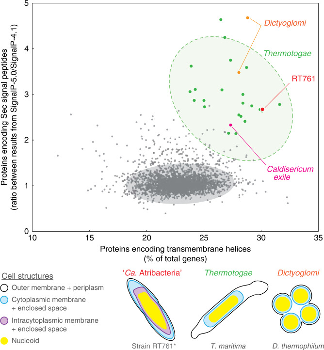
A key feature that differentiates prokaryotic cells from eukaryotes is the absence of an intracellular membrane surrounding the chromosomal DNA. Here, we isolate a member of the ubiquitous, yet-to-be-cultivated phylum 'Candidatus Atribacteria' (also known as OP9) that has an intracytoplasmic membrane apparently surrounding the nucleoid. The isolate, RT761, is a subsurface-derived anaerobic bacterium that appears to have three lipid membrane-like layers, as shown by cryo-electron tomography. Our observations are consistent with a classical gram-negative structure with an additional intracytoplasmic membrane. However, further studies are needed to provide conclusive evidence for this unique intracellular structure. The RT761 genome encodes proteins with features that might be related to the complex cellular structure, including: N-terminal extensions in proteins involved in important processes (such as cell-division protein FtsZ); one of the highest percentages of transmembrane proteins among gram-negative bacteria; and predicted Sec-secreted proteins with unique signal peptides. Physiologically, RT761 primarily produces hydrogen for electron disposal during sugar degradation, and co-cultivation with a hydrogen-scavenging methanogen improves growth. We propose RT761 as a new species, Atribacter laminatus gen. nov. sp. nov. and a new phylum, Atribacterota phy. nov.


Conflict of interest statement

The authors declare no competing interests.

Figures

Fig. 1
Fig. 1. Morphology and membrane structure in RT761 cells showing the presence of three lipid membrane-like layers (LMLs) with the innermost layer surrounding the nucleoid.
a Phase-contrast microscopy. b Scanning electron microscopy. ce Cryo-electron tomography (also see Supplementary Movies 1 and 2). c The original slice picture is shown in Supplementary Fig. 4. Black arrowheads indicate the outer (1), middle (2), and inner (3) LMLs. White arrowheads indicate the 2.2 nm thick layer (1) and faint layers (2). d White arrow indicates inner LML invagination. e 3D-rendered reconstruction of the cell in d. (also see Supplementary Movie 4). Color code: outer LML, orange; middle LML, blue; inner LML, yellow; ribosome, green. f Transmission electron micrograph of a thin section of RT761 cells. N nucleoid. Scale bars, 5.0 μm (a), 1.5 μm (b), 0.2 μm (c), 0.1 μm (d, e), and 0.5 μm (f).
Fig. 2
Fig. 2. Confocal-laser microscopy showing the localization of DNA and RNA within the intracytoplasmic membrane structure. DNA, RNA, and membrane lipids were stained by Hoechst (blue), SYTO RNAselect (green) and FM4-64 (red), respectively.
Outlines of the cell from a are included in all panels. a Phase contrast image. bd Confocal-laser images. eh Image overlays. i Line profiles of fluorescence intensity plotted longitudinally along white arrow in h. Membrane staining maxima are indicated by arrowheads in h. Source data are provided as a Source Data file. Scale bars, 1 μm.
Fig. 3
Fig. 3. Unique genomic compositions of membrane-related features observed for phyla with unique cell structures.
The horizontal axis shows the genomic proportion proteins encoding transmembrane helices. The vertical axis shows the ratio of proportions of proteins encoding Sec signal peptides estimated by SignalP-5.0 and SignalP-4.1. RT761 (red), type strains from Thermotogae (green), Dictyoglomi (orange), Caldiserica (pink), and other gram-negative type strains (gray) are plotted (3502 genomes downloadable from the Joint Genome Institute Integrated Microbial Genomes and Microbiomes database). For Thermotogae and other gram-negative type strains, 95% (green) and 99.9% (gray) confidence ellipses are shown respectively. Source data are provided as a Source Data file. (Bottom) Cell structures of select species are shown for “Ca. Atribacteria”, Thermotogae, and Dictyoglomi. The illustrations indicate the outer membrane (black), cytoplasmic membrane (blue), intracytoplasmic membrane (purple), and nucleoid (yellow). *For RT761, the shown schematic requires further investigation to conclude the identity/role of each layer.

Comment in

References

    1. Hug LA, et al. A new view of the tree of life. Nat. Microbiol. 2016;1:16048. doi: 10.1038/nmicrobiol.2016.48. - DOI - PubMed
    1. Brown CT, et al. Unusual biology across a group comprising more than 15% of domain Bacteria. Nature. 2015;523:208–211. doi: 10.1038/nature14486. - DOI - PubMed
    1. He X, et al. Cultivation of a human-associated TM7 phylotype reveals a reduced genome and epibiotic parasitic lifestyle. Proc. Natl Acad. Sci. USA. 2015;112:244–249. doi: 10.1073/pnas.1419038112. - DOI - PMC - PubMed
    1. Dodsworth JA, et al. Single-cell and metagenomic analyses indicate a fermentative and saccharolytic lifestyle for members of the OP9 lineage. Nat. Commun. 2013;4:1854. doi: 10.1038/ncomms2884. - DOI - PMC - PubMed
    1. Nobu MK, et al. Phylogeny and physiology of candidate phylum ‘Atribacteria’ (OP9/JS1) inferred from cultivation-independent genomics. ISME J. 2016;10:273–286. doi: 10.1038/ismej.2015.97. - DOI - PMC - PubMed

Publication types