. 2020 Dec 14;3(1):766.
doi: 10.1038/s42003-020-01419-w.

Distortion of the bilayer and dynamics of the BAM complex in lipid nanodiscs

Affiliations

Distortion of the bilayer and dynamics of the BAM complex in lipid nanodiscs

Matthew G Iadanza et al. Commun Biol. .

Abstract

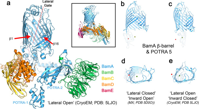
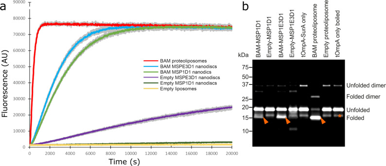
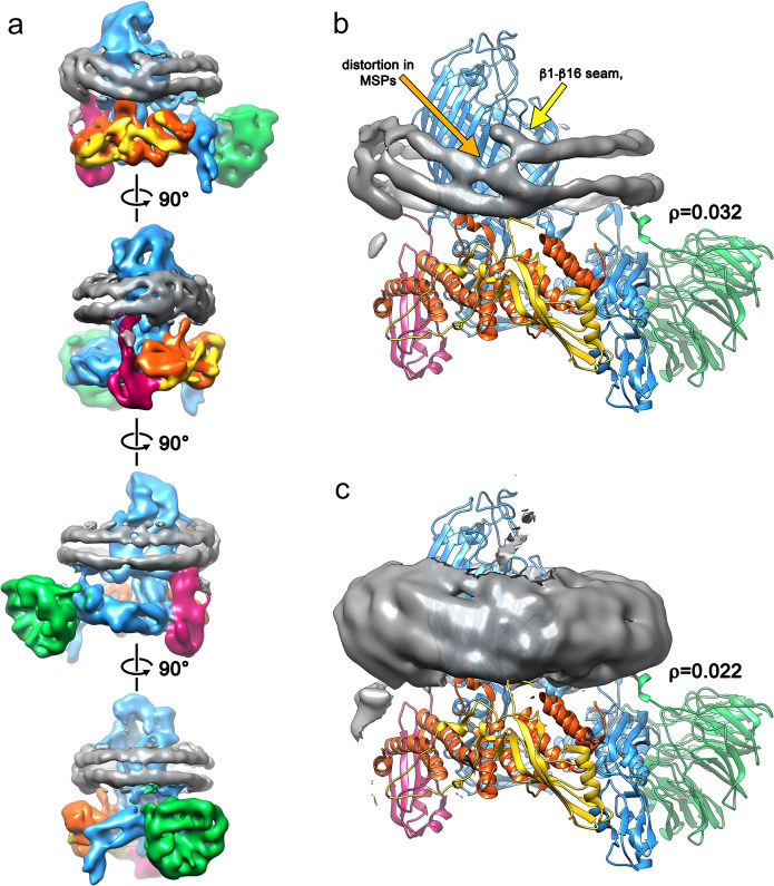
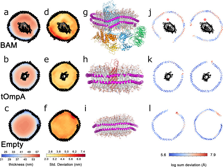
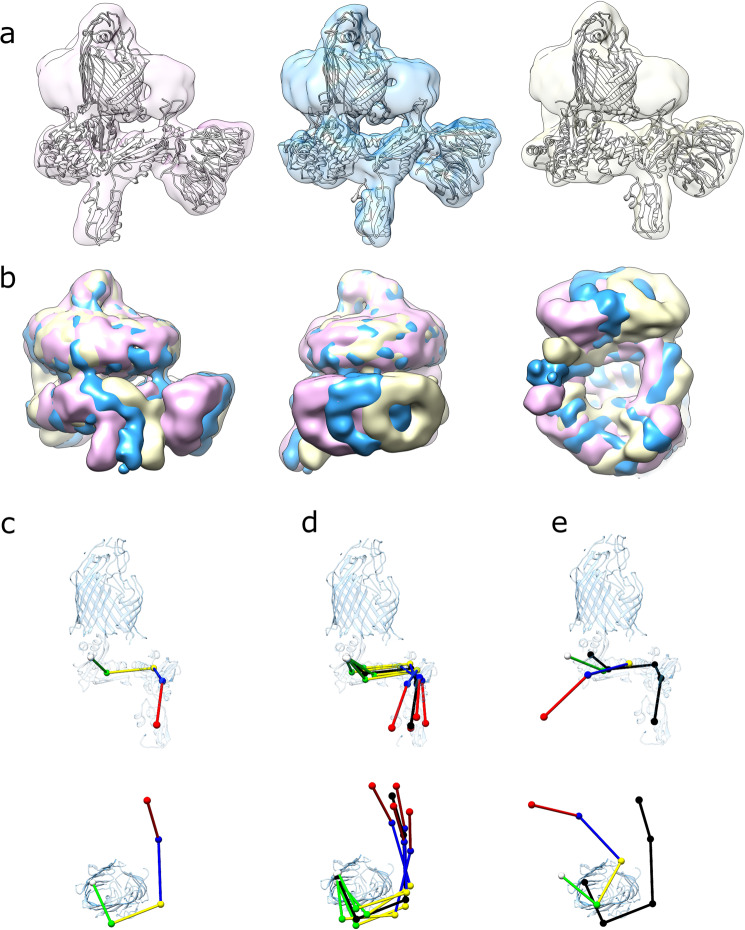
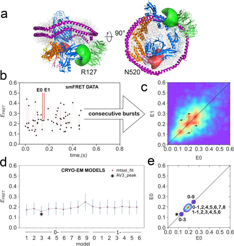
The β-barrel assembly machinery (BAM) catalyses the folding and insertion of β-barrel outer membrane proteins (OMPs) into the outer membranes of Gram-negative bacteria by mechanisms that remain unclear. Here, we present an ensemble of cryoEM structures of the E. coli BamABCDE (BAM) complex in lipid nanodiscs, determined using multi-body refinement techniques. These structures, supported by single-molecule FRET measurements, describe a range of motions in the BAM complex, mostly localised within the periplasmic region of the major subunit BamA. The β-barrel domain of BamA is in a 'lateral open' conformation in all of the determined structures, suggesting that this is the most energetically favourable species in this bilayer. Strikingly, the BAM-containing lipid nanodisc is deformed, especially around BAM's lateral gate. This distortion is also captured in molecular dynamics simulations, and provides direct structural evidence for the lipid 'disruptase' activity of BAM, suggested to be an important part of its functional mechanism.


Conflict of interest statement

The authors declare no competing interests.

Figures

Fig. 1
Fig. 1. Structure of BAM highlighting its lateral gate.
a Cryo-EM structure (PDB 5LJO) of detergent (DDM)-solubilised BAM in the ‘lateral open’ conformation. The β-strands 1 and 16 and the lateral gate are indicated. Inset: the crystal structure (PDB 5D0O) of BAM in the ‘lateral closed’ conformation. b The conformation of the BamA β-barrel and position of POTRA5 in the ‘lateral closed’ X-ray structure (pdb 5D0O) and c ‘lateral open’ cryo-EM structure (pdb 5LJO). The centres of mass of POTRA5 for both conformations are marked with red (lateral closed) and green (lateral-open) spheres in each panel. d, e Bottom (periplasmic) view of the d lateral closed and e lateral-open conformations of BAM showing the different shape of the β-barrel. As in panels b and c, centres of mass for POTRA5 in both conformations are marked in each panel. POTRA domains 1–4 and the lipoproteins (BamB–E) have been removed in panels (b) through (e) for clarity.
Fig. 2
Fig. 2. Comparison of the catalytic activity of BAM in proteoliposomes and nanodiscs.
a Fluorescence detected from cleavage of a fluorescent OmpT substrate in BAM-containing proteoliposomes (red), BAM in MSPE3D1 (blue) or MSP1D1 nanodiscs (green), empty nanodiscs (purple and dark green for MSPE3D1 and MSP1D1, respectively) and empty liposomes (i.e. lacking BAM) (yellow). For each sample, the coloured line represents the mean fluorescence, whilst grey lines represent the minimum and maximum values for three replicates. b Gel shift assay measuring BAM-catalysed folding of tOmpA. Folded tOmpA is indicated by red arrows, whilst a contaminating band, present in all samples, is marked with a red asterisk. In this assay, tOmpA is labelled with Alexa Fluor 488, enabling the folding of the substrate to be visualised using fluorescence detection, without interference from protein bands arising from BamABCDE and SurA.
Fig. 3
Fig. 3. The cryo-EM structure of BAM reveals distortion of the lipid nanodisc MSPs.
a Cryo-EM map of the consensus structure of the BAM-containing MSP1D1 nanodisc with all five BAM subunits and nanodisc density colour coded as in Fig. 1 and the nanodisc density in grey. Each panel represents a 90° rotation around the axis shown. The cryo-EM density for the membrane-scaffold proteins is shown with the fitted atomic model of the BAM complex at low contour level (ρ = 0.032; panel b) and high contour level (ρ = 0.022; panel c) showing distortion of the MSP from the expected planar geometry adjacent to the BamA lateral gate.
Fig. 4
Fig. 4. Molecular dynamics simulations of protein-containing and empty lipid nanodiscs.
Mean thickness of lipid bilayers over the course of a 1-µs simulation for BAM-containing (a), tOmpA-containing (b) or empty (c) MSP1D1 nanodiscs. The standard deviation of bilayer thickness over the 1-μs simulation for BAM-containing (d), tOmpA-containing (e) or empty (f) MSP1D1 nanodiscs. Residues in the lateral gate are indicated in white. The 3D structures of the final frame of each simulation, coloured as in Fig. 1, with the MSP in magenta for BAM-containing (g), tOmpA-containing (h) or empty (i) MSP1D1 nanodiscs. Deviation from planarity for the upper (left) and lower (right) MSP in BAM-containing (j), tOmpA-containing (k) or empty (l) MSP1D1 nanodiscs. In a, b, d, e, j and k the mean positions of the α-carbons of the MSPs and β-barrels of the inserted proteins are represented by black dots. Strands β1 and β16, which make up the lateral gate of BamA, are indicated with a red asterisk in (j).
Fig. 5
Fig. 5. Cryo-EM structures generated by separation along eigenvectors 0 and 1.
a EM maps of (left to right) conformations 0–1, 0–5 and 0–9, with fitted models. Each reconstruction is aligned on the β-barrel of BamA illustrating the difference in orientation of the bottom half of the complex. b EM maps of conformations 0–1 (pink), 0–5 (gold) and 0–9 (blue) aligned on the β-barrel of BamA and overlaid. c Positions of centres of mass for POTRA domains 1 through 5 (red, blue, yellow, green and white, respectively) in the BAM-nanodisc consensus structure. d Positions of the POTRA domains in the four extremes of the ensemble (0–1, 0–9, 1–2 and 1–8, respectively, coloured) compared with the consensus structure (black). e Comparison of the positions of the POTRA domains in the ‘lateral closed’ crystal structure (5D0O, coloured) with the BAM-nanodisc consensus structure (black).
Fig. 6
Fig. 6. Comparison of experimentally measured and predicted smFRET values of BAM in MSP1D1 nanodiscs.
a Structure of a dye-labelled double-cysteine variant of BAM (R127C/N520C) predicted from the previous MD simulations. Green and red regions represent possible space filled by Alexa Fluor 488 and Dylight 594 maleimide, respectively. b Representative time trace showing EFRET values for single molecules. c Experimental 2D transfer-efficiency contour plot (RASP analysis, see Methods) showing the measured EFRET between the dyes for pairs of consecutive bursts E0 and E1 with dt < 30 ms. Density on the diagonal line indicates persistent conformational states, while off-diagonal density indicates transitions between those states that occur on a 1–30-ms timescale (curved arrows). d Predicted EFRET for each of the 16 cryo-EM-derived model structures; each point represents the maximum of the predicted distribution as shown for selected structures in Supplementary Fig. 13a. e Projections of the predicted EFRET into a 2D plot for direct comparison with the experimental data (panel b) showing that the static pattern of density along the diagonal could be recapitulated by the 16 cryo-EM-derived model structures. An alternative modelling approach afforded a better match for the data in the case of one structure (0–3 black star).

References

    1. Noinaj N, Gumbart JC, Buchanan SK. The beta-barrel assembly machinery in motion. Nat. Rev. Microbiol. 2017;15:197–204. doi: 10.1038/nrmicro.2016.191. - DOI - PMC - PubMed
    1. Konovalova A, Kahne D, Silhavy T. Outer membrane biogenesis. Annu. Rev. Microbiol. 2017;71:539–556. doi: 10.1146/annurev-micro-090816-093754. - DOI - PMC - PubMed
    1. Voulhoux R, Bos M, Geurtsen J, Mols M, Tommassen J. Role of a highly conserved bacterial protein in outer membrane protein assembly. Science. 2003;299:262–265. doi: 10.1126/science.1078973. - DOI - PubMed
    1. Schiffrin B, Brockwell DJ, Radford SE. Outer membrane protein folding from an energy landscape perspective. BMC Biol. 2017;15:123–139. doi: 10.1186/s12915-017-0464-5. - DOI - PMC - PubMed
    1. Hagan CL, Wzorek JS, Kahne D. Inhibition of the beta-barrel assembly machine by a peptide that binds BamD. Proc. Natl Acad. Sci. USA. 2015;112:2011–2016. doi: 10.1073/pnas.1415955112. - DOI - PMC - PubMed

Publication types