Review
. 2021 Mar;17(3):135-156.
doi: 10.1038/s41582-020-00428-x. Epub 2020 Dec 14.

Recovery from disorders of consciousness: mechanisms, prognosis and emerging therapies

Affiliations
Review

Recovery from disorders of consciousness: mechanisms, prognosis and emerging therapies

Brian L Edlow et al. Nat Rev Neurol. 2021 Mar.

Abstract

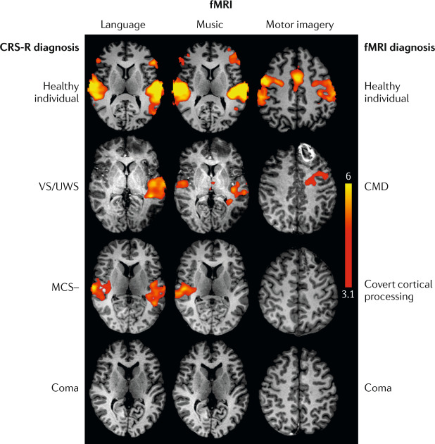
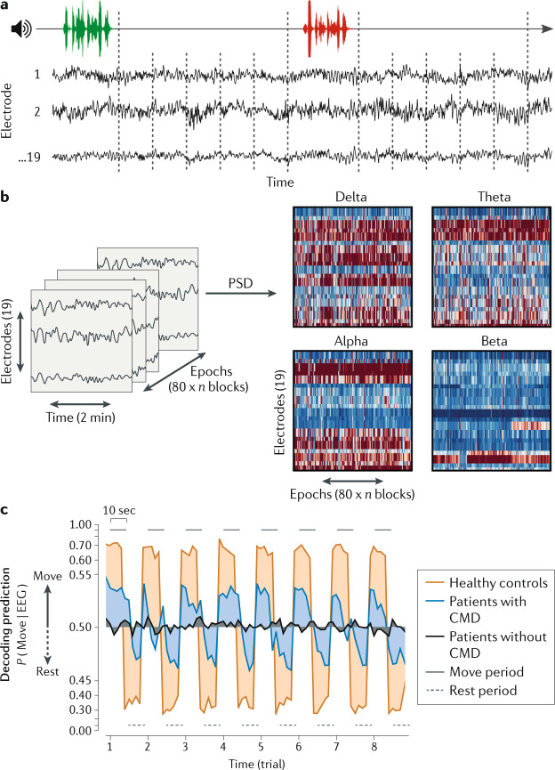
Substantial progress has been made over the past two decades in detecting, predicting and promoting recovery of consciousness in patients with disorders of consciousness (DoC) caused by severe brain injuries. Advanced neuroimaging and electrophysiological techniques have revealed new insights into the biological mechanisms underlying recovery of consciousness and have enabled the identification of preserved brain networks in patients who seem unresponsive, thus raising hope for more accurate diagnosis and prognosis. Emerging evidence suggests that covert consciousness, or cognitive motor dissociation (CMD), is present in up to 15-20% of patients with DoC and that detection of CMD in the intensive care unit can predict functional recovery at 1 year post injury. Although fundamental questions remain about which patients with DoC have the potential for recovery, novel pharmacological and electrophysiological therapies have shown the potential to reactivate injured neural networks and promote re-emergence of consciousness. In this Review, we focus on mechanisms of recovery from DoC in the acute and subacute-to-chronic stages, and we discuss recent progress in detecting and predicting recovery of consciousness. We also describe the developments in pharmacological and electrophysiological therapies that are creating new opportunities to improve the lives of patients with DoC.


Conflict of interest statement

The authors declare no competing interests.

Figures

Fig. 1
Fig. 1. Multidimensional assessment of consciousness.
During recovery from coma, patients are evaluated for overt cognition and motor function using the Coma Recovery Scale — Revised (CRS-R). This evaluation enables the classification of patients into the groups illustrated in green, with the exception of patients who emerge from a minimally conscious state with language (MCS+) to a confusional state, in whom the Confusion Assessment Protocol (CAP) is used to differentiate between a confusional state, cognitive dysfunction and full recovery. In patients with no behavioural evidence of language function, functional MRI (fMRI) or EEG evidence of command-following (active) indicates cognitive motor dissociation, fMRI or EEG responses within an association cortex during language or music stimuli (passive) indicate covert cortical processing, and an absence of fMRI or EEG responses indicates a true negative fMRI/EEG classification. Patients with behavioural evidence of language are classified as false negatives if there are no fMRI or EEG responses, and as true positives if there are fMRI and EEG responses. CLIS, complete locked-in syndrome; LIS, locked-in syndrome; MCS–, minimally conscious state without language; VS/UWS, vegetative state/unresponsive wakefulness syndrome. *Patients with LIS are identified by the presence of consistent purposeful movements, typically vertical eye movements, and a reliable movement-based communication system. Patients with LIS who demonstrate inconsistent movements would not be distinguishable from patients with CLIS, cognitive dysfunction, confusional state or MCS. Some patients with LIS are able to communicate via assistive communication devices. Adapted with permission from ref., OUP.
Fig. 2
Fig. 2. Diffusion tractography detects acute disconnection of the ascending arousal network.
a,b | Post-mortem diffusion tractography scans showing that the brainstem and thalamic nuclei of the ascending arousal network are extensively interconnected in a control individual (part a), but completely disconnected in a patient who died in the intensive care unit 3 days after a coma-inducing severe traumatic brain injury (part b). Scans shown from a posterior perspective, with fibre tracts colour-coded according to the brainstem nucleus from which they originate: purple, pedunculotegmental nucleus (PTg); yellow, parabrachial complex (PBC); turquoise, dorsal raphe (DR); dark blue, locus coeruleus (LC); green, median raphe (MnR); and pink, ventral tegmental area (VTA). Fibre tracts in the control, but not in the patient with coma, connect with the intralaminar nuclei (central lateral nuclei (CL) and centromedian/parafascicular complex (CEM/Pf)) and the reticular nuclei (Ret) of the thalamus. Two midbrain haemorrhages (Haem) in the patient with coma are rendered in red. c,d | Ascending arousal network connectograms for the control (part c) and the patient with coma (part d). Brainstem nuclei are listed on the outside of the circle, and their subcortical targets are shown on the inside of the circle. Lines indicate connections between network nodes, with line thickness being proportional to the number of tracts visualized. Brainstem connections with thalamic and basal forebrain nuclei were disrupted in the patient with coma (part d), but connections with the hypothalamus were partially preserved. BNM/SI, nucleus basalis of Meynert/substantia innominata; DBB, diagonal band of Broca; IL, intralaminar nuclei of thalamus; LDTg, laterodorsal tegmental nucleus; LHA, lateral hypothalamic area; mRt, mesencephalic reticular formation; PAG, periaqueductal grey; PnO, pontis oralis; PV, paraventricular nucleus of the thalamus; SUM, supramammillary nucleus of the hypothalamus; TM, tuberomammillary nucleus of the hypothalamus. Parts a and b reprinted with permission from ref., OUP.
Fig. 3
Fig. 3. Mapping loss of consciousness to a common brain network using the human connectome.
A novel technique termed ‘lesion network mapping’ was used to test whether lesions producing prolonged loss of consciousness (LoC) map to a distributed brain network or a single brain region. a | A sample of 171 individuals who experienced penetrating traumatic brain injuries and underwent head CT was classified into three groups: no LoC, brief (<1 day) LoC and prolonged (>1 day) LoC. Lesions were transformed onto a common brain template. Representative lesions from three individuals are shown in red. b | The brain regions functionally connected to each lesion location were identified using a database of 1,000 resting state functional MRI scans from healthy controls. c | Using voxel-wise ordinal logistic regression, the investigators determined that a lesion’s connectivity with a specific region of the brainstem tegmentum (blue) was the strongest predictor of LoC. d | The network of cortical regions functionally connected to the identified brainstem region (blue) visualized alongside representative lesions (semi-transparent red). The lesions that caused no LoC show no overlap with the functional connectivity map whereas the lesions that caused prolonged LoC do overlap with the functional connectivity map. These findings suggest that lesions causing prolonged LoC injure a widely distributed brain network that is functionally connected to the brainstem tegmentum, known to contain consciousness-promoting arousal nuclei. Adapted from ref., CC BY 4.0 (https://creativecommons.org/licenses/by/4.0/).
Fig. 4
Fig. 4. Task-based functional MRI detects cognitive motor dissociation in the intensive care unit.
Functional MRI (fMRI) results for language, music and motor imagery paradigms for a healthy individual and patients with a broad range of behavioural diagnoses after acute severe traumatic brain injury. The second row shows scans from a patient with a behavioural diagnosis of vegetative state/unresponsive wakefulness syndrome (VS/UWS) but fMRI evidence of command-following, resulting in an fMRI diagnosis of cognitive motor dissociation (CMD). The third row shows scans from a patient with a behavioural diagnosis of minimally conscious state without language (MCS–) but fMRI evidence of association cortex activation to language and music stimuli, resulting in an fMRI diagnosis of covert cortical processing. The patients diagnosed with CMD, covert cortical processing and coma all recovered consciousness, communication and functional independence, highlighting the prognostic limitations and confounders associated with fMRI in the intensive care unit (for example, the comatose patient was on a propofol drip during the scan). Thus, a negative fMRI result should not be interpreted as a reliable indicator of poor outcome. fMRI data are shown as Z-statistic images thresholded at cluster-corrected Z scores of 3.1 (inset colour bar) and superimposed upon T1-weighted axial images. CRS-R, Coma Recovery Scale — Revised. Reprinted with permission from ref., OUP.
Fig. 5
Fig. 5. EEG detection of cognitive motor dissociation in the intensive care unit predicts 1-year functional recovery.
Task-based EEG data were acquired in a sample of 104 patients with severe brain injuries of various aetiologies in the intensive care unit. a | EEG was recorded for 10 s following alternating instructions of “keep opening and closing your right hand” (green) and “stop opening and closing your right hand” (red). b | Power spectral density (PSD) analysis was applied to the EEG recorded from each electrode in four frequency bands: delta (1–3 Hz), theta (4–7 Hz), alpha (8–13 Hz) and beta (14–30 Hz). c | Resulting features were used to train and test a machine learning algorithm (support vector machine). Classification performance for a given recording was assessed as the area under the receiver-operating characteristic curve. Decoding prediction is represented on the y axis, reflecting the EEG response corresponding to a “keep moving” or a “stop moving” instruction. Lines represent group averages of the decoding prediction curves of healthy volunteers and patients with or without cognitive motor dissociation (CMD). This study found that intensive care unit patients with acute CMD had a higher likelihood of functional recovery at 1 year post injury than patients without CMD. Reprinted from ref., Claassen, J. et al. Detection of brain activation in unresponsive patients with acute brain injury. 380, 2497–2505. Copyright © (2019) Massachusetts Medical Society. Reprinted with permission.

References

    1. Giacino JT, Fins JJ, Laureys S, Schiff ND. Disorders of consciousness after acquired brain injury: the state of the science. Nat. Rev. Neurol. 2014;10:99–114. - PubMed
    1. Giacino JT, et al. Practice guideline update recommendations summary: disorders of consciousness: report of the Guideline Development, Dissemination, and Implementation Subcommittee of the American Academy of Neurology; the American Congress of Rehabilitation Medicine; and the National Institute on Disability, Independent Living, and Rehabilitation Research. Neurology. 2018;91:450–460. - PMC - PubMed
    1. Teasdale G, Jennett B. Assessment of coma and impaired consciousness. A practical scale. Lancet. 1974;2:81–84. - PubMed
    1. Laureys S, et al. Unresponsive wakefulness syndrome: a new name for the vegetative state or apallic syndrome. BMC Med. 2010;8:68. - PMC - PubMed
    1. Jennett B, Plum F. Persistent vegetative state after brain damage. A syndrome in search of a name. Lancet. 1972;1:734–737. - PubMed

Publication types

MeSH terms