. 2021 Jan;28(1):29-37.
doi: 10.1038/s41594-020-00530-0. Epub 2020 Dec 14.

Structures of radial spokes and associated complexes important for ciliary motility

Affiliations

Structures of radial spokes and associated complexes important for ciliary motility

Miao Gui et al. Nat Struct Mol Biol. 2021 Jan.

Abstract

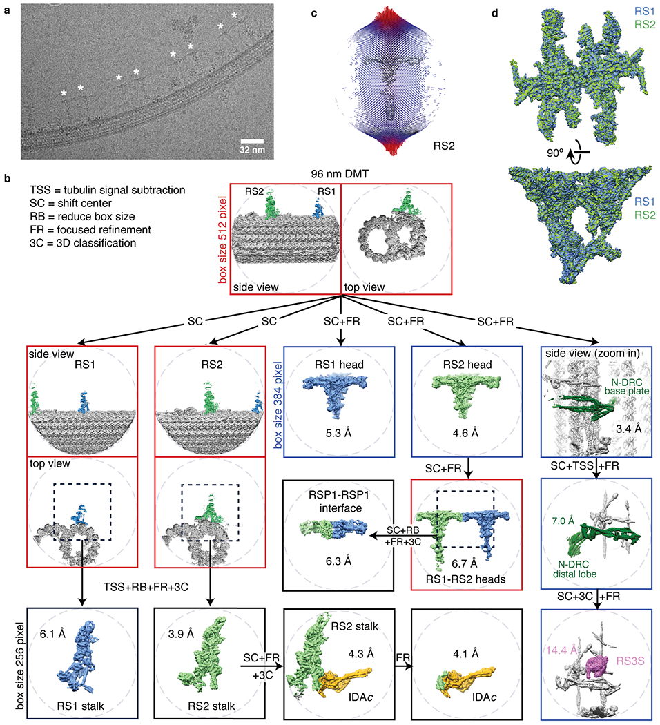
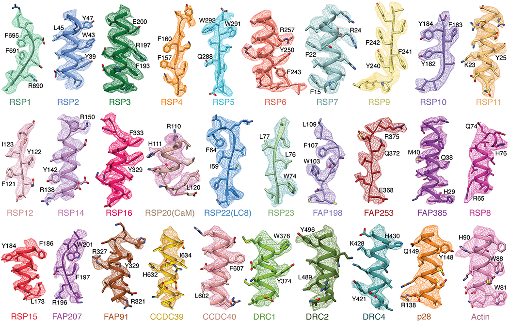
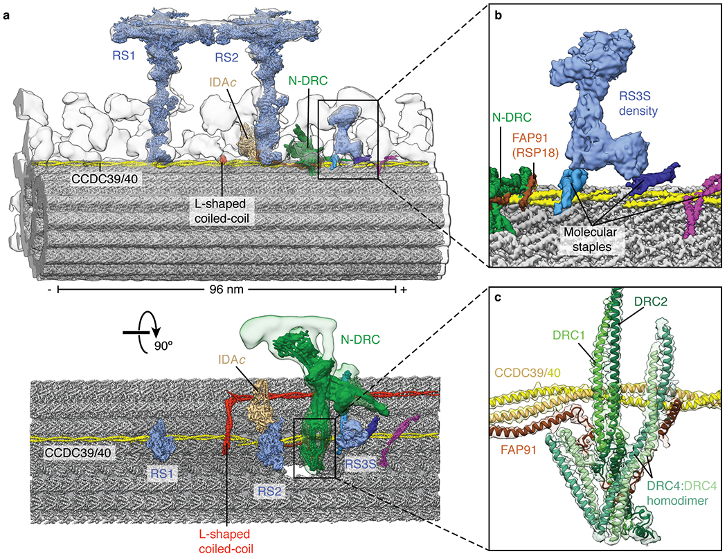
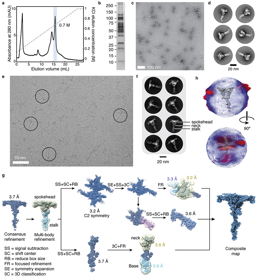
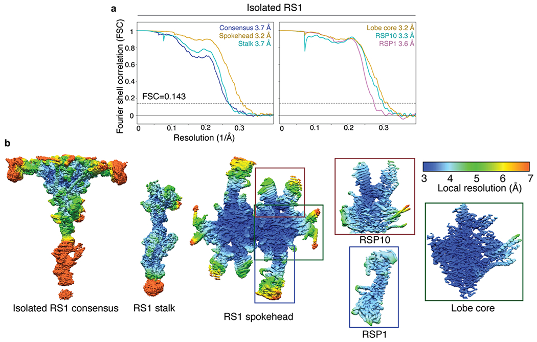
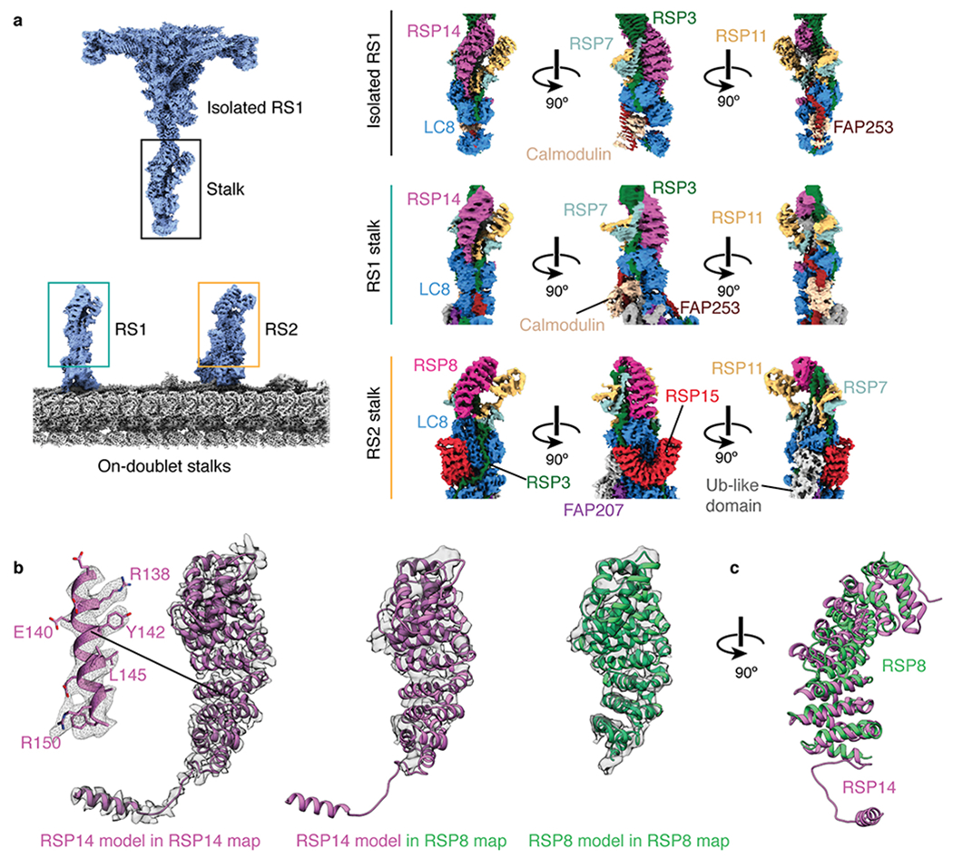
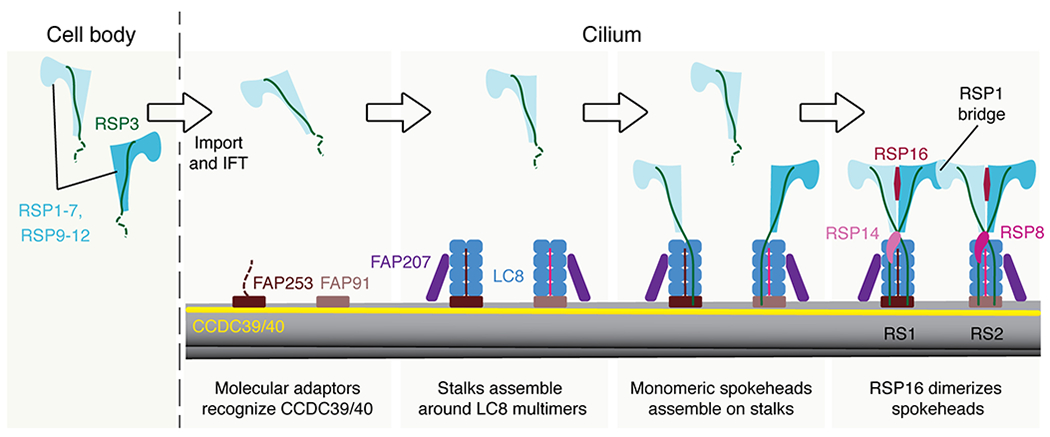
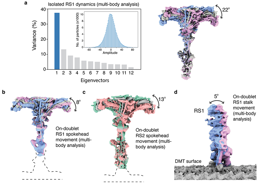
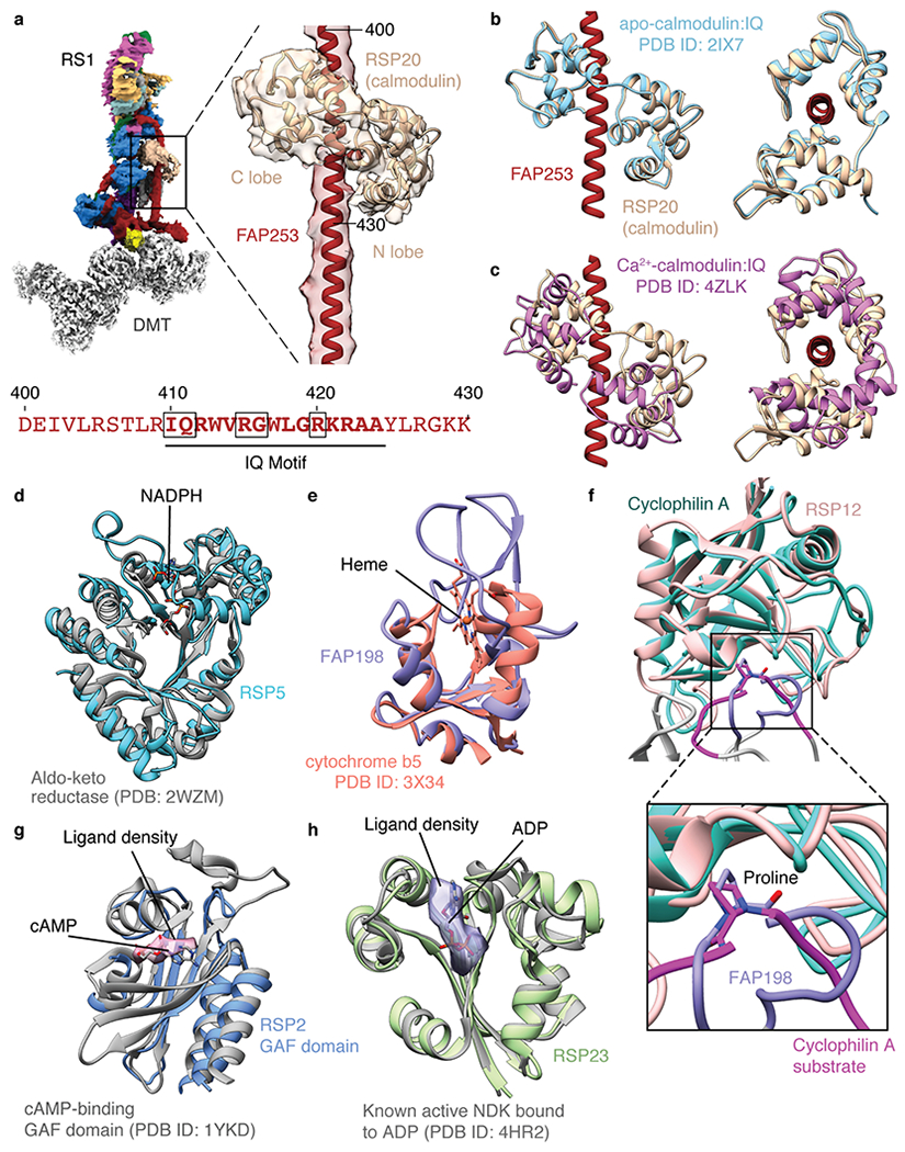
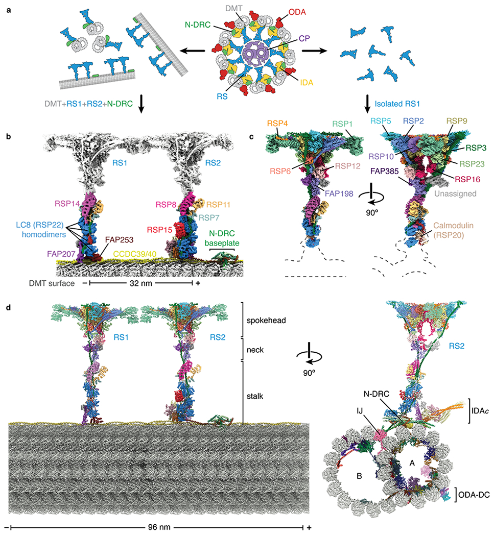
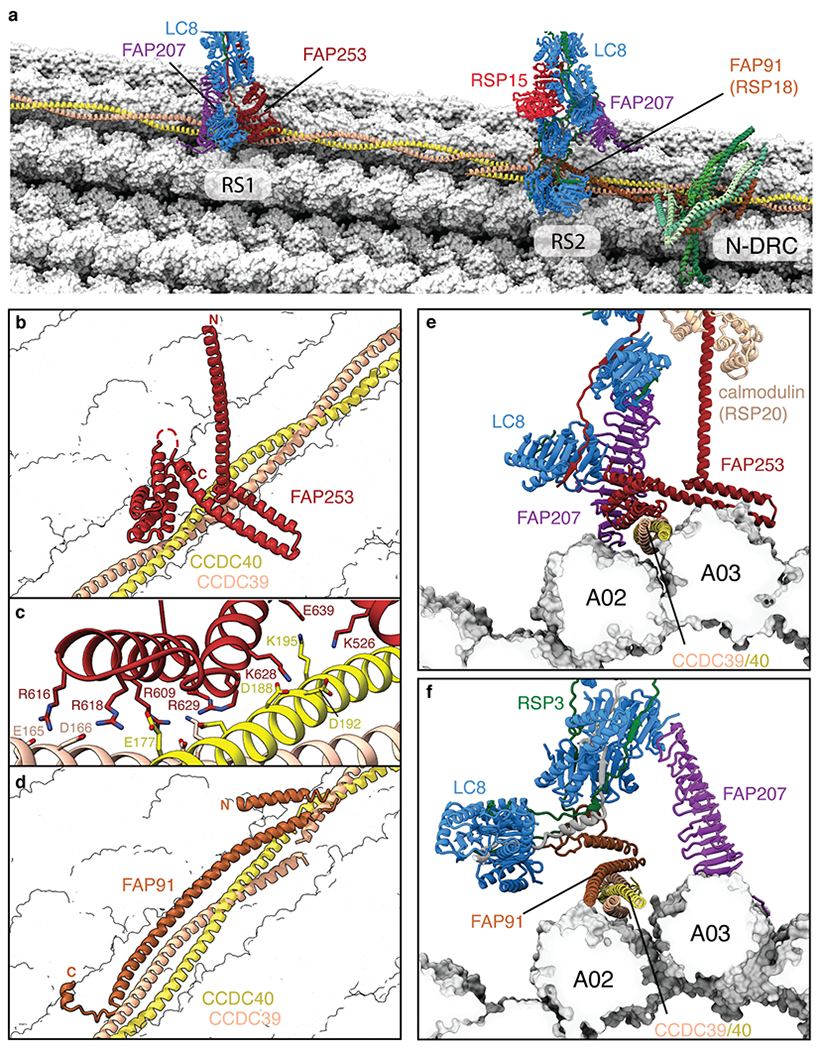
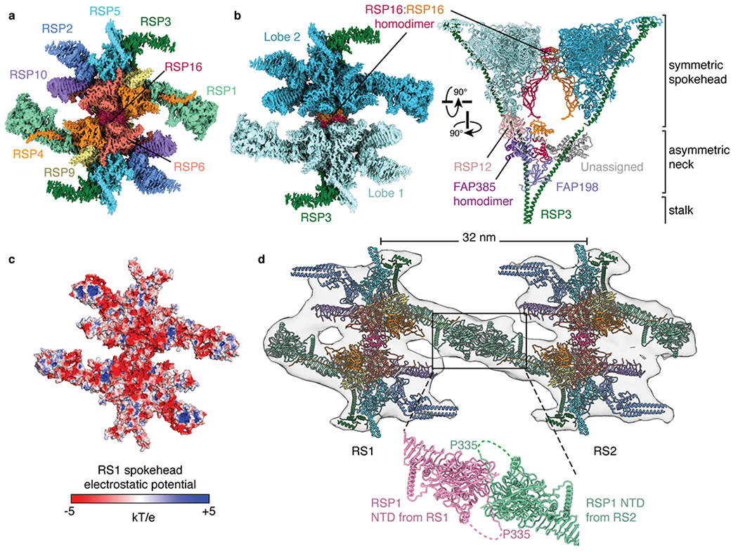
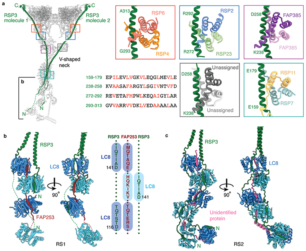
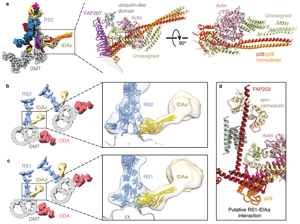
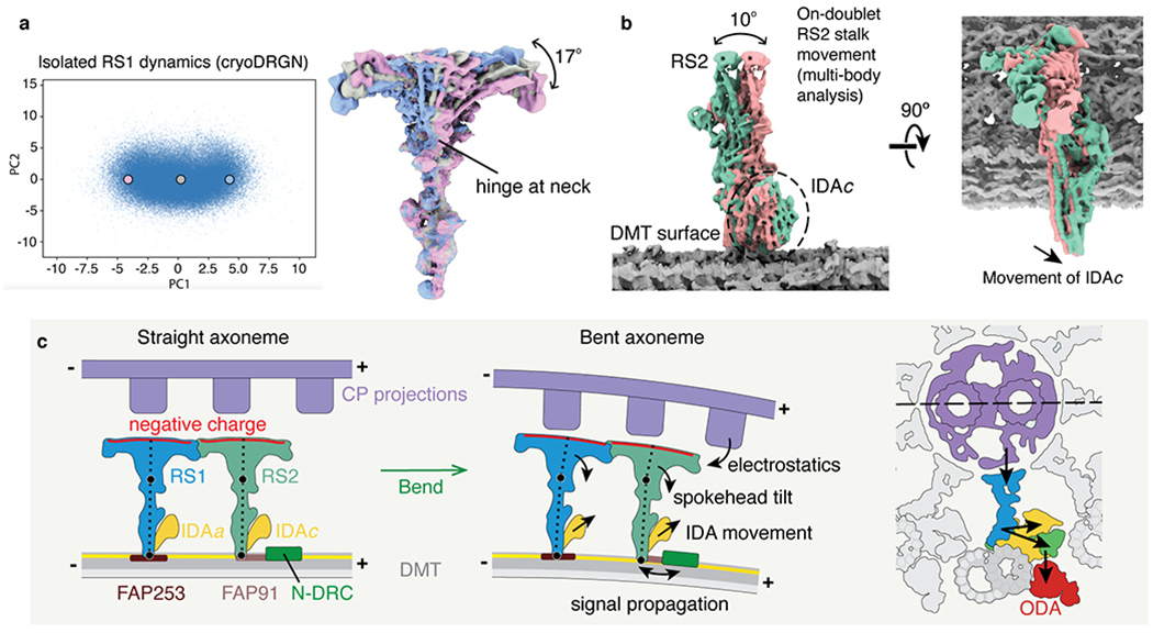
In motile cilia, a mechanoregulatory network is responsible for converting the action of thousands of dynein motors bound to doublet microtubules into a single propulsive waveform. Here, we use two complementary cryo-EM strategies to determine structures of the major mechanoregulators that bind ciliary doublet microtubules in Chlamydomonas reinhardtii. We determine structures of isolated radial spoke RS1 and the microtubule-bound RS1, RS2 and the nexin-dynein regulatory complex (N-DRC). From these structures, we identify and build atomic models for 30 proteins, including 23 radial-spoke subunits. We reveal how mechanoregulatory complexes dock to doublet microtubules with regular 96-nm periodicity and communicate with one another. Additionally, we observe a direct and dynamically coupled association between RS2 and the dynein motor inner dynein arm subform c (IDAc), providing a molecular basis for the control of motor activity by mechanical signals. These structures advance our understanding of the role of mechanoregulation in defining the ciliary waveform.


Conflict of interest statement

Competing Interests

The authors declare no competing interests.

Figures

Extended Data Fig. 1 |
Extended Data Fig. 1 |. Data collection and processing for the on-doublet mechanoregulatory complexes.
a, Section of an electron micrograph showing radial spokes (marked with an asterisk) bound to a doublet microtubule in vitreous ice. b, Processing scheme used to generate reconstructions of mechanoregulatory complexes bound to doublet microtubules (DMT, gray). To resolve various structural features with 96-nm periodicity (RS1 spokehead/stalk, RS2 spokehead/stalk, RSP1-RSP1 interface, RS3S, IDAc, or N-DRC baseplate/lobe), it was necessary to use a combination of tubulin signal subtraction (TSS), shifting the center (SC) of coordinates to the feature of interest, focused refinement (FR) and 3D classification without alignment (3C). When possible, the box size was reduced (RB) to 256 or 384 instead of 512 pixels to facilitate data processing. c, Angular distribution of the particle views used for reconstruction of on-doublet RS2. Similar distributions were obtained for on-doublet RS1. The height of the cylinders, colored from blue to red, represents the number of particles. The final density map of RS2 is shown in gray. d, Superimposition of the on-doublet RS1 and RS2 spokeheads confirms that they have identical structure.
Extended Data Fig. 2 |
Extended Data Fig. 2 |. Global and local resolution of on-doublet mechanoregulatory complexes.
a, Fourier shell correlation (FSC) curves calculated between masked independent half maps for on-doublet structures. Left panel, FSC curves are shown for focused refinements of the RS1 spokehead and stalk, RS2 spokehead and stalk, and the RSP1-RSP1 dimer. Right panel, FSC curves for the base of RS1, the RS2-IDAc complex, IDAc, and the N-DRC baseplate. The nominal resolution was estimated using the FSC = 0.143 criterion (dashed line). b, Density maps for on-doublet structures colored by local resolution. Only the maps used for model building are shown. The local resolution is colored from 3 to 7 Å.
Extended Data Fig. 3 |
Extended Data Fig. 3 |. Map quality.
Examples of map density for all 30 non-tubulin proteins identified in this study. The first 19 proteins show density from isolated RS1 contoured at 0.009-0.013. The remaining 11 proteins (starting from RSP8) show density from on-doublet maps contoured at 0.020-0.031. Landmark residues are labeled. Note that the sidechains of RSP20 (calmodulin) and RSP8 are not well resolved and are truncated in the deposited model.
Extended Data Fig. 4 |
Extended Data Fig. 4 |. Single-particle cryo-EM maps docked into a subtomogram average of the axoneme.
a, Two views showing the single-particle cryo-EM maps of RS1, RS2, RS3S, N-DRC, and IDAc docked into the subtomogram average of the 96-nm repeat of the Chlamydomonas axoneme (EMD-6872). The subtomogram average is shown as a gray isosurface. b, Zoom-in view showing the map of RS3S. Density for RS3S is recovered in 25% of the particles following 3D classification (Extended Data Fig. 1b). RS3S interacts with two molecular staples of unknown identity. c, Zoom-in view showing the model and map for the N-DRC baseplate. Three N-DRC subunits (DRC1, DRC2, and DRC4) can be unambiguously identified. FAP91 interacts with all three.
Extended Data Fig. 5 |
Extended Data Fig. 5 |. Data collection and processing for isolated RS1.
a, Chromatogram showing the elution of RS1 from an anion-exchange column using a KCl gradient. The peak fraction containing RS1 is highlighted and elutes at ~0.7 M KCl. b, Silver-stained SDS-PAGE gel showing the purity of isolated RS1 following anion-exchange chromatography. The molecular weights of markers (in kDa) are indicated on the left. The result of mass spectrometry analysis of this sample is given in Supplementary Data 1. c, Section of a negative-stain electron micrograph showing homogeneous and monodisperse radial spokes. d, Selected two-dimensional class averages of particles selected from negative-stain electron micrographs. e, Section of an electron micrograph showing radial spokes in vitreous ice. Particles showing the characteristic T-shaped projection of radial spokes are circled. f, Selected two-dimensional class averages of radial spokes showing well defined spokeheads but nebulous density for the stalk consistent with flexibility at the neck. g, Schematic showing the processing of the isolated RS1 data. Following a consensus refinement, the spokehead and stalk were independently refined. The twofold rotational symmetry of the spokehead was exploited to improve the map quality. Further masked refinement was used to improve the flexible projections of the spokehead and the base and neck of the stalk. These individual maps were recombined to generate a final composite cryo-EM map. h, Angular distribution of the particle views used for the consensus reconstruction of isolated RS1. The height of the cylinders, colored from blue to red, represents the number of particles. The final density map of RS1 is shown in gray.
Extended Data Fig. 6 |
Extended Data Fig. 6 |. Global and local resolution of isolated RS1.
a, FSC curves calculated between masked independent half maps for isolated RS1. Left panel, FSC curves are shown for the consensus refinement of isolated RS1, focused refinement of the stalk, and focused refinement of the spokehead after applying C2 symmetry. Right panel, FSC curves for focused refinements of three subdomains of a single lobe of the RS1 spokehead. The colors of the curves match the masks used in Extended Data Fig. 5g. The nominal resolution was estimated using the FSC = 0.143 criterion (dashed line). b, Density maps for the consensus refinement of isolated RS1 and various focused refinements colored by local resolution. The local resolution is colored from 3 to 7 Å.
Extended Data Fig. 7 |
Extended Data Fig. 7 |. The stalks of RS1 and RS2.
a, The stalk of the isolated radial spoke is consistent with the on-doublet stalk of RS1 only. FAP253, RSP14, and calmodulin are present in the stalk of RS1 but not RS2. RSP8, RSP15, and an unidentified ubiquitin (Ub)-like domain are present in the stalk of RS2 but not RS1. LC8, FAP207, and RSP3 are common to both RS1 and RS2 but adopt different conformations. The RSP7/11 heterodimer is similar in both radial spokes. b, RSP14 and RSP8 are structurally similar armadillo proteins present in different radial spokes. Left, RSP14 was identified in the stalk of RS1 based on well-defined sidechain density. Middle, the model of RSP14 is incompatible with the density of the armadillo protein in RS2, indicating that they are different proteins with similar folds. Right, a model for RSP8 built into the RS2 density. c, Superposition of the atomic models for RSP8 and RSP14.
Extended Data Fig. 8 |
Extended Data Fig. 8 |. Model of radial spoke assembly.
Proposed model of radial spoke assembly. Monomeric spokehead lobes, comprising RSP1-7 and RSP9-12, assemble in the cell body,– before being imported into the cilium by intraflagellar transport (IFT),. In the cilium, the axonemal doublet microtubules are bound by the CCDC39/40 coiled coil. Specific sequences within the coiled coil are recognized by molecular adaptors FAP253 and FAP91 that establish the binding sites for RS1 and RS2. These molecular adaptors recruit LC8 and FAP207, although the arrangement of these elements is different in the two stalks. RSP3 in the precursor binds the LC8 multimers (Fig. 4b), helping dock the spokehead lobe onto the preassembled stalks. Two lobes can bind a single stalk. Binding of RSP16 is presumably a relatively late step that dimerizes the lobes. At a similar time, RS-specific proteins bind; RSP14 to RS1 and RSP8 to RS2.
Extended Data Fig. 9 |
Extended Data Fig. 9 |. Dynamics of radial spokes by multi-body analysis.
a, Multi-body analysis of isolated RS1. Left, the contributions of all eigenvectors to the variance. The first eigenvector accounts for 37% of all variability. Inset, the unimodal histogram of amplitudes along the first eigenvector indicates continuous motion. Right, the density maps at the extremes and middle show the same tilting of the spokehead relative to the stalk as observed by the neural-network approach in Fig. 6a. b, Multi-body analysis of on-doublet RS1 shows the same direction of spokehead tilt as isolated RS1. c, Multi-body analysis of on-doublet RS2 shows that the spokeheads of both radial spokes tilt in similar directions to similar extents. d, Multi-body analysis of the movement of the RS1 stalk with respect to the doublet microtubule (DMT) surface.
Extended Data Fig. 10 |
Extended Data Fig. 10 |. Potential chemical modulation of radial spokes.
a, Calmodulin binds the IQ motif of FAP253 at the base of RS1. Below, sequence of FAP253 residues 400-430 showing the presence of an IQ motif (emboldened with motif-defining residues boxed). b, Structural comparison of calmodulin bound to FAP253 with apo-calmodulin bound to an IQ motif from myosin V (PDB 2IX7). c, Structural comparison of calmodulin bound to FAP253 with Ca2+-calmodulin bound to an IQ motif from myosin 5a (PDB 4ZLK). d, The structure of RSP5 resembles an NADPH-dependent aldo-keto reductase domain (PDB 2WZM). However, the NADPH binding site of RSP5 is absent and filled by two loops (residues 393-414 and 468-484 of RSP5). e, The N-terminal domain of FAP198 closely resembles heme-binding cytochrome b5 (PDB 3X34). However, no heme is observed bound to FAP198, and the putative heme-binding site is occluded by a loop of FAP198 (residues 89-95). f, RSP12 structurally resembles cylophilin-type peptidyl-prolyl cis-trans isomerase (PDB 1AK4). The putative substrate-binding site of RSP12 is occupied by a loop of FAP198 (residues 96-105), which positions a proline (P99) in the active site. g, Atomic model of the GAF domain from RSP2 superposed with the model of a cAMP-bound GAF domain (PDB 1YKD). Unexplained density in the RSP2 GAF domain (pink, contoured at 0.01) is observed in the cAMP binding pocket, but the resolution is insufficient to assign it as a cyclic nucleotide. The cAMP ligand from PDB 1YKD is shown for comparison. h, Atomic model of RSP23 superposed with an active, ADP-bound nucleoside diphosphate kinase (NDK; PDB 4HR2). Many of the active site residues are conserved. Potential density for a bound nucleotide to RSP23 is observed in the on-doublet map of RS1 (purple, contoured at 0.017) but not in the isolated RS1 map. The ADP ligand from PDB 4HR2 is shown for comparison.
Fig. 1 |
Fig. 1 |. Structures of radial spokes on and off doublet microtubules.
a, Schematic representation showing biochemical fragmentation of the Chlamydomonas axoneme (viewed in cross-section; center) into mechanoregulator-bound doublet microtubules (left) and isolated radial spokes (right). The axoneme consists of a central pair of microtubules (CP, purple) surrounded by doublet microtubules (DMT; gray) bound by radial spokes (RS; blue), nexin-dynein regulatory complexes (N-DRC; green), inner dynein arm (IDA; yellow), and outer dynein arm (ODA; red). b, Composite density map for on-doublet RS1 and RS2. The maps of the stalks are colored by subunit. The neck and spokehead, which are less well resolved, are colored gray. c, Orthogonal views of a composite density map for isolated RS1 with the map colored by subunit. Dashed lines indicate the positions of the stalks and doublet microtubule which are not present in the reconstruction. d, Orthogonal views of an atomic model for the 96-nm repeat of the doublet microtubule. The model combines atomic models of the doublet-bound stalks of RS1 and RS2, and the stalk, neck, and spokehead of isolated RS1 with model of the doublet microtubule (PDB 6U42). Individual subunits are colored according to panels b and c. In panels b and d, the minus (−) and plus (+) ends of the doublet microtubule are indicated at the ends of the scale bar.
Fig. 2 |
Fig. 2 |. Structural basis for the microtubule docking and longitudinal periodicity of radial spokes.
a, Overview showing the bases of radial spokes 1 and 2 (RS1 and RS2) bound to the doublet microtubule. The radial spoke subunits (LC8, FAP91, FAP207, FAP253, and RSP15), the CCDC39/40 molecular ruler, and the subunits of the nexin-dynein regulatory complex (N-DRC) baseplate are shown as cartoons. Tubulin is shown in surface representation. b, The binding site for RS1 is determined by recognition of CCDC39/40 by FAP253. c, Zoom-in view showing that the interface between FAP253 and CCDC39/40 is dominated by a network of salt bridges. d, The binding site for RS2 is determined by FAP91 (RSP18) which forms a triple coiled coil with CCDC39/40. e, Cross-section view of the RS1-doublet microtubule interaction showing recognition of CCDC39/40 by FAP253. FAP207 bridges LC8 with protofilament A02. f, Cross-section view of the RS2-doublet microtubule interaction showing recognition of CCDC39/40 by FAP91. FAP207 bridges LC8 with protofilament A03.
Fig. 3 |
Fig. 3 |. Structure of the radial spokehead.
a, View of the top of the radial spokehead. Composite map colored by subunit. b, Two views of the radial spokehead. The two symmetric lobes of the spokehead (colored different shades of blue) are dimerized by a homodimer of RSP16 and flanked by the α-helices of RSP3. The symmetric spokehead sits on a V-shaped asymmetric neck containing FAP198, FAP385, RSP12, and the N-terminal domains of RSP16. c, Atomic model of the radial spokehead colored by electrostatic potential. d, Atomic models of the radial spokes docked into an isosurface rendering of the subtomogram average of the Chlamydomonas axoneme (EMD-6872). The two radial spokes interact through the N-terminal domains (NTD) of RSP1. The zoom-in shows the model of the on-doublet interaction between the NTDs of RSP1 from RS1 and RS2.
Fig. 4 |
Fig. 4 |. Interactions between RSP3 and radial spoke proteins.
a, Left, atomic model of RS1 with its two molecules of RSP3 colored green. Colored boxes indicate the binding sites on RSP3 of five different dimers. The partially boxed proximal region of the stalk is shown in detail in panel b. Right, atomic models of the interactions between RSP3 and different dimers. The sequence alignment shows amphipathic helices of RSP3 with hydrophobic residues recognized by the various dimers colored red. The four-helix bundle of RSP7/RSP11 belongs to the RIIa dimerization/docking domain and the four-helix bundles of RSP4/RSP6, RSP2/RSP23, RSP385/RSP385 and the unassigned dimer belong to Dpy-30 domain (which has an additional helix at the N-terminus compared with RIIa dimerization/docking domain). b, Left, orthogonal views of the interactions of RSP3 and FAP253 with the LC8 homodimers in RS1. Other subunits have been omitted for clarity. Right, a schematic showing the resolved LC8-binding motifs of FAP253 and RSP3. c, Orthogonal views of the interactions between RSP3 and the LC8 homodimers in RS2. An unassigned protein (pink) also interacts with the β-edges of LC8. Other subunits have been omitted for clarity.
Fig. 5 |
Fig. 5 |. IDA subforms a and c dock onto the bases of radial spokes.
a, Left, composite map showing the densities for the base of radial spoke 2 (RS2), inner dynein arm subform c (IDAc), and the doublet microtubule (DMT). The maps of RS2 and IDAc are colored by subunit. Right, atomic model of IDAc showing the interaction with FAP207 and an unidentified ubiquitin-like domain of RS2. The unassigned helices of IDAc may correspond to the tail of the dynein heavy chain. b, Left, slice through the subtomogram average of the Chlamydomonas axoneme (EMD-6872) with RS2 colored blue, IDAc colored yellow, outer dynein arm (ODA) colored red, and the DMT colored gray. Zoomed-in view showing the models of RS2 and IDAc (colored blue and yellow, respectively) docking into the subtomogram average. c, Left, slice through the subtomogram average of the Chlamydomonas axoneme (EMD-6872) with radial spoke 1 (RS1) colored blue, inner dynein arm subform a (IDAa) colored yellow, ODA colored red, and the DMT colored gray. Zoomed-in view showing the atomic models of RS1 and IDAc (colored blue and yellow, respectively) docking into the subtomogram average. The putative interface between IDAa and RS1 is distinct from the interface between RS2 and IDAc. d, Details of the expected interface between RS1 and IDAa based on docking the model of IDAc into the subtomogram average map of IDAa (EMD-6872).
Fig. 6 |
Fig. 6 |. Molecular basis for the control of IDA motor activity by mechanical signals.
a, Conformational dynamics of isolated radial spoke 1 (RS1) inferred from deep neural networks. Left, Principle component (PC) analysis projection of latent space. Density maps were generated at three points along PC1. Right, Density maps generated from these 3 points were aligned on the stalk and show tilting of the spokehead relative to the stalk. The hinge lies in the neck. b, Multi-body analysis of the movement of the RS2 stalk and IDAc with respect to the doublet microtubule (DMT) surface. The DMT, RS2, and IDAc were separately masked and treated as individual bodies free to move relative to one another. Movement of IDAc corresponds with the movement of the RS2 stalk. c, A model for mechanoregulation of ciliary motility. As the axoneme bends, the projections of the central pair (CP) are brought closer to the spokeheads. An increase in the electrostatic force with decreasing distance causes the radial spoke to tilt at two hinge points (indicated with black circles). As the movement of IDAc is coupled to the movement of RS2, tilting of the radial spoke changes the orientation of the IDA. IDAa may likewise be coupled to the movement of RS1. Signals may propagate from RS2 to N-DRC through FAP91 and the CCDC39/40 coiled coil and from the N-DRC to the ODAs via the outer–inner dynein linkers.

References

    1. Ginger ML, Portman N & McKean PG Swimming with protists: perception, motility and flagellum assembly. Nat Rev Micro 6, 838–850 (2008). - PubMed
    1. Bustamante-Marin XM & Ostrowski LE Cilia and Mucociliary Clearance. Cold Spring Harbor Perspectives in Biology 9, a028241 (2017). - PMC - PubMed
    1. Lyons RA, Saridogan E & Djahanbakhch O The reproductive significance of human Fallopian tube cilia. Hum. Reprod. Update 12, 363–372 (2006). - PubMed
    1. Faubel R, Westendorf C, Bodenschatz E & Eichele G Cilia-based flow network in the brain ventricles. Science 353, 176–178 (2016). - PubMed
    1. Nonaka S et al. Randomization of left-right asymmetry due to loss of nodal cilia generating leftward flow of extraembryonic fluid in mice lacking KIF3B motor protein. Cell 95, 829–837 (1998). - PubMed

Publication types

MeSH terms