Review
. 2020 Jul 16:27:177-190.
doi: 10.1016/j.jare.2020.07.006. eCollection 2021 Jan.

Targeting hydrogen sulphide signaling in breast cancer

Affiliations
Review

Targeting hydrogen sulphide signaling in breast cancer

Rana Ahmed Youness et al. J Adv Res. .

Abstract

Introduction: Hydrogen sulphide (H2S) has been established as a key member of the gasotransmitters family that recently showed a pivotal role in various pathological conditions including cancer.

Objectives: This study investigated the role of H2S in breast cancer (BC) pathogenesis, on BC immune recognition capacity and the consequence of targeting H2S using non-coding RNAs.

Methods: Eighty BC patients have been recruited for the study. BC cell lines were cultured and transfected using validated oligonucleotide delivery system. Gene and protein expression analysis was performed using qRT-PCR, western blot and flow-cytometry. In-vitro analysis for BC hallmarks was performed using MTT, BrdU, Modified Boyden chamber, migration and colony forming assays. H2S and nitric oxide (NO) levels were measured spectrophotometrically. Primary natural killer cells (NK cells) and T cell isolation and chimeric antigen receptor transduction (CAR T cells) were performed using appropriate kits. NK and T cells cytotoxicity was measured. Finally, computational target prediction analysis and binding confirmation analyses were performed using different software and dual luciferase assay kit, respectively.

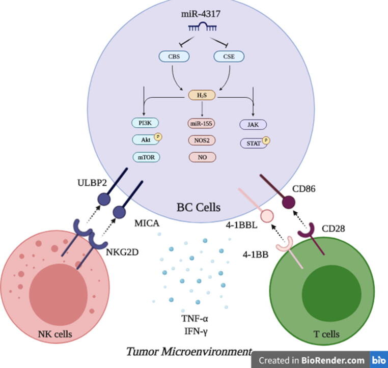
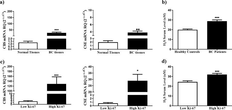
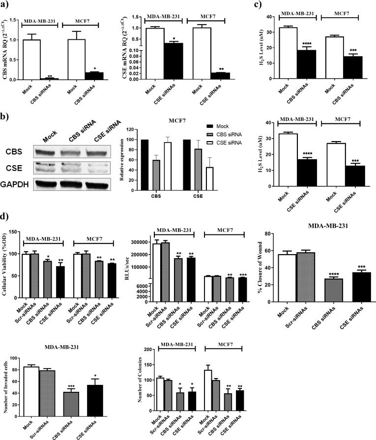
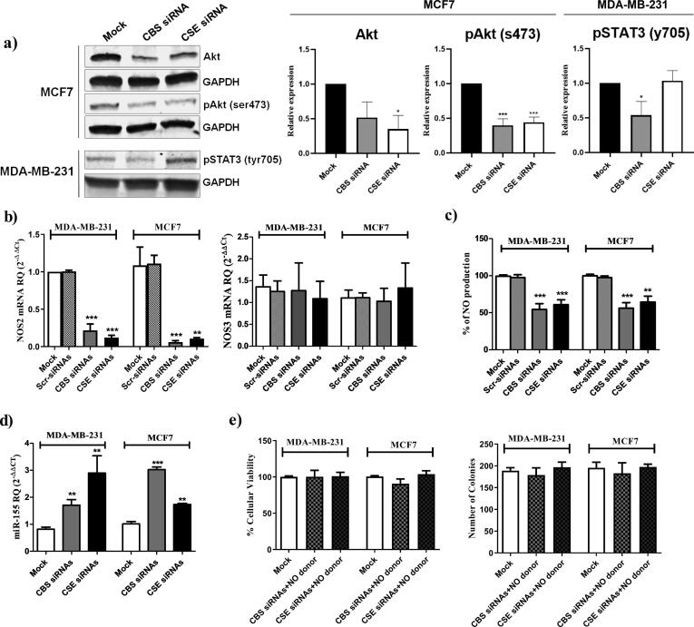
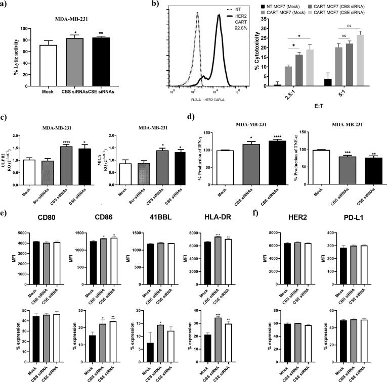
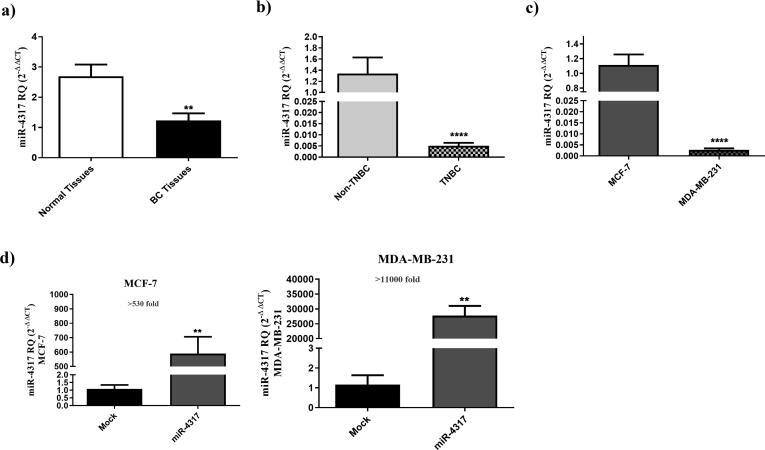
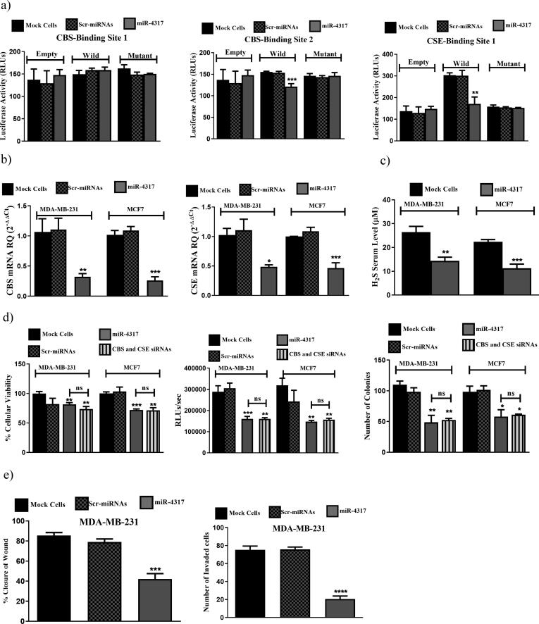
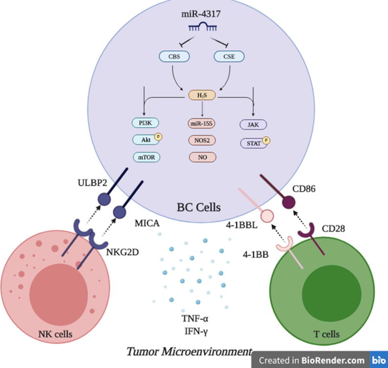
Results: The H2S synthesizing enzymes, cystathionine β-synthase (CBS) and cystathionine γ-lyase (CSE), exhibited elevated levels in the clinical samples that correlated with tumor proliferation index. Knock-down of CBS and CSE in the HER2+ BC and triple negative BC (TNBC) cells resulted in significant attenuation of BC malignancy. In addition to increased susceptibility of HER2+ BC and TNBC to the cytotoxic activity of HER2 targeting CAR T cells and NK cells, respectively. Transcriptomic and phosphoprotein analysis revealed that H2S signaling is mediated through Akt in MCF7, STAT3 in MDA-MB-231 and miR-155/ NOS2/NO signaling in both cell lines. Lastly, miR-4317 was found to function as an upstream regulator of CBS and CSE synergistically abrogates the malignancy of BC cells.

Conclusion: These findings demonstrate the potential role of H2S signaling in BC pathogenesis and the potential of its targeting for disease mitigation.

Keywords: 41BBL, 41BB Ligand; 51Cr-release, Chromium release assay; BC, Breast Cancer; Breast cancer; CAR T cells; CAR, Chimeric antigen receptor; CBS, Cystathionine β-synthase; CD80, Cluster of differentiation 80; CD86, Cluster of differentiation 86; CSE, Cystathionine γ-lyase; CTL, Cytotoxic T lymphocyte; H2S, Hydrogen sulphide; HCC, Hepatocellular carcinoma; HLA-DR, Human Leukocytic antigen DR; Hydrogen sulphide; IFN-γ, Interferon gamma; KD, Knock down; LDH, Lactate dehydrogenase Assay; MICA/B, MHC class I polypeptide-related sequence A/B; NK, Natural killer; NKG2D, Natural Killer Group 2D; NO, Nitric oxide; NOS2, Inducible nitric oxide synthase-2; NOS3, Endothelial nitric oxide synthase-3; Natural killer cells; Nitric oxide; PD-L1, Programmed death-ligand 1; PI3K/AKT signaling pathway; Scr-miRNAs, Scrambled microRNAs; Scr-siRNAs, Scrambled siRNAs; TNBC, Triple negative breast cancer; TNF-α, Tumor necrosis factor-α; ULBP2/5/6, UL16 binding protein 2/5/6; miR-155/NOS2/NO signaling pathway; miR-4317; miRNA, MicroRNA; ncRNAs, Non-coding RNAs; siRNAs, Small interfering RNAs.


Conflict of interest statement

The authors declare that they have no known competing financial interests or personal relationships that could have appeared to influence the work reported in this paper.

Figures

None
Graphical abstract
Fig. 1
Fig. 1
Overexpression of CBS and CSE enzymes in BC tissues. (a) CBS and CSE expression profiles were analyzed in breast tissues isolated from 80 BC patients using qRT- PCR, normalized to 18 s rRNA as an internal control, and compared to normal BC tissues from the same patients (b) Serum H2S was compared between BC patients and healthy volunteers. (c) CBS and CSE expression was compared according to the tumor proliferation index ki-67. (d) Serum H2S levels were compared according to the tumor proliferative index ki-67.
Fig. 2
Fig. 2
CBS and CSE sequence specific siRNAs downregulate RNA, protein and H2S production levels and impact BC malignant properties. The expression of CBS and CSE transcripts and protein levels in mock and siRNAs transfected MDA-MB-231 and MCF7 cells were determined using (a) qRT-PCR 48 h post-transfection and (b) western blot analysis 72 h post-transfection (c) H2S levels were determined in the supernatant of MDA-MB-231 and MCF7 after silencing of CBS and CSE (d) Knocking down of CBS and CSE using CBS and CSE siRNAs resulted in a marked repression of cellular viability assessed by MTT assay (top left), cellular proliferation assessed using BrdU incorporation assay (top middle), migration capacity measured by wound healing assay (top right), invasion capacity measured by Boyder chamber assay (bottom left) and colony formation ability (bottom right).
Fig. 3
Fig. 3
CBS/CSE silencing modulates PI3K/Akt, JAK/STAT3 and miR-155/NOS2/NO signaling pathways. (a) The protein expression of Akt, phosho-Akt (serine 473) in mock or siRNA transfected MCF7 and phosphor-STAT3 (tyrosine 705) in mock or siRNA transfected MDA-MB-231 was determined using western blot 72 h post transfection. Relative expression plots were normalized to the internal control GAPDH, then compared to the mock ± standard deviation from 3 separate experiments (b) the RNA trnascripts of NOS2 and NOS3 in mock and siRNAs transfected MDA-MB-231 and MCF7 cells determined by qRT-PCR 48 h post-transfection. (c) NO levels were determined in the supernatant of MDA-MB-231 and MCF7 after silencing of CBS and CSE. (d) The expression pattern of miR-155 normalized to RNU6B as an internal control in mock, CBS siRNAs and CSE siRNAs MDA-MB-231 and MCF7 cells were determined by qRT-PCR 48 h post-transfection. (e) Cellular viability and colony forming ability of MDA-MB-231 and MCF-7 cells co-treated with a NO donor (NaNO2) and CBS or CSE siRNA.
Fig. 4
Fig. 4
Impact of silencing CBS and CSE on immune cell mediated cytotoxicity, immune ligand expression and cytokines release. (a) NK cell mediated cytotoxicity was tested via co-culturing experiments that were done between mock, CBS/CSE KDMDA-MB-231 and primary NK cells using LDH assay. (b) T cell mediated cytotoxicity was tested using HER2 targeting CAR T cells. CAR transduction rate was tested using a recombinant human ErbB2/Her2 Fc chimeric protein + PE-conjugated goat anti-human IgG (Fc gamma-specific) andcompared to non-transduced (NT) T cells. CAR T cell mediated cytotoxicity was tested by a chromium (51Cr) release assay. CAR T cells were incubated with MCF7 cells at effector to target ratios of 2.5:1 and 5:1 for 6 h. Cytotoxicity is presented as percentage normalized to triton-X induced cytotoxicity.(c) ULBP2 and MICA were analyzed on the mRNA transcripts level in mock, CBS or CSE KD MDA-MB-231 using qRT-PCR 48 h post-transfection. (d) The supernatant of mock, CBS/CSE KD MDA-MB-231 was screened for IFN-γ and TNF-α production using ELISA. (e) CD80, CD86, 41BBL and HLA-DR surface expression on mock, CBS or CSE KD MCF7, represented as percentage expression and median fluorescence intensity, were analyzed using flow cytometry 72 h post-transfection (f) HER2 and PD-L1 surface expression on mock, CBS/CSE KD MCF7, represented as percentage expression and median fluorescence intensity, was analyzed using flow cytometry 72 h post-transfection.
Fig. 5
Fig. 5
miR-4317 is downregulated in TNBC clinical samples and cell line. (a) miR-4317 expression profile was analyzed in tumor tissues from 80 BC patients and compared to patient matched normal tissues using qRT-PCR, and normalized to RNU6B as an internal control. (b) Repressed expression levels of miR-4317 in TNBC patients compared to non-TNBC patients, (c) Reduced expression levels of miR-4317 in MDA-MB-231 cell line when compared to MCF7 cells. The expression of miR-4317 in MDA-MB-231 and MCF7 cells was determined by qRT-PCR 48 h post-transfection. (d) Levels of miR-4317 showed more than 530 folds increase in miR-4317 plasmid transfected MCF7 cells compared to mock cells. miR-4317 plasmid transfected MDA-MB-231 cells showed more than 11,000 folds increase in the expression level of miR-4317 when compared to mock cells, determined by qRT-PCR 48 h post-transfection.
Fig. 6
Fig. 6
miR-4317 co-targets CBS and CSE simultaneously mitigating BC malignant properties. (a) Luciferase reporter assay for binding confirmation of miR-4317 on 3′UTR of CBS and CSE. miR-4317-CBS first binding site construct showed no inhibition of luciferase activity. miR-4317-CBS second binding site construct showed significant reduction of luciferase activity in cells co-transfected with miR-4317 mimics plus WT construct compared to mock and scr-miRNAs transfected cells transfected with the WT construct alone. Likewise, for the miR-4317-CSE binding site construct, luciferase activity was also significantly repressed in cells co-transfected with miR-4317 mimics plus WT construct compared to mock and scr-miRNAs transfected cells transfected with the WT construct alone. (b) Down-regulation of CBS and CSE expression by miR-4317 mimics in MDA-MB-231 and MCF7 cells. (c) Reduced H2S levels produced from BC cells upon transfection with miR-4317 mimics. (d) miR-4317 mimics reduced cellular viability (left), cellular proliferation (middle), and colony formation ability (right) in MDA-MB-231 and MCF7. (e) miR-4317 mimics resulted in reduction in migration (left) and invasion (right) capacities in MDA-MB-231.
Fig. 7
Fig. 7
. Targeting H2S synthesizing machinery in BC cells. This is a conclusive descriptive figure highlighting the major roles of endogenously produced H2S in BC cells. Mechanistically, PI3K/Akt/mTOR, miR-155/NOS2/NO, JAK/STAT can be drawn downstream CBS and CSE induced H2S in BC cells. Functionally, this results in abrogation of BC hallmarks, potentiating NK cells and cytotoxic T cells activity and alleviating the immune suppressive microenvironment in BC tumors. Dual Targeting of CBS and CSE in BC cells was achieved using the novel tumor suppressor miR-4317.

References

    1. Waks A.G., Winer E.P. Breast cancer treatment: a review. JAMA. 2019;321(3):288–300. - PubMed
    1. Bray F. Global cancer statistics 2018: GLOBOCAN estimates of incidence and mortality worldwide for 36 cancers in 185 countries. CA Cancer J Clin. 2018;68(6):394–424. - PubMed
    1. Nielsen D.L. Efficacy of HER2-targeted therapy in metastatic breast cancer. Monoclonal antibodies and tyrosine kinase inhibitors. Breast. 2013;22(1):1–12. - PubMed
    1. Denkert C. Molecular alterations in triple-negative breast cancer-the road to new treatment strategies. Lancet. 2017;389(10087):2430–2442. - PubMed
    1. Youness R.A., Gad M.Z. Long non-coding RNAs: Functional regulatory players in breast cancer. Noncoding RNA Res. 2019;4(1):36–44. - PMC - PubMed