. 2020 Dec 7;6(12):e05574.
doi: 10.1016/j.heliyon.2020.e05574. eCollection 2020 Dec.

Visualizing cellular heterogeneity by quantifying the dynamics of MAPK activity in live mammalian cells with synthetic fluorescent biosensors

Affiliations

Visualizing cellular heterogeneity by quantifying the dynamics of MAPK activity in live mammalian cells with synthetic fluorescent biosensors

Min Ma et al. Heliyon. .

Abstract

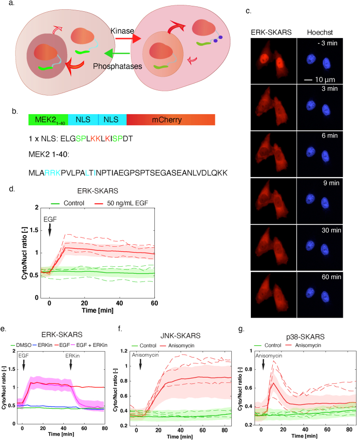
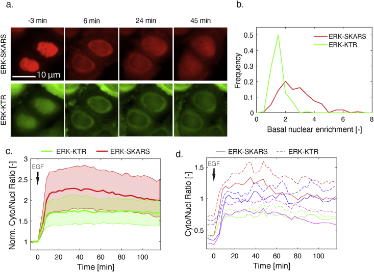
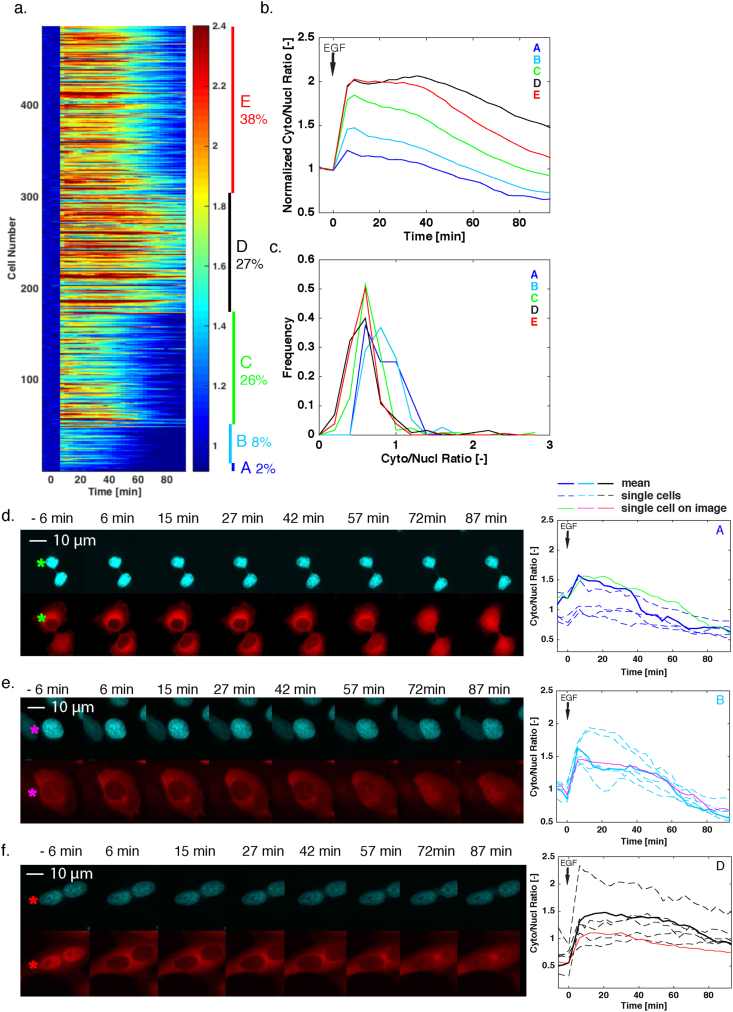
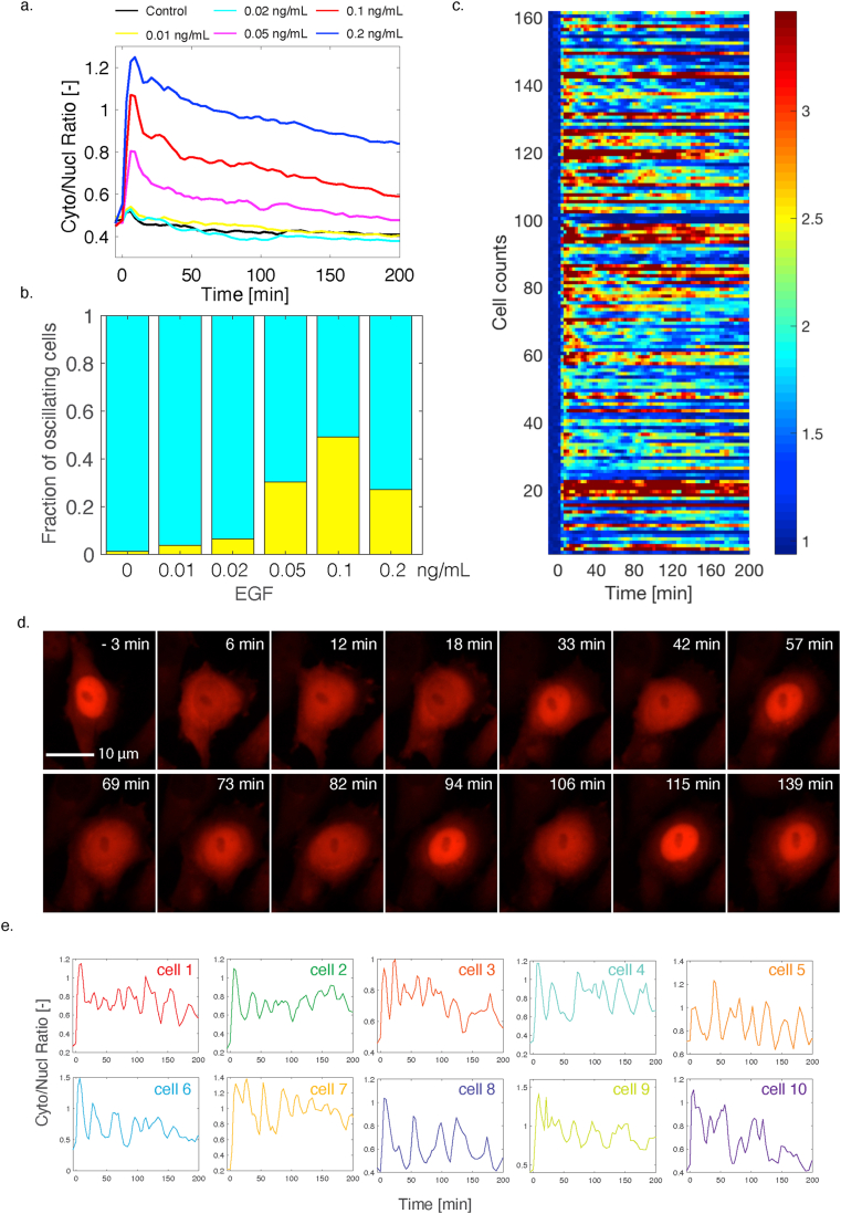
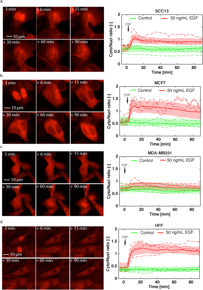
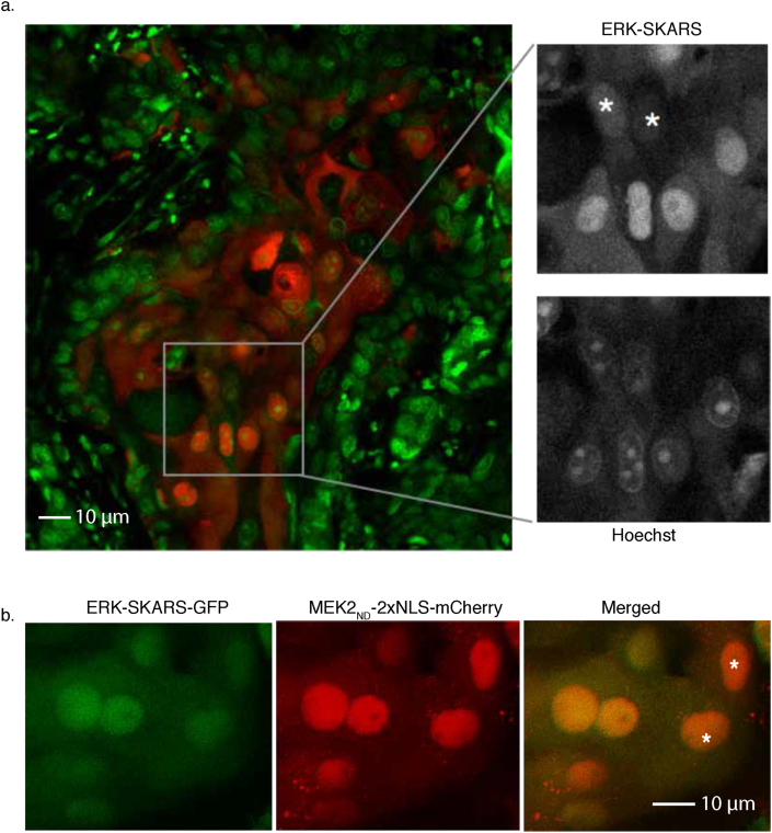
Mitogen-Activated Protein Kinases (MAPKs) control a wide array of cellular functions by transducing extracellular information into defined biological responses. In order to understand how these pathways are regulated, dynamic single cell measurements are highly needed. Fluorescence microscopy is well suited to perform these measurements. However, more dynamic and sensitive biosensors that allow the quantification of signaling activity in living mammalian cells are required. We have engineered a synthetic fluorescent substrate for human MAPKs (ERK, JNK and p38) that relocates from the nucleus to the cytoplasm when phosphorylated by the kinases. We demonstrate that this reporter displays an improved response compared to other relocation biosensors. This assay allows to monitor the heterogeneity in the MAPK response in a population of isogenic cells, revealing pulses of ERK activity upon a physiological EGFR stimulation. We show applicability of this approach to the analysis of multiple cancer cell lines and primary cells as well as its application in vivo to developing tumors. Using this ERK biosensor, dynamic single cell measurements with high temporal resolution can be obtained. These MAPK reporters can be widely applied to the analysis of molecular mechanisms of MAPK signaling in healthy and diseased state, in cell culture assays or in vivo.

Keywords: Biochemistry; Cancer research; Cell biology; Fluorescent biosensor; Live-cell imaging; MAPK signaling; Single cells; Systems biology.


Conflict of interest statement

The authors declare no conflict of interest.

Figures

Figure 1
Figure 1
Principle and development of synthetic kinase activity relocation sensor (SKARS) to monitor MAPK activity in mammalian cells. a. Scheme of the SKARS relocation process. When the kinase is inactive, the NLS is functional and the sensor accumulates in the nucleus. When the kinase is active, it phosphorylates the SKARS, which relocates into the cytoplasm. b. The ERK-SKARS contains three domains: the ERK docking site (MEK2 1–40), the two Nuclear Localization Sequences (NLS), and the fluorescent protein for visualization. Residues involved in interaction, nuclear import or phosphorylation are shown in cyan, red and green, respectively. c. Representative microscopy images of HeLa cells expressing the ERK-SKARS cells and stimulated with EGF (50 ng/ml) for 1 h period. In the red channel, the ERK-SKARS translocates from the nucleus to the cytoplasm. Nuclei are identified by a Hoechst staining. d. After quantification of the time-lapse movies, the ratio of the average cytoplasmic fluorescence of the average nuclear fluorescence (Cyto/Nucl ratio) is plotted as function of time. For all similar figures in this paper, the solid lines represent the median of the cell population and the shaded area the 25 and 75 percentiles of the population. The dotted lines represent a few single-cell traces extracted from the cell population. More than hundred single cells measured were plotted in the graph. The red curve represents HeLa cell treated with 50 ng/ml EGF (Number of cells: Nc = 340) and the green curve, mock-treated control cells (Nc = 449). e. Cyto/Nucl ratios of HeLa cells expressing the ERK-SKARS exposed to EGF stimulation (50 ng/ml) and ERK inhibition (FR 180204, 50 ng/ml) are plotted as function of time. EGF and ERK inhibitor were added at the time points indicated by the arrows. f. and g. HeLa cells expressing the JNK-SKARS (f) and the p38-SKARS (g) were treated with (red) and without (green) Anisomycin (50 ng/ml). The Cyto/Nucl ratio is plotted as function of time.
Figure 2
Figure 2
Direct comparison of ERK-SKARS and ERK-KTR in the same cells. a. Representative microscopy images of single HeLa cells carrying ERK-SKARS (red) and ERK-KTR (green) exposed to stimulation (EGF 50 ng/ml) and imaged at indicated time points. b. Histogram of SKARS and KTR nuclear enrichments before (Nucl/Cyto) of HeLa cells expressing ERK-SKARS (red) and ERK-KTR (green) before stimulation (mean of three first time points). c. Median (solid line) and 25- to 75-percentile (area) of the Cyto/Nucl ratios from ERK-SKARS (red) and ERK-KTR (green) upon stimulation by 10 ng/ml EGF. The single cell traces were normalized to the basal level (the mean of the first 3 three time points) (Nc = 234). d. Single cell traces of the Cyto/Nucl ratios for ERK-SKARS (solid line) and ERK-KTR (dashed line) were plotted as function of time. Each color represents the measurements from one single cell, and both reporter traces for each cell are displayed.
Figure 3
Figure 3
Cellular heterogeneity in ERK activity upon stimulation with high concentration of EGF. a. Heat map of the response of individual cells to a stimulation with EGF (50 ng/ml) in single clonal HeLa cells expressing the ERK-SKARS. Each line represents the cytoplasmic to nuclear ratio of one single cell normalized to the first three time points before stimulation. The cells were sorted using k-mean clustering (See Methods). The different sub-populations are identified by the five colored bars on the right of the map. b. Median of the normalized Cyto/Nucl ratios from the indicated subgroups identified in the population. c. Histograms of the basal Cyto/Nucl ratios measured in the five subgroups. d., e. and f. Representative microscopy images of HeLa cells expressing the ERK-SKARS from three different sub-populations (A, B, D). The right panel displays the Cyto/Nucl measurements of five single cells from that sub-population (dashed lines). The thin solid line corresponds to the single cell identified in the microscopy images on the left with an asterisk. The thick solid line represents the mean response from the sub-population.
Figure 4
Figure 4
Monitoring of ERK activity pulses upon low doses of EGF. a. Median Cyto/Nucl ratio of ERK-SKARS in more than 400 single cells from a clonal HeLa cell population stimulated with low doses of EGF and monitored for more than 3 h b. Fraction of cells identified in the experiment from panel 4a that display three pulses or more in ERK activity. c. Heat map of the Cyto/Nucl ratio measured upon 0.1 ng/ml EGF treatment in HeLa cells. Only cells where three or more pulses were identified are displayed in this graph. d. Microscopy images of HeLa cell displaying pulses in ERK activation upon 0.1 ng/ml EGF stimulation. e. Various examples of single cell traces displaying fluctuations in ERK activation upon 0.1 ng/ml EGF treatment.
Figure 5
Figure 5
SKARS measurements performed in cancer and primary cell lines. a to d. Left panels show representative microscopy images of SCC13 (a), MCF7 (b), MDA-MB231 (c), HFF (Human Foreskin Fibroblast) (d) cells expressing the ERK-SKARS and stimulated with EGF (50 ng/ml). The right panels show the Cyto/Nucl ratio in the respective cell lines. Quantifications of the time-lapse movies for samples treated with EGF are plotted in red and control samples in green. For each trace, more than 100 single cells measurements were combined to generate the graphs.
Figure 6
Figure 6
Application of SKARS for kinase activity quantification in tissue. a. Confocal images of HeLa cells expressing ERK-SKARS (red) in tumor implanted in a mouse ear. The green channel represents the nuclei of the cancerous and normal tissues, which were stained with Hoechst. The inset is a magnification of the image to display cells with different levels of nuclear accumulation of relative to the nuclear staining. Asterisk highlight cells with higher ERK activity. b. Confocal images of SCC13 cells expressing ERK-SKARS-GFP (MEK2DS, green) and MEK2ND-2xNLS-mCherry (red) in tumor implanted inside of mouse ears. Asterisk highlight cells with higher ERK activity.

References

    1. Altschuler S.J., Wu L.F. Cellular heterogeneity: do differences make a difference? Cell. 2010;141:559–563. - PMC - PubMed
    1. Ferrell J.E., Machleder E.M. The biochemical basis of an all-or-none cell fate switch in Xenopus oocytes. Science. 1998;280:895–898. - PubMed
    1. Purvis J.E. p53 dynamics control cell fate. Science. 2012;336:1440–1444. - PMC - PubMed
    1. Nandagopal N. Dynamic ligand discrimination in the notch signaling pathway. Cell. 2018;172:869–880. e19. - PMC - PubMed
    1. Cohen P. The regulation of protein function by multisite phosphorylation--a 25 year update. Trends Biochem. Sci. 2000;25:596–601. - PubMed