. 2020 Dec 15:9:e60812.
doi: 10.7554/eLife.60812.

A prefrontal-bed nucleus of the stria terminalis circuit limits fear to uncertain threat

Affiliations

A prefrontal-bed nucleus of the stria terminalis circuit limits fear to uncertain threat

Lucas R Glover et al. Elife. .

Abstract

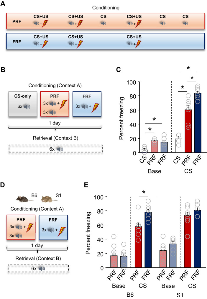
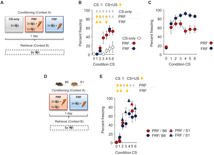
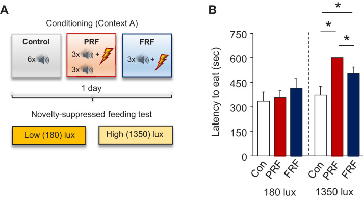
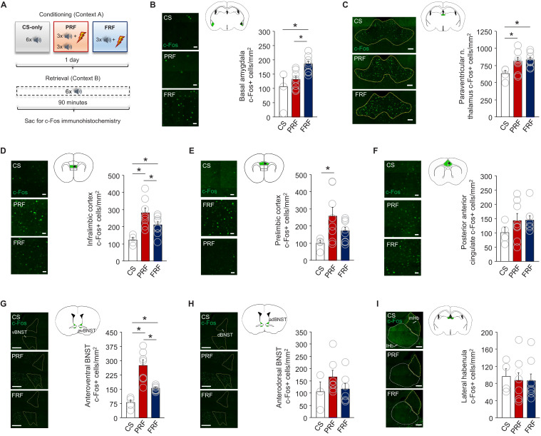
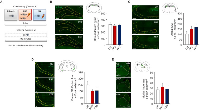
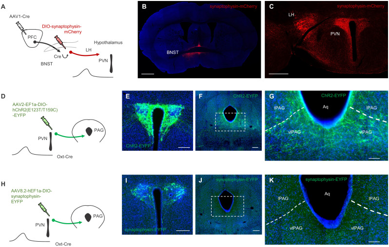
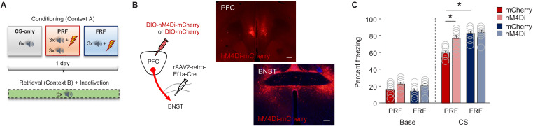
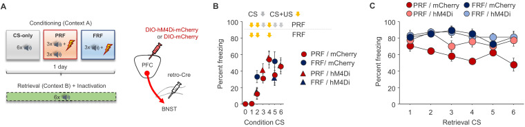
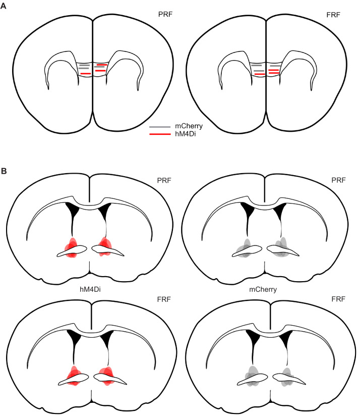
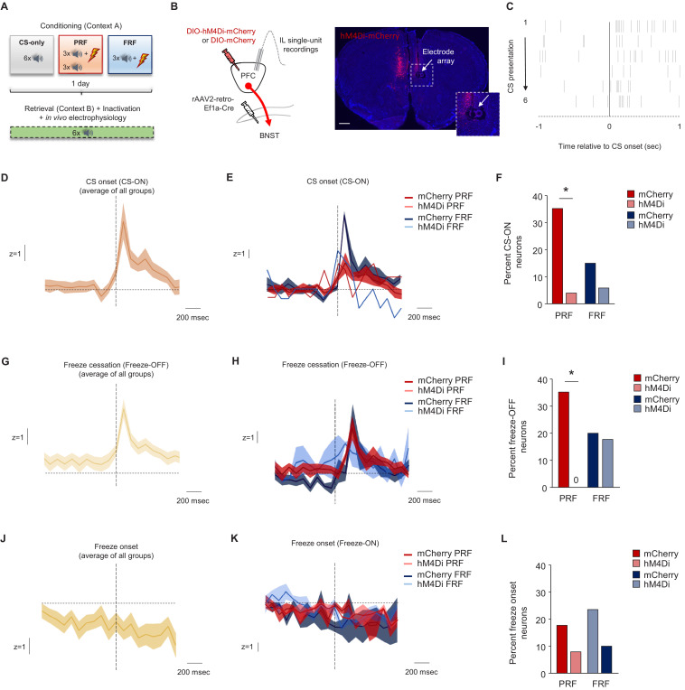
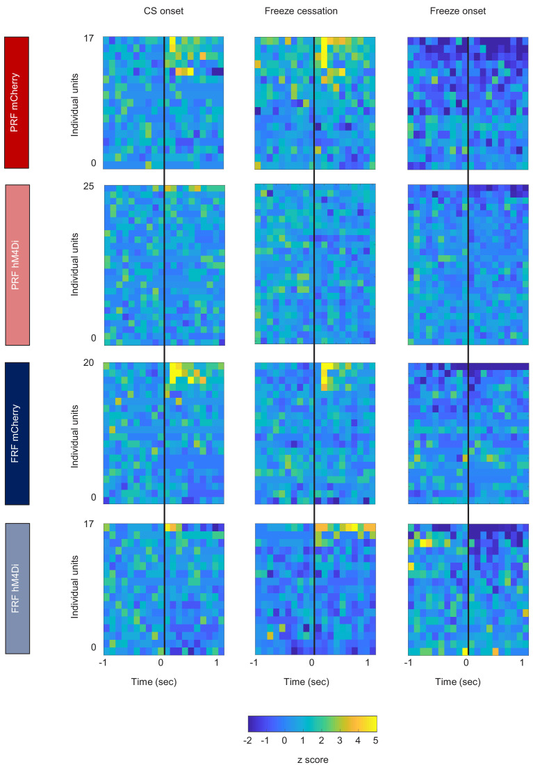
In many cases of trauma, the same environmental stimuli that become associated with aversive events are experienced on other occasions without adverse consequence. We examined neural circuits underlying partially reinforced fear (PRF), whereby mice received tone-shock pairings on half of conditioning trials. Tone-elicited freezing was lower after PRF conditioning than fully reinforced fear (FRF) conditioning, despite an equivalent number of tone-shock pairings. PRF preferentially activated medial prefrontal cortex (mPFC) and bed nucleus of the stria terminalis (BNST). Chemogenetic inhibition of BNST-projecting mPFC neurons increased PRF, not FRF, freezing. Multiplexing chemogenetics with in vivo neuronal recordings showed elevated infralimbic cortex (IL) neuronal activity during CS onset and freezing cessation; these neural correlates were abolished by chemogenetic mPFC→BNST inhibition. These data suggest that mPFC→BNST neurons limit fear to threats with a history of partial association with an aversive stimulus, with potential implications for understanding the neural basis of trauma-related disorders.

Keywords: amygdala; anxiety; fear; mouse; neuroscience; ptsd; stress; trauma.

Plain language summary

While walking home alone late one night, you hear footsteps behind you. Your heart starts to beat faster as you wonder whether someone might be following you. Being able to identify and evade threats is essential for survival. A key part of this process is learning to recognize signals that predict potential danger: the sound of footsteps behind you, for example. But many such cues are unreliable. The person behind you might simply be heading in the same general direction as you. And if you spend too much time and energy responding to such false alarms, you may struggle to complete other essential tasks. To be useful, responses to cues that signal potential threats must thus be proportionate to the likelihood that danger is actually present. By studying threat detection in mice, Glover et al. have identified a brain circuit that helps ensure that this is the case. Two groups of mice learned to fear a tone that predicted the delivery of a mild footshock. In one group of animals, the tone was followed by a shock on every trial (it was said to be ‘fully reinforced’). But in the other group, the tone was followed by a shock on only 50% of trials (‘partially reinforced’). After training, both groups of mice froze whenever they heard the tone – freezing being a typical fear response in rodents. But the animals trained with the partially reinforced tone showed less freezing than their counterparts in the fully reinforced group. Moreover, freezing in response to the partially reinforced tone was accompanied by activity in a specific neural pathway connecting the frontal part of the brain to an area called the bed nucleus of the stria terminalis. Inhibiting this pathway made mice respond to the partially reinforced tone as though it had been reinforced on every trial. This suggests that activity in this pathway helps dampen responses to unpredictable threat cues. In people with anxiety disorders, cues that become associated with unpleasant events can trigger anxiety symptoms, even if the association is unreliable. The findings of Glover et al. suggest that reduced activity of circuits that constrain excessive responses to threats might contribute to anxiety disorders.


Conflict of interest statement

LG, KM, MB, SS, NR, SO, TY, AP, MN, LH, AH No competing interests declared

Figures

Figure 1.
Figure 1.. Lower freezing during retrieval of partially reinforced fear; effects of genetic strain.
(A) Schematic depiction of experimental procedure for assessing, in B6 mice, PRF and FRF, along with CS-only controls. (B) Schematic depiction of experimental procedure for assessing, in B6 mice, PRF and FRF retrieval in a novel context (context B) and the conditioning context (context A) (C) Lower CS-related freezing during retrieval in PRF mice than in FRF mice. Higher baseline and CS-related freezing in PRF and FRF mice relative to CS-only controls (n = 4–8 mice per group). (D) Schematic depiction of experimental procedure for assessing PRF and FRF retrieval in the B6 and S1 genetic strains. (E) Lower CS-related freezing during retrieval in PRF than in FRF in B6, not S1, mice (n = 7–8 mice per group/strain). Data are means ± SEM. *p<0.05.
Figure 1—figure supplement 1.
Figure 1—figure supplement 1.. Freezing during conditioning.
(A) Schematic depiction of experimental procedure for assessing, in B6 mice, PRF and FRF, along with CS-only controls. (B) No group differences in CS-related freezing during conditioning (n = 4–8 mice per group). (C) No difference in CS-related freezing between FRF and PRF mice during retrieval, broken down by CS presentation (n = 8 mice per group). (D) Schematic depiction of experimental procedure for assessing PRF and FRF retrieval in the B6 and S1 genetic strains. (E) No group differences in CS-related freezing during conditioning (n = 7–8 mice per group/strain). Data are means ± SEM.
Figure 1—figure supplement 2.
Figure 1—figure supplement 2.. Increased latency to feed in the NSF test after PRF.
(A) Schematic depiction of experimental procedure for assessing behavior on the novelty-suppressed feeding (NSF) test after PRF or FRF conditioning, along with context-exposed controls. (B) Longer latencies to eat under high, but not low, illumination in PRF and FRF versus controls and in PRF versus FRF. n = 8 mice per group. Data are means ± SEM. *p<0.05.
Figure 2.
Figure 2.. PRF preferentially activates subregions of mPFC and BNST.
(A) Schematic depiction of experimental procedure for assessing ex vivo neuronal regional activity (via c-Fos immunohistochemistry) after PRF or FRF retrieval, along with CS-only controls. Representative images and c-Fos+ cell count differences for basal amygdala (B), paraventricular nucleus of the thalamus (C), infralimbic cortex (D), prelimbic cortex (E), posterior portion of the anterior cingulate cortex (F), anteroventral BNST (G), anterodorsal BNST (H), and lateral habenula (I). For corresponding behavioral data, see Figure 1B. Scale bars = 30 µm (B,D–F), 100 µm (C,I), 300 µm (G,H). n = 4–8 mice per group. Data are means ± SEM. *p<0.05.
Figure 2—figure supplement 1.
Figure 2—figure supplement 1.. Ex vivo neuronal regional activity correlates of PRF.
(A) Schematic depiction of experimental procedure for assessing ex vivo neuronal regional activity (via c-Fos immunohistochemistry) after PRF or FRF retrieval. Representative images and c-Fos+ cell counts for the dorsal dentate gyrus (B), dorsal CA3 (C), and ventral CA1/subiculum (D) regions of the hippocampus and the medial habenula (E). There were no group differences. Scale bars = 100 µm. n = 4–8 mice per group. Data are means ± SEM.
Figure 2—figure supplement 2.
Figure 2—figure supplement 2.. Connectivity between mPFC, BNST, and downstream targets.
(A) Schematic depiction of viral strategy to label mPFC inputs to BNST neurons and their onward projections to the hypothalamus. (B) Representative image of synaptophysin-mCherry labeling in BNST neurons receiving mPFC input (scale bar = 1000 µm). (C) Representative image of synaptophysin-mCherry labeling in mPFC-innervated BNST neuronal projections in hypothalamus (scale bar = 500 µm). (D) Schematic depiction of viral strategy to label PVN oxytocin cell inputs to the PAG in Oxt-Cre mice. (E) Representative image of ChR2-EYFP labeling in the PVN (scale bar = 200 µm). Low (F) and high (G) magnification images of ChR2-EYFP labeling in the PAG (scale bars = 200 µm and 100 µm, respectively). (H) Schematic depiction of viral strategy to label PVN oxytocin cell inputs to the PAG in Oxt-Cre mice. (I) Representative image of synaptophysin-EYFP labeling in the PVN (scale bar = 200 µm). Low (J) and high (K) magnification images of synaptophysin-EYFP labeling in the PAG (scale bars = 200 µm and 100 µm, respectively). Note: LH = Lateral Hypothalamus; PVN = paraventricular nucleus of the hypothalamus; IPAG = lateral periaqueductal gray; vlPAG = ventrolateral periaqueductal gray; Aq = aqueduct.
Figure 3.
Figure 3.. Inhibition of mPFC→BNST neurons increases PRF.
(A) Schematic depiction of experimental procedure for assessing effects of chemogenetic inhibition of mPFC→BNST neurons during retrieval. (B) Cartoon of viral strategy and representative images of hM4Di–mCherry labeling in BNST neurons receiving mPFC input (scale bars = 200 µm). (C) Lower CS-related freezing during retrieval in PRF mice than in FRF mice transfected with mCherry, not hHM4Di. Data are means ± SEM. *p<0.05.
Figure 3—figure supplement 1.
Figure 3—figure supplement 1.. Freezing during conditioning prior to mPFC→BNST inhibition on retrieval.
(A) Schematic depiction of viral strategy to selectively inhibit BNST-projecting mPFC neurons during retrieval. (B) Freezing increased across CS trials, irrespective of virus group. (C) Trial-by-trial breakdown of freezing during each CS of retrieval indicated a non-significant trend for decreasing freezing across trials in the mCherry PRF group. n = 8–9 mice group/virus. Freezing data are means ± SEM.
Figure 3—figure supplement 2.
Figure 3—figure supplement 2.. Electrode placements and virus localization for combined chemogenetic/single-unit recordings.
Estimated location of tips of the electrodes at the center of the multi-array in the IL of PRF (A) and FRF (B) mice. Estimated extent of virus, as indicated by mCherry expression, in the BNST of PRF (C) and FRF (D) mice (darker shading represents areas of greater overlap across mice).
Figure 3—figure supplement 3.
Figure 3—figure supplement 3.. CS and freezing-related IL unit activity and effects of mPFC→BNST inhibition.
(A) Schematic depiction of experimental procedure for in vivo IL single-unit recordings, combined with chemogenetic inhibition of mPFC→BNST neurons during retrieval (n = 8–9 mice per group/virus). (B) Cartoon of viral strategy and representative image of hM4Di-mCherry labeling in the mPFC and electrode array placement in the IL (scale bar = 500 µm). (C) Raster plot of a representative IL unit firing in response to CS onset (CS-ON neuron). Baseline-normalized population trace of CS-ON neuronal activity during retrieval; average of all groups (D) and split by group (E). (F) Higher percentage of CS-ON units during PRF than FRF retrieval in mice transfected with mCherry, not hHM4Di (n = 17 recorded units in PRF/mCherry, n = 25 units in PRF/hM4Di, n = 20 units in FRF/mCherry, n = 17 units in FRF/hM4Di, from three mice per group/virus). Baseline-normalized population trace of freeze cessation (Freeze-OFF neurons) IL unit activity during retrieval; average of all groups (G) and split by group (H) (n = 3 CS units in PRF/mCherry, n = 6 units in PRF/hM4Di, n = 1 units in FRF/mCherry, n = 1 units in FRF/hM4Di). (I) Higher percentage of Freeze-OFF units during PRF than FRF retrieval in mice transfected with mCherry, not hHM4Di (n = 17 recorded units in PRF/mCherry, n = 25 units in PRF/hM4Di, n = 20 units in FRF/mCherry, n = 17 units in FRF/hM4Di, from three mice per group/virus, n = 4 Freeze-OFF units in PRF/mCherry, n = 6 units in PRF/hM4Di, n = 3 units in FRF/mCherry, n = 0 units in FRF/hM4Di). Baseline-normalized population trace of freeze onset (Freeze-ON neurons) IL unit activity during retrieval; average of all groups (J) and split by group (K). (L) No differences in the percentage of Freeze-OFF units during retrieval between groups (n = 17 units in PRF/mCherry, n = 25 units in PRF/hM4Di, n = 20 units in FRF/mCherry, n = 17 units in FRF/hM4Di, from three mice per group/virus, n = 4 Freeze-ON units in PRF/mCherry, n = 6 units in PRF/hM4Di, n = 4 units in FRF/mCherry, n = 2 units in FRF/hM4Di). Data are means ± SEM. *p<0.05.
Figure 3—figure supplement 4.
Figure 3—figure supplement 4.. Heat maps illustrating IL unit activity.
(A) Heat plots of unit activity aligned to CS onset (left columns), freeze cessation (center columns), and freeze onset (right columns). The same data shown as peri-event histograms and % event-related activity can be found in Figure 3—figure supplement 3.

References

    1. Asok A, Draper A, Hoffman AF, Schulkin J, Lupica CR, Rosen JB. Optogenetic silencing of a corticotropin-releasing factor pathway from the central amygdala to the bed nucleus of the stria terminalis disrupts sustained fear. Molecular Psychiatry. 2018;23:914–922. doi: 10.1038/mp.2017.79. - DOI - PMC - PubMed
    1. Bjorni M, Rovero NG, Yang ER, Holmes A, Halladay LR. Phasic signaling in the bed nucleus of the stria terminalis during fear learning predicts within- and across-session cued fear expression. Learning & Memory. 2020;27:83–90. doi: 10.1101/lm.050807.119. - DOI - PMC - PubMed
    1. Blanchard DC, Blanchard RJ. Innate and conditioned reactions to threat in rats with amygdaloid lesions. Journal of Comparative and Physiological Psychology. 1972;81:281–290. doi: 10.1037/h0033521. - DOI - PubMed
    1. Bloodgood DW, Sugam JA, Holmes A, Kash TL. Fear extinction requires infralimbic cortex projections to the basolateral amygdala. Translational Psychiatry. 2018;8:60. doi: 10.1038/s41398-018-0106-x. - DOI - PMC - PubMed
    1. Breitfeld T, Bruning JE, Inagaki H, Takeuchi Y, Kiyokawa Y, Fendt M. Temporary inactivation of the anterior part of the bed nucleus of the stria terminalis blocks alarm pheromone-induced defensive behavior in rats. Frontiers in Neuroscience. 2015;9:321. doi: 10.3389/fnins.2015.00321. - DOI - PMC - PubMed

Publication types