Review
. 2021 Mar 15;42(2):101-132.
doi: 10.1210/endrev/bnaa032.

Revisiting the Complexity of GLP-1 Action from Sites of Synthesis to Receptor Activation

Affiliations
Review

Revisiting the Complexity of GLP-1 Action from Sites of Synthesis to Receptor Activation

Brent A McLean et al. Endocr Rev. .

Abstract

Glucagon-like peptide-1 (GLP-1) is produced in gut endocrine cells and in the brain, and acts through hormonal and neural pathways to regulate islet function, satiety, and gut motility, supporting development of GLP-1 receptor (GLP-1R) agonists for the treatment of diabetes and obesity. Classic notions of GLP-1 acting as a meal-stimulated hormone from the distal gut are challenged by data supporting production of GLP-1 in the endocrine pancreas, and by the importance of brain-derived GLP-1 in the control of neural activity. Moreover, attribution of direct vs indirect actions of GLP-1 is difficult, as many tissue and cellular targets of GLP-1 action do not exhibit robust or detectable GLP-1R expression. Furthermore, reliable detection of the GLP-1R is technically challenging, highly method dependent, and subject to misinterpretation. Here we revisit the actions of GLP-1, scrutinizing key concepts supporting gut vs extra-intestinal GLP-1 synthesis and secretion. We discuss new insights refining cellular localization of GLP-1R expression and integrate recent data to refine our understanding of how and where GLP-1 acts to control inflammation, cardiovascular function, islet hormone secretion, gastric emptying, appetite, and body weight. These findings update our knowledge of cell types and mechanisms linking endogenous vs pharmacological GLP-1 action to activation of the canonical GLP-1R, and the control of metabolic activity in multiple organs.

Keywords: brain; cardiovascular; diabetes; gastrointestinal tract; islets; obesity; receptor.


Figures

Graphical Abstract
Graphical Abstract
Figure 1.
Figure 1.
GLP-1 source, GLP-1R labeling and GLP-1R signaling in pancreatic islets. (A) Potential sources of GLP1 in the islets, as well as paracrine signaling pathways. Proglucagon products from α-cells that activate islet GLP-1Rs include glucagon and potentially GLP-1. The biological relevance of GLP-1 produced by α-cells remains an unresolved line of investigation. (B) GLP1R labelling is observed at the membrane in β-cells. Predominantly cytoplasmic labelling is nonspecific unless the GLP1R is agonist-bound in which case some surface and punctate intracellular staining is detected due to GLP1R internalization. (C) Major GLP1R signaling pathways reported in primary rodent and human beta cells or islets. It is well established that GLP-1R signals through cAMP pathways, including PKA and EPAC2. β-arrestin 1 has been proposed to regulate GLP-1R signaling; however, the mechanisms of this pathway remain unresolved. (D) Actions mediated by the GLP-1R in pancreatic islets. ADCY, adenylate cyclase, GLP1R, Glucagon-like peptide-1 receptor; PKA, protein kinase A; EPAC2, exchange protein directly activated by cAMP 2; INS, insulin SST, somatostatin.
Figure 2.
Figure 2.
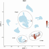
Expression of GLP1R in 9940 human pancreatic islet endocrine cells. The UMAP (Uniform Manifold Approximation and Projection) plots were created to visualize gene expression in each cell. The x-axis and y-axis indicate the overall transcriptional difference of each cells arbitrated as “distance” in a 2-dimensional space; the closer the 2 cells on the plot, the more similar their transcriptomes are, and in turn they are more likely to share a lineage. The expression scales are continual and log2 normalized, ranging from light blue (low expression) to dark red (high expression) in color, Cell types were classified based on markers genes INS and MAFA (mature β cells), GCG (α cells), SST (δ cells), and PPY (PP cells). Count matrices were accessed on September 19, 2020, and aggregated from the scRNA-seq datasets reported by Segerstolpe et al. (37) (GEO accession number GSE73727), Enge et al. (220) (GEO accession number GSE81547), and Camunas-Soler et al. (38) (GEO accession number GSE124742). Human gene nomenclature: INS, insulin; GCG, glucagon; SST, somatostatin; PPY, pancreatic polypeptide Y; MAFA, V-maf musculoaponeurotic fibrosarcoma oncogene homologue A.
Figure 3.
Figure 3.
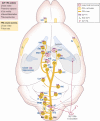
GLP-1 receptors (GLP-1Rs) and brain-derived GLP-1 in the central nervous system. Within the CNS GLP-1 is produced by PPG neurons (light yellow) and distributed to their axon terminals ready for synaptic release (dark green). Names of brain areas that receive PPG innervation and express GLP-1 receptors are given as abbreviations in blue. Systemically distributed GLP-1 receptor agonists (GLP-1RAs) are depicted as red circles and access the circumventricular organs with a leaky blood–brain barrier, and the ventricles, but not the brain parenchyma. The olfactory bulb and the piriform cortex (Pir) form areas that express GLP-1Rs and harbor PPG neurons that project locally only. Abbreviations: AH, anterior hypothalamus; AP, area postrema; ARC, arcuate nucleus; Barr, Barrington’s nucleus; BNST, bed nucleus of the stria terminalis; CAA, central autonomic area (lamina X); caud Hipp, caudal ventral hippocampus; CeA, central nucleus of the amygdala; DMH, dorsomedial hypothalamus; DMNX, dorsal vagal motornucleus; GrO, granule cell layer of the olfactory bulb; IML, intermediolateral nucleus; IRT, intermediate reticular nucleus; LC; locus coeruleus; NAc, nucleus accumbens; NTS, nucleus tractus solitarius; OVLT, organum vasculosum of the lamina terminalis; PAG, periaqueductal grey; PBN, parabrachial nucleus; Pir, piriform cortex; PVN, paraventricular nucleus; PVT, paraventricular thalamus; Sep, lateral septum; SFO, subfornical organ; RPa, raphe pallidus; VLM, ventrolateral medulla; VMPO, ventromedial posterior nucleus.
Figure 4.
Figure 4.
GLP-1 receptor expression in heart and blood vessels within select organs and actions of GLP-1 associated with these organ and cell types. The arrow depicts the relative levels of GLP-1R expression deduced from human gene expression databases such as the Genotype Tissue Expression portal.
Figure 5.
Figure 5.
Expression of mouse Glp1r in 40 449 murine endothelial cells. Count matrices were accessed on September 19, 2020, and reported by Kalucka et al. (221) (ArrayExpress ID E-MTAB-8077).
Figure 6.
Figure 6.
Coexpression of the proglucagon gene (Gcg) and enteroendocrine genes in 411 murine small intestine L cells. Count matrices were accessed on September 19, 2020, and retrieved from the scRNA-seq datasets reported by Gehart et al. (167) (GEO accession number GSE113561). Mouse gene nomenclature: Pyy, peptide YY; Gip, glucose-dependent insulinotropic polypeptide; Nts, neurotensin; Tph1, tryptophan hydroxylase 1; Sst, somatostatin; Ghrl, ghrelin; Sct, secretin; Cck, cholecystokinin.
Figure 7.
Figure 7.
Expression of Glp1r in 4974 murine ileal and colonic enteric neurons. Enteric neuron subtypes types were classified based on neuropeptide genes Adcyap1 (PACAP neurons and Gal (galanin neurons). Count matrices were accessed on September 19, 2020, and aggregated from the scRNA-seq datasets reported by Drokhlyansky et al (183) (Broad Institute Single Cell Portal SCP1038). Mouse gene nomenclature: Adcyap1, pituitary adenylate cyclase activating polypeptide or PACAP; Gal, galanin.
Figure 8.
Figure 8.
Schematic depiction of major targets of GLP-1 action, and the cell types within key organs that express the GLP-1 Receptor (GLP1R). The GLP-1 peptide is produced in and shown adjacent to the intestine, pancreas, and brain, the 3 major sites of GCG expression. EC, endothelial cells; IEL, intestinal intraepithelial lymphocytes; SA node, sinoatrial node; VSMC, vascular smooth muscle cell.

Comment in

  • GLP-1 receptor - Do we really know what we're looking at?
    Homolak J, Babic Perhoc A, Knezovic A, Osmanovic Barilar J, Salkovic-Petrisic M. Homolak J, et al. Acta Histochem. 2021 Jul;123(5):151732. doi: 10.1016/j.acthis.2021.151732. Epub 2021 May 18. Acta Histochem. 2021. PMID: 34015646 No abstract available.

References

    1. Drucker DJ. Evolving concepts and translational relevance of enteroendocrine cell biology. J Clin Endocrinol Metab. 2016;101(3):778-786. - PubMed
    1. Bell GI, Sanchez-Pescador R, Laybourn PJ, Najarian RC. Exon duplication and divergence in the human preproglucagon gene. Nature. 1983;304(5924):368-371. - PubMed
    1. Heinrich G, Gros P, Lund PK, Bentley RC, Habener JF. Pre-proglucagon messenger ribonucleic acid: nucleotide and encoded amino acid sequences of the rat pancreatic complementary deoxyribonucleic acid. Endocrinology. 1984;115(6):2176-2181. - PubMed
    1. Drucker DJ, Habener JF, Holst JJ. Discovery, characterization, and clinical development of the glucagon-like peptides. J Clin Invest. 2017;127(12):4217-4227. - PMC - PubMed
    1. Sandoval DA, D’Alessio DA. Physiology of proglucagon peptides: role of glucagon and GLP-1 in health and disease. Physiol Rev. 2015;95(2):513-548. - PubMed

Publication types

Substances