

Skin pigmentation and its control: From ultraviolet radiation to stem cells
- PMID: 33320376
- PMCID: PMC8218595
- DOI: 10.1111/exd.14260
Skin pigmentation and its control: From ultraviolet radiation to stem cells
Abstract
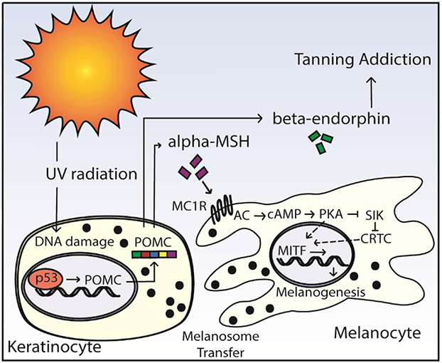
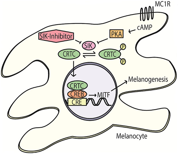
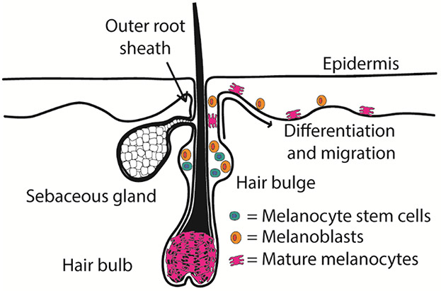
In the light of substantial discoveries in epithelial and hair pigmentation pathophysiology, this review summarizes the current understanding of skin pigmentation mechanisms. Melanocytes are pigment-producing cells, and their key regulating transcription factor is the melanocyte-specific microphthalmia-associated transcription factor (m-MITF). Ultraviolet (UV) radiation is a unique modulator of skin pigmentation influencing tanning pathways. The delayed tanning pathway occurs as UVB produces keratinocyte DNA damage, causing p53-mediated expression of the pro-opiomelanocortin (POMC) gene that is processed to release α-melanocyte-stimulating hormone (α-MSH). α-MSH stimulates the melanocortin 1 receptor (MC1R) on melanocytes, leading to m-MITF expression and melanogenesis. POMC cleavage also releases β-endorphin, which creates a neuroendocrine pathway that promotes UV-seeking behaviours. Mutations along the tanning pathway can affect pigmentation and increase the risk of skin malignancies. MC1R variants have received considerable attention, yet the allele is highly polymorphic with varied phenotypes. Vitiligo presents with depigmented skin lesions due to autoimmune destruction of melanocytes. UVB phototherapy stimulates melanocyte stem cells in the hair bulge to undergo differentiation and upwards migration resulting in perifollicular repigmentation of vitiliginous lesions, which is under sophisticated signalling control. Melanocyte stem cells, normally quiescent, undergo cyclic activation/differentiation and downward migration with the hair cycle, providing pigment to hair follicles. Physiological hair greying results from progressive loss of melanocyte stem cells and can be accelerated by acute stress-induced, sympathetic driven hyperproliferation of the melanocyte stem cells. Ultimately, by reviewing the pathways governing epithelial and follicular pigmentation, numerous areas of future research and potential points of intervention are highlighted.
Keywords: MC1R; hair greying; melanocyte stem cells; perifollicular repigmentation; tanning.
© 2020 John Wiley & Sons A/S. Published by John Wiley & Sons Ltd.
Conflict of interest statement
Figures
References
-
- Boissy RE, Nordlund JJ. Molecular basis of congenital hypopigmentary disorders in humans: a review. Pigment Cell Res. 1997;10(1-2):12–24. - PubMed
-
- Cochran AJ. The incidence of melanocytes in normal human skin. J Invest Dermatol. 1970;55(1):65–70. - PubMed
-
- Tarnowski WM. Ultrastructure of the epidermal melanocyte dense plate. J Invest Dermatol. 1970;55(4):265–268. - PubMed
-
- Nishimura EK, Jordan SA, Oshima H, et al. Dominant role of the niche in melanocyte stem-cell fate determination. Nature. 2002;416(6883):854–860. - PubMed
-
- Nishimura EK, Granter SR, Fisher DE. Mechanisms of hair graying: incomplete melanocyte stem cell maintenance in the niche. Science. 2005;307(5710):720–724. - PubMed
Publication types
MeSH terms
Substances
Grants and funding
LinkOut - more resources
Full Text Sources
Medical
Research Materials
Miscellaneous
