. 2021 Jun 1;203(11):1419-1430.
doi: 10.1164/rccm.202005-1761OC.

Circulating BMP9 Protects the Pulmonary Endothelium during Inflammation-induced Lung Injury in Mice

Affiliations

Circulating BMP9 Protects the Pulmonary Endothelium during Inflammation-induced Lung Injury in Mice

Wei Li et al. Am J Respir Crit Care Med. .

Abstract

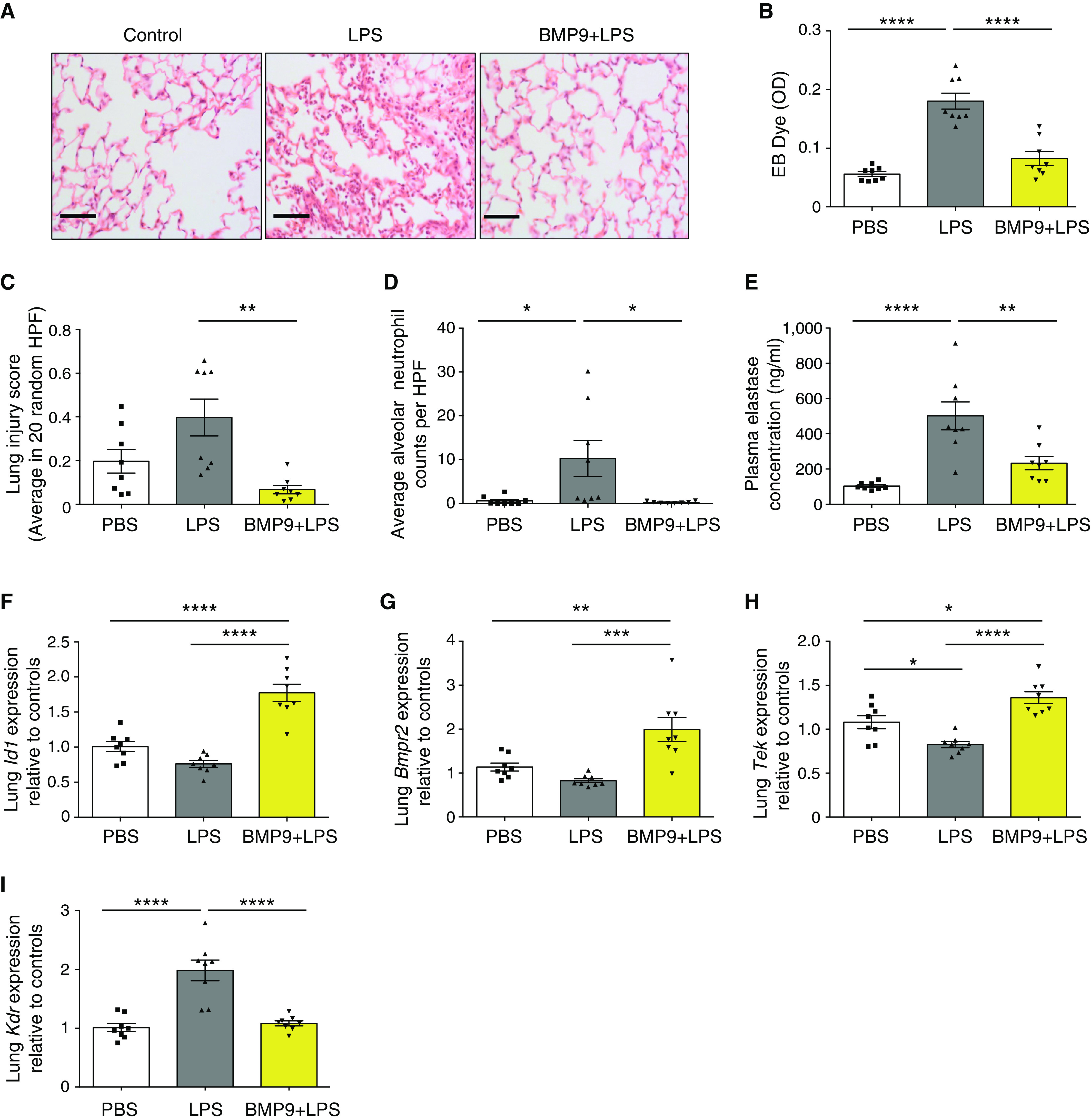
Rationale: Pulmonary endothelial permeability contributes to the high-permeability pulmonary edema that characterizes acute respiratory distress syndrome. Circulating BMP9 (bone morphogenetic protein 9) is emerging as an important regulator of pulmonary vascular homeostasis. Objectives:To determine whether endogenous BMP9 plays a role in preserving pulmonary endothelial integrity and whether loss of endogenous BMP9 occurs during LPS challenge. Methods: A BMP9-neutralizing antibody was administrated to healthy adult mice, and lung vasculature was examined. Potential mechanisms were delineated by transcript analysis in human lung endothelial cells. The impact of BMP9 administration was evaluated in a murine acute lung injury model induced by inhaled LPS. Levels of BMP9 were measured in plasma from patients with sepsis and from endotoxemic mice. Measurements and Main Results: Subacute neutralization of endogenous BMP9 in mice (N = 12) resulted in increased lung vascular permeability (P = 0.022), interstitial edema (P = 0.0047), and neutrophil extravasation (P = 0.029) compared with IgG control treatment (N = 6). In pulmonary endothelial cells, BMP9 regulated transcriptome pathways implicated in vascular permeability and cell-membrane integrity. Augmentation of BMP9 signaling in mice (N = 8) prevented inhaled LPS-induced lung injury (P = 0.0027) and edema (P < 0.0001). In endotoxemic mice (N = 12), endogenous circulating BMP9 concentrations were markedly reduced, the causes of which include a transient reduction in hepatic BMP9 mRNA expression and increased elastase activity in plasma. In human patients with sepsis (N = 10), circulating concentratons of BMP9 were also markedly reduced (P < 0.0001). Conclusions: Endogenous circulating BMP9 is a pulmonary endothelial-protective factor, downregulated during inflammation. Exogenous BMP9 offers a potential therapy to prevent increased pulmonary endothelial permeability in lung injury.

Keywords: BMP signaling in endothelial cells; BMP9; lung injury; pulmonary endothelium.


Figures

Figure 1.
Figure 1.
Neutralizing endogenous BMP9 (bone morphogenetic protein 9) results in lung vascular leak and neutrophil extravasation. (A) A schematic diagram indicating the treatment regimen. (B) Inhibiting endogenous BMP9 activity leads to lung vascular leak. Representative images of the lungs (left), showing the Evans Blue (EB)-stained lungs from LPS- and anti-BMP9–treated animals. Quantification of EB content in the lungs (right). N = 6 per group. (C and D) Anti-BMP9 treatment leads to an increase in the perivascular adventitial area, similar to LPS treatment (black arrows). (C) Representative pictures of hematoxylin and eosin–stained lung section. Scale bars, 100 μM. (D) Adventitial area in 20 random high-power fields (HPF), with its correlation to the EB content in the lung shown on the right; the Spearman correlation test was used. (E) Anti-BMP9 treatment increases alveolar neutrophil counts, revealed by myeloperoxidase staining. Scale bars, 50 μM. The counts were the mean of six random HPF per animal. Arrows point to neutrophils. (F) BMP9 activity in plasma measured by ID1-gene induction in human pulmonary artery endothelial cells. Serum-starved human pulmonary artery endothelial cells were treated with 1% plasma samples for 1 hour before cells were harvested for quantitative RT-PCR analysis of ID1 gene induction. The operator was blinded to the treatment samples. For all panels, data are shown as means ± SEMs. Two-tailed Mann-Whitney tests were used to compare LPS treatment with PBS treatment and anti-BMP9 treatment with IgG treatment. *P < 0.05, **P < 0.01, and ***P < 0.001. LPS1 = LPS at 1.5 mg/kg; LPS2 = LPS at 3 mg/kg; n.s. < not significant; OD < optical density of absorbance measurements; PBS = phosphate-buffered saline.
Figure 2.
Figure 2.
BMP9 (bone morphogenetic protein 9) signaling regulates genes involved in endothelial-cell integrity. (A) A volcano plot of microarray transcriptional analysis of BMP9-regulated genes. Serum-starved human pulmonary artery endothelial cells (hPAECs) were treated with 0.4 ng/ml (GF-domain concentration) pro-BMP9 (prodomain-bound form of BMP9) for 5 hours before cells were harvested for microarray analysis using the Human Gene 2.1 ST array (Affymetrix). Four independent hPAEC lines were used. Data were processed using package Oligo in R (R Foundation for Statistical Computing) (55) and normalized using robust multichip analysis (56), and comparisons were performed using the limma package (57). Resulting P values were corrected for multiple testing using the false discovery rate (58). Hits with adjusted. P values of less than 0.05 are shown in red, and those with adjusted P values not reaching statistical significance shown in light blue. (B) Validation of microarray results using quantitative RT-PCR. BMP9 signaling regulates mRNA expression of AQP1 (aquaporin-1), KDR (VEGFR2), and TEK (Tie2) in hPAECs. N = 5. (C) Changes of gene expression in hPAECs after inhibition of BMP9 activity in fetal bovine serum with a neutralizing anti-BMP9 antibody. hPAECs were grown in endothelial basal medium with 2% fetal bovine serum and treated with IgG control or anti-BMP9 antibody (both at 20 μg/ml) for 3 hours (for TEK) or 5 hours (for AQP1 and KDR) before cells were harvested for RNA extraction and quantitative RT-PCR analysis. N = 5. (D) BMP9 treatment suppresses VEGFR2 total proteins. Serum-starved hPAECs were treated with pro-BMP9 (0.4 ng/ml GF-domain concentration) for 5 hours (N = 6). Three independent treatments were run on the same Western blot and are shown. Quantification was performed using ImageJ (Wayne Rasband, National Institutes of Health), and loading was corrected by β actin controls. Changes upon BMP9 treatment relative to PBS controls were calculated and shown as means ± SEMs. A two-tailed Wilcoxon test was used. (E) BMP9 regulates AQP1, KDR and TEK expressions in human pulmonary microvascular endothelial cells (hPMECs). (F) In hPMECs, anti-BMP9 treatment leads to enhanced apoptosis measured using Caspase 3/7 Glo assay. (G) Anti-BMP9 treatment in hPMECs causes enhanced monolayer permeability measured by HRP–Transwell assay as described previously (8). For B, C, E, F, and G, means ± SEMs are shown, and two-tailed Mann-Whitney tests were used. *P < 0.05 and **P < 0.01. adj. = adjusted; B2M = β2-microglobulin; BMPR2 = BMP receptor type 2; FC = fold change; GF = growth factor; HRP = horseradish peroxidase; MW = molecular weight; PBS = phosphate-buffered saline.
Figure 3.
Figure 3.
BMP9 (bone morphogenetic protein 9) prevents vascular leak and lung injury in inhaled LPS–challenged mice and involves TEK (Tie2) and KDR (VEGFR2). (A) Representative images of hematoxylin and eosin–stained lung tissues. Mice were challenged intranasally with LPS (at 20 μg/mouse) for 24 hours before lungs were harvested for immunohistological examination. Scale bars, 100 μM. (B) BMP9 prevented vascular leak measured by EB dye retained in the lungs. (C) BMP9 protected acute lung injury induced by inhaled LPS. Lung injury scores were based on 20 HPF per animal as per protocol from the American Thoracic Society Workshop report (27) (more details can be found in the Methods in the online supplement). (D) Administration of BMP9 prevented the extravasation of neutrophils into the alveolar space. Neutrophils were counted from the hematoxylin and eosin–stained slides on the basis of the shape of the cells and nuclei. (E) Administration of BMP9 prevented the increase of plasma elastase after inhaled-LPS challenge. (FI) Lung mRNA expression measured by quantitative RT-PCR. RPL32 was used as the housekeeping gene. The operator was blinded to the treatment in this experiment. For all panels, means ± SEMs are shown; one-way ANOVA and Tukey’s post hoc test were used. *P < 0.05, **P < 0.01, ***P < 0.001, and ****P < 0.0001. Bmpr2 = BMP receptor type 2; EB = Evans Blue; HPF = high-power fields; OD = optical density of absorbance measurements; PBS = phosphate-buffered saline.
Figure 4.
Figure 4.
Endogenous BMP9 (bone morphogenetic protein 9) is reduced in endotoxemic mice and patients with sepsis. (A) Circulating BMP9 concentrations are significantly reduced in a murine endotoxemia model. Mice were treated with 2 mg/kg of LPS intraperitoneally for 18 hours before plasma samples were taken for BMP9 measurement (N = 12). Data are shown as means ± SEMs. A two-tailed unpaired t test was used. (B) Dynamic changes in circulating BMP9 after LPS-induced inflammation. Mice were treated with 2 mg/kg of LPS intraperitoneally and killed at 0, 0.75, 3, 6, 18, and 24 hours (N = 6 per group). Three animals were treated with PBS at each time point and used as control animals. Concentrations of BMP9 in plasma were measured by ELISA, normalized to controls. (CE) Dynamic changes of liver mRNA expression relative to controls after LPS challenge. Data were analyzed using the ΔΔCt method, using RPL32 as the housekeeping gene. (F) Changes in plasma elastase (red line) and AAT (alpha-1 antitrypsin; black line) protein concentrations relative to controls during endotoxemia. The actual control value for AAT is 3.44 ± 0.09 mg/ml, and the actual control value for elastase is 128.6 ± 26.8 ng/ml. (G) Plasma BMP9 concentrations from patients with SIRS or sepsis are significantly lower than those from healthy control subjects. In measurements for BG, means ± SEMs are shown, and one-way ANOVA and the Dunnett post hoc test against controls were used. *P < 0.05, **P < 0.01, ***P < 0.001, and ****P < 0.0001. PBS = phosphate-buffered saline; SIRS = systemic inflammatory response syndrome.
Figure 5.
Figure 5.
BMP9 (bone morphogenetic protein 9) is a substrate for NE (neutrophil elastase). (A) BMP9 is a direct substrate of elastase. Purified pro-BMP9 (prodomain-bound form of BMP9) was incubated with recombinant HNE (human NE) or trypsin at indicated concentration (% w/w) in phosphate-buffered saline overnight, and the mixture was fractionated by sodium dodecyl sulfate–polyacrylamide gel electrophoresis under reducing conditions and visualized by Coomassie Blue staining. (B) A schematic diagram illustrating the generation of supernatants from activated neutrophils. Neutrophils were isolated from the peripheral blood of healthy volunteers and incubated under hypoxia for 4 hours before priming with GM-CSF and activation with fMLP as described previously (29). (C and D) NE is the major protease cleaving pro-BMP9 in the activated neutrophil supernatant. Pro-BMP9 was incubated with the supernatant from activated neutrophils in the presence or absence of a panel of PI overnight, and the mixture was fractionated by sodium dodecyl sulfate–polyacrylamide gel electrophoresis under reducing conditions and visualized by Coomassie Blue staining. A representative gel from four independent experiments is shown in C (two parts of the same gels are shown), and the quantification of BMP9 bands from four experiments are shown in D. Means ± SEMs are shown, and a two-tailed, Mann-Whitney test was used. *P < 0.05. AAT = alpha-1 antitrypsin; EDTA = ethylenediaminetetraacetic acid; fMLP = formylmethionylleucylphenylalanine; MW = molecular weight; ns = not significant; P = prodomain; PI = protease inhibitors.

Comment in

References

    1. Millar FR, Summers C, Griffiths MJ, Toshner MR, Proudfoot AG. The pulmonary endothelium in acute respiratory distress syndrome: insights and therapeutic opportunities. Thorax . 2016;71:462–473. - PubMed
    1. Komarova YA, Kruse K, Mehta D, Malik AB. Protein interactions at endothelial junctions and signaling mechanisms regulating endothelial permeability. Circ Res . 2017;120:179–206. - PMC - PubMed
    1. David L, Mallet C, Mazerbourg S, Feige JJ, Bailly S. Identification of BMP9 and BMP10 as functional activators of the orphan activin receptor-like kinase 1 (ALK1) in endothelial cells. Blood . 2007;109:1953–1961. - PubMed
    1. Scharpfenecker M, van Dinther M, Liu Z, van Bezooijen RL, Zhao Q, Pukac L, et al. BMP-9 signals via ALK1 and inhibits bFGF-induced endothelial cell proliferation and VEGF-stimulated angiogenesis. J Cell Sci . 2007;120:964–972. - PubMed
    1. Upton PD, Davies RJ, Trembath RC, Morrell NW. Bone morphogenetic protein (BMP) and activin type II receptors balance BMP9 signals mediated by activin receptor-like kinase-1 in human pulmonary artery endothelial cells. J Biol Chem . 2009;284:15794–15804. - PMC - PubMed

Publication types

MeSH terms

Substances