. 2021 Jan 25;6(2):e143474.
doi: 10.1172/jci.insight.143474.

Identification of Wee1 as a target in combination with avapritinib for gastrointestinal stromal tumor treatment

Affiliations

Identification of Wee1 as a target in combination with avapritinib for gastrointestinal stromal tumor treatment

Shuai Ye et al. JCI Insight. .

Abstract

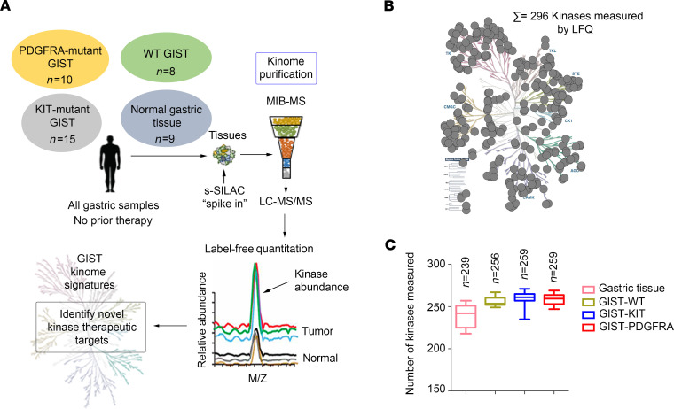
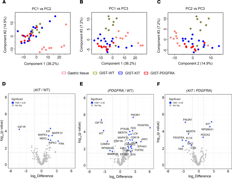
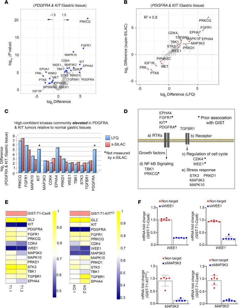
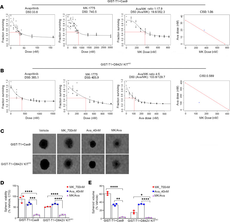
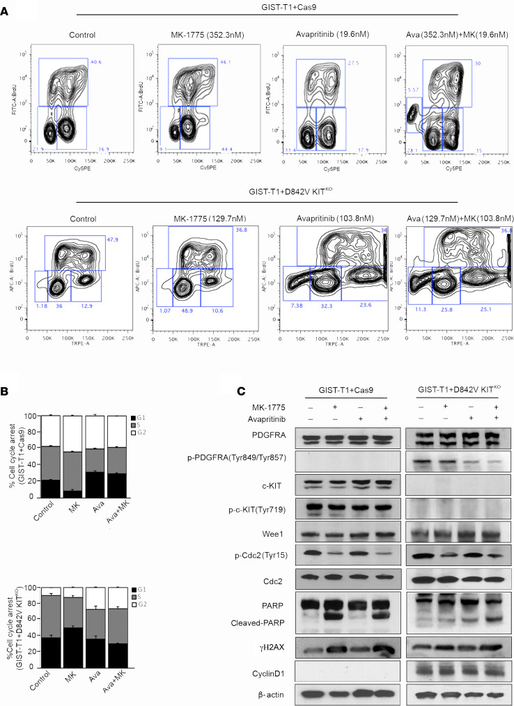
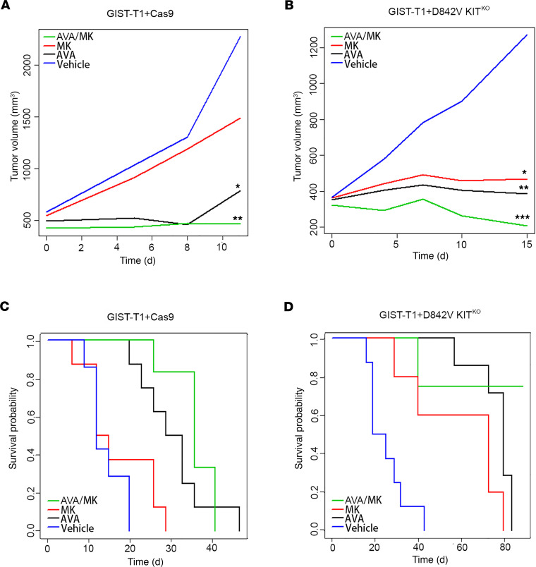
Management of gastrointestinal stromal tumors (GISTs) has been revolutionized by the identification of activating mutations in KIT and PDGFRA and clinical application of RTK inhibitors in advanced disease. Stratification of GISTs into molecularly defined subsets provides insight into clinical behavior and response to approved targeted therapies. Although these RTK inhibitors are effective in most GISTs, resistance remains a significant clinical problem. Development of effective treatment strategies for refractory GISTs requires identification of novel targets to provide additional therapeutic options. Global kinome profiling has the potential to identify critical signaling networks and reveal protein kinases essential in GISTs. Using multiplexed inhibitor beads and mass spectrometry, we explored the majority of the kinome in GIST specimens from the 3 most common molecular subtypes (KIT mutant, PDGFRA mutant, and succinate dehydrogenase deficient) to identify kinase targets. Kinome profiling with loss-of-function assays identified an important role for G2/M tyrosine kinase, Wee1, in GIST cell survival. In vitro and in vivo studies revealed significant efficacy of MK-1775 (Wee1 inhibitor) in combination with avapritinib in KIT mutant and PDGFRA mutant GIST cell lines as well as notable efficacy of MK-1775 as a monotherapy in the engineered PDGFRA mutant line. These studies provide strong preclinical justification for the use of MK-1775 in GIST.

Keywords: Cancer; Oncology; Therapeutics.


Conflict of interest statement

Conflict of interest: MVM has served as a member of the scientific steering committee for the phase I and phase III studies of avapritinib in GIST; her institution has received funding for the conduct of these clinical trials. She has also received honoraria as a scientific advisor to Blueprint Medicines. MCH received honoraria from Novartis; has served in advisory or consultancy roles for MolecularMD, Novartis, Blueprint Medicines, and Deciphera; and has provided expert testimony for Novartis. He has the following patents: “Methods of Detecting a Neoplasia Associated with an Activating Platelet Derived Growth Factor Receptor Alpha Mutation” (US patent 7,595,154 B2; 2009); “Nucleic Acids Encoding Platelet Derived Growth Factor Alpha Activating Mutations” (US patent 7,875,710; 2011); and “Nucleic Acids Encoding Platelet Derived Growth Factor Alpha Polypeptides Comprising Activating Mutations” (US patent 8,202,969; 2012). His institution receives royalties for these patents, which were licensed by Novartis. RD received research support from Blueprint Medicines.

Figures

Figure 1
Figure 1. Characterizing the GIST kinome in primary tumors using MIB-MS to identify therapeutic targets.
(A) Schematic of experimental approach. MIB-MS was used to quantify the kinase abundance in patients with GISTs (untreated, gastric primary GIST from 3 molecular subtypes: KIT mutant, n = 15; PDGFRA mutant, n = 10; WT GIST, n = 8; and normal gastric tissue, n = 9) to map the proteomic landscape of the kinome and identify targets. Kinase levels in tissues were determined using a combination of LFQ and s-SILAC. (B) Kinome tree depicts fraction of kinome quantitated by MIB-MS and frequency across 42 samples measured. (C) Average number of kinases detected by MIB-MS profiling broken down by tissue type. GIST, gastrointestinal stromal tumor; MIB-MS, multiplexed inhibitor beads and mass spectrometry; LFQ, label-free quantitation; s-SILAC, super-SILAC.
Figure 2
Figure 2. Mapping the distinct kinome signatures among GIST subtypes.
(A–C) PCA, including PC1 vs. PC2 (A), PC1 vs.PC3 (B), and PC2 vs.PC3 (C) of MIB-MS in 3 GIST subtypes (KIT mutant, blue; PDGFRA mutant, red; WT, green) and normal gastric tissue (pink). (D) Volcano plot comparisons of KIT mutant vs. WT, (E) PDGFRA mutant vs. WT, and (F) KIT mutant vs. PDGFRA mutant GIST MIB-MS kinome profiles. Differences in kinase log2 LFQ intensities among tumors and normal tissues determined by paired t test Benjamini-Hochberg adjusted P values at FDR of <0.05 using Perseus software. PCA, principal component analysis; GIST, gastrointestinal stromal tumor; MIB-MS, multiplexed inhibitor beads and mass spectrometry; LFQ, label-free quantitation.
Figure 3
Figure 3. Targeting the mutant-GIST kinome signature identifies WEE1 as candidate target.
(A) Volcano plot comparisons of KIT mutant and PDGFRA mutant GIST vs. normal gastric tissue MIB-MS kinome profiles. Differences in kinase log2 LFQ intensities among tumors and normal tissues determined by paired t test Benjamini-Hochberg adjusted P values at FDR <0.05 using Perseus software. (B) Scatter plot depicts overlap in kinases elevated or reduced determined by LFQ or s-SILAC. Regression analysis (R2) among quantitative methods was performed in Perseus software. Differential expressed kinases commonly identified by LFQ and s-SILAC quantitation (FDR <0.05) are labeled. (C) Bar graph depicts high-confident kinases log2 LFQ z scores overexpressed in mutant-GIST determined by LFQ and/or s-SILAC quantitation (FDR <0.05). (D) Associated pathways/functions of kinases overexpressed in KIT mutant and PDGFRA mutant GIST vs. normal tissues determined by quantitative MIB-MS profiling. (E) Heatmap depicting viability scores for siRNA library screen targeting high-confident kinases elevated in KIT mutant and PDGFRA mutant GIST in GIST-T1+Cas9 and GIST-T1+D842V KITKO cell lines as measured by Cell Titer Blue assay. siGL2 was negative control, viability score = 1.0. Two independent replicates were performed per cell line. (F) Quantitative RT-PCR confirmed >70% knockdown of Wee1 (top) and MAP3K3 (bottom) mRNA in both cell lines. Expression levels were normalized to HPRT. Data represent mean ± SD. GIST, gastrointestinal stromal tumor; MIB-MS, multiplexed inhibitor beads and mass spectrometry; LFQ, label-free quantitation; s-SILAC, super-SILAC.
Figure 4
Figure 4. MK-1775 and avapritinib have enhanced combination on in vitro GIST cell growth.
Panels 1 and 2 show dose response curves for single agents (avapritinib, MK-1775) in GIST-T1+Cas9 (A) and GIST-T1+D842V KITKO (B) cell lines. Red box indicates estimation of LD50 concentration for each single drug. Panel 3 shows dose response curve representing increasing series of combinations in GIST-T1+Cas9 (A) and GIST-T1+D842V KITKO (B) cell lines. Red box indicates estimation of LD50 concentration for combination of drugs. Panel 4 shows single point (blue) on isobole curve for 50% kill. Red line indicates 50% isobole for strictly additive effect. CILD50 in GIST-T1+Cas9 is 1.06 and not found in the synergistic triangle (region below the red line) (A). CILD50 is 0.589 in GIST-T1+D842V KITKO and is found within the synergistic triangle (B). Representative images of GIST-T1+Cas9 and GIST-T1+D842V KITKO spheroids after 120-hour treatment at indicated concentrations (C). Bars represent average viability ± SEM after 120-hour treatment at indicated drug concentrations for GIST-T1+Cas9 and GIST-T1+D842V KITKO spheroids as a percentage of vehicle-treated spheroids (D). Bars represent the average spheroid volume ± SEM of GIST-T1+Cas9 and GIST-T1+D842V KITKO spheroids as a percentage of vehicle-treated spheroids (E). All spheroid data were analyzed using GraphPad Prism, with comparisons of treatment groups performed in 1-way ANOVA and post hoc comparisons made using Bonferroni’s multiple comparisons method; *P = 0.0165, **P = 0.0046, ***P = 0.0008, ****P ≤ 0.0001. GIST, gastrointestinal stromal tumor.
Figure 5
Figure 5. Mechanism of MK-1775 and avapritinib combination in KIT-dependent and –independent GIST cell lines.
(A) Representative flow cytometry plots and (B) quantification of BrdU incorporation in GIST-T1+Cas9 (upper panel) treated with 352.3 nM MK-1775, 19.6 nM avapritinib and combination for 72 hours. Statistically significant differences were observed between the following comparisons: for G1 arrest, vehicle vs. avapritinib (P < 0.0009) and vehicle vs. avapritinib/MK-1775 (P < 0.0002); for G2 arrest, vehicle vs. MK-1775 (P < 0.005). (A) Representative flow cytometry plots and (B) quantification of BrdU incorporation in GIST-T1-D842V+ KITKO treated (bottom panel) with 129.7 nM MK-1775, 103.8 nM avapritinib and combination for 72 hours. Statistically significant differences were observed between the following comparisons: for G1 arrest, vehicle vs. MK-1775 (P < 0.0001); for G2 arrest, vehicle vs. avapritinib (P < 0.005), vehicle vs. avapritinib/MK-1775 (P < 0.0002). Data represent mean ± SD. (C) Immunoblot assays of WCEs from GIST-T1+Cas9 (KIT-dependent) and GIST-T1+D842V KITKO (KIT-independent) cell lines treated as in A and B. Equal concentrations (45–90 μg) of WCE from each sample were subjected to immunoblotting with specific antibodies, as indicated. β-Actin served as a loading control. GIST, gastrointestinal stromal tumor; WCE, whole cell extract.
Figure 6
Figure 6. The combination of MK-1775 and avapritinib significantly inhibits GIST growth in vivo and improves disease-specific survival.
(A) Statistically significant decreases in the rate of GIST-T1+Cas9 xenograft tumor growth were observed due to treatment with avapritinib (*P = 0.05, black) and avapritinib+MK-1775 combination (**P = 0.002, green) compared with vehicle group (blue) on day 11. (B) Statistically significant decreases in the rate of GIST-T1+D842V KITKO xenograft tumor growth were observed due to treatment with avapritinib (**P = 0.002) and MK-1775 (*P = 0.02) and avapritinib+MK-1775 (***P ≤ 0.0002) compared with vehicle group on day 15. Smoothed tumor growth curves (tumor volume vs. time) were computed for each treatment using the lowess smoother in the R statistical language. (C) Kaplan-Meier estimate of the probability of disease-specific survival of GIST-T1+Cas9 xenografts. Statistically significant differences (even after adjusting for multiple testing) in disease-specific survival were observed between the following comparisons: vehicle vs. avapritinib (P < 0.0001); vehicle vs. avapritinib/MK-1775 (P < 0.0001); and MK-1775 vs. avapritinib/MK-1775 (P < 0.0001). (D) Kaplan-Meier estimate of the probability of disease-specific survival of GIST-T1+D842V KITKO xenografts. Statistically significant differences (even after adjusting for multiple testing) in disease-specific survival were observed between the following comparisons: vehicle vs. MK-1775 (P = 0.01); vehicle vs. avapritinib (P < 0.0001); vehicle vs. avapritinib/MK-1775 (P < 0.0001); MK-1775 vs. avapritinib/MK-1775 (P = 0.01); and avapritinib vs. avapritinib/MK-1775 (P = 0.02). The overall test is also significant (P < 0.0001). GIST, gastrointestinal stromal tumor.

References

    1. Goettsch WG, et al. Incidence of gastrointestinal stromal tumours is underestimated: results of a nation-wide study. Eur J Cancer. 2005;41(18):2868–2872. doi: 10.1016/j.ejca.2005.09.009. - DOI - PubMed
    1. Corless CL, et al. Gastrointestinal stromal tumours: origin and molecular oncology. Nat Rev Cancer. 2011;11(12):865–878. doi: 10.1038/nrc3143. - DOI - PubMed
    1. Belinsky MG, et al. Overexpression of insulin-like growth factor 1 receptor and frequent mutational inactivation of SDHA in wild-type SDHB-negative gastrointestinal stromal tumors. Genes Chromosomes Cancer. 2013;52(2):214–224. doi: 10.1002/gcc.22023. - DOI - PMC - PubMed
    1. Killian JK, et al. Recurrent epimutation of SDHC in gastrointestinal stromal tumors. Sci Transl Med. 2014;6(268):268ra177. doi: 10.1126/scitranslmed.3009961. - DOI - PMC - PubMed
    1. Debiec-Rychter M, et al. KIT mutations and dose selection for imatinib in patients with advanced gastrointestinal stromal tumours. Eur J Cancer. 2006;42(8):1093–1103. doi: 10.1016/j.ejca.2006.01.030. - DOI - PubMed

Publication types

MeSH terms