Comparison of the within-reader and inter-vendor agreement of left ventricular circumferential strains and volume indices derived from cardiovascular magnetic resonance imaging
- PMID: 33320865
- PMCID: PMC7737975
- DOI: 10.1371/journal.pone.0242908
Comparison of the within-reader and inter-vendor agreement of left ventricular circumferential strains and volume indices derived from cardiovascular magnetic resonance imaging
Abstract
Purpose: Volume indices and left ventricular ejection fraction (LVEF) are routinely used to assess cardiac function. Ventricular strain values may provide additional diagnostic information, but their reproducibility is unclear. This study therefore compares the repeatability and reproducibility of volumes, volume fraction, and regional ventricular strains, derived from cardiovascular magnetic resonance (CMR) imaging, across three software packages and between readers.
Methods: Seven readers analysed 16 short-axis CMR stacks of a porcine heart. Endocardial contours were manually drawn using OsiriX and Simpleware ScanIP and repeated in both softwares. The images were also contoured automatically in Circle CVI42. Endocardial global, apical, mid-ventricular, and basal circumferential strains, as well as end-diastolic and end-systolic volume and LVEF were compared.
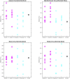
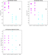
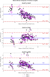
Results: Bland-Altman analysis found systematic biases in contour length between software packages. Compared to OsiriX, contour lengths were shorter in both ScanIP (-1.9 cm) and CVI42 (-0.6 cm), causing statistically significant differences in end-diastolic and end-systolic volumes, and apical circumferential strain (all p<0.006). No differences were found for mid-ventricular, basal or global strains, or left ventricular ejection fraction (all p<0.007). All CVI42 results lay within the ranges of the OsiriX results. Intra-software differences were found to be lower than inter-software differences.
Conclusion: OsiriX and CVI42 gave consistent results for all strain and volume metrics, with no statistical differences found between OsiriX and ScanIP for mid-ventricular, global or basal strains, or left ventricular ejection fraction. However, volumes were influenced by the choice of contouring software, suggesting care should be taken when comparing volumes across different software.
Conflict of interest statement
The authors have declared that no competing interests exist.
Figures


Similar articles
-
Regional left ventricular endocardial strains estimated from low-dose 4DCT: Comparison with cardiac magnetic resonance feature tracking.Med Phys. 2022 Sep;49(9):5841-5854. doi: 10.1002/mp.15818. Epub 2022 Jul 6. Med Phys. 2022. PMID: 35751864 Free PMC article.
-
Comparison of left ventricular strains and torsion derived from feature tracking and DENSE CMR.J Cardiovasc Magn Reson. 2018 Sep 13;20(1):63. doi: 10.1186/s12968-018-0485-4. J Cardiovasc Magn Reson. 2018. PMID: 30208894 Free PMC article.
-
A dual propagation contours technique for semi-automated assessment of systolic and diastolic cardiac function by CMR.J Cardiovasc Magn Reson. 2009 Aug 13;11(1):30. doi: 10.1186/1532-429X-11-30. J Cardiovasc Magn Reson. 2009. PMID: 19674481 Free PMC article.
-
Accuracy and reproducibility of quantitation of left ventricular function by real-time three-dimensional echocardiography versus cardiac magnetic resonance.Am J Cardiol. 2008 Sep 15;102(6):778-83. doi: 10.1016/j.amjcard.2008.04.062. Epub 2008 Jul 9. Am J Cardiol. 2008. PMID: 18774006
-
Left ventricular global myocardial strain assessment comparing the reproducibility of four commercially available CMR-feature tracking algorithms.Eur Radiol. 2018 Dec;28(12):5137-5147. doi: 10.1007/s00330-018-5538-4. Epub 2018 Jun 5. Eur Radiol. 2018. PMID: 29872912
Cited by
-
Acute regional changes in myocardial strain may predict ventricular remodelling after myocardial infarction in a large animal model.Sci Rep. 2021 Sep 15;11(1):18322. doi: 10.1038/s41598-021-97834-y. Sci Rep. 2021. PMID: 34526592 Free PMC article.
-
Deep learning automates detection of wall motion abnormalities via measurement of longitudinal strain from ECG-gated CT images.Front Cardiovasc Med. 2022 Dec 15;9:1009445. doi: 10.3389/fcvm.2022.1009445. eCollection 2022. Front Cardiovasc Med. 2022. PMID: 36588550 Free PMC article.
-
MRI-based strain measurements reflect morphological changes following myocardial infarction: A study on the UK Biobank cohort.J Anat. 2023 Jan;242(1):102-111. doi: 10.1111/joa.13787. Epub 2022 Dec 9. J Anat. 2023. PMID: 36484568 Free PMC article.
References
-
- Jennings RB G. C., “Structural changes in myocardium during acute ischemia.,” Circ. Res., vol. 34, pp. 156–168, 1974. - PubMed
Publication types
MeSH terms
Grants and funding
LinkOut - more resources
Full Text Sources
Medical

