Review
. 2021 Jul:83:100936.
doi: 10.1016/j.preteyeres.2020.100936. Epub 2020 Dec 13.

Targeting complement components C3 and C5 for the retina: Key concepts and lingering questions

Affiliations
Review

Targeting complement components C3 and C5 for the retina: Key concepts and lingering questions

Benjamin J Kim et al. Prog Retin Eye Res. 2021 Jul.

Abstract

Age-related macular degeneration (AMD) remains a major cause of legal blindness, and treatment for the geographic atrophy form of AMD is a significant unmet need. Dysregulation of the complement cascade is thought to be instrumental for AMD pathophysiology. In particular, C3 and C5 are pivotal components of the complement cascade and have become leading therapeutic targets for AMD. In this article, we discuss C3 and C5 in detail, including their roles in AMD, biochemical and structural aspects, locations of expression, and the functions of C3 and C5 fragments. Further, the article critically reviews developing therapeutics aimed at C3 and C5, underscoring the potential effects of broad inhibition of complement at the level of C3 versus more specific inhibition at C5. The relationships of complement biology to the inflammasome and microglia/macrophage activity are highlighted. Concepts of C3 and C5 biology will be emphasized, while we point out questions that need to be settled and directions for future investigations.

Keywords: Age-related macular degeneration; C3; C5; Complement; Geographic atrophy; Retina.


Figures

Fig. 1.
Fig. 1.
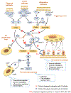
Overview of the complement system. The complement cascade involves many protein interactions that occur within the plasma, on cell surfaces, and within cells. It begins with the classical, lectin, and alternative pathways. The three pathways converge on C3, and C3 mediates multiple functions. Cleavage of C5 brings about the terminal lytic pathway with formation of the membrane attack complex. Shown on the right are key components of the C3 breakdown pathway, including C3 fragments, the complement receptors, and associated functions. This portion of the schematic intends to illustrate the multiplicity of interactions between C3 fragments and their receptors on a generic cellular scaffold and does not imply the concomitant presence of all complement receptors on the same cell type. Shown on the left are the anaphylatoxins (C3a and C5a), their receptors, and associated functions. Red X’s highlight pathways blocked by C3 inhibition, and black X’s highlight pathways blocked by C5 inhibition. Convertases are shown in yellow boxes. Important interactions are discussed in more detail within the text. For simplicity, the schematic does not depict intracellular or extrinsic protease-mediated routes of complement activation, but these important aspects of complement biology are discussed in the text. Abbreviations: MBL, mannose-binding lectin; MASPs, mannose-binding lectin associated serine proteases; FP, factor properdin; FB, factor B; FD, factor D; FI, factor I; CR, complement receptor; C3aR, C3a receptor; CSaR1, C5a receptor 1; C5aR2, C5a receptor 2; MAC, membrane attack complex; RCA, regulator of complement activation; MCP, membrane cofactor protein; DAF, decay accelerating factor.
Fig. 2.
Fig. 2.
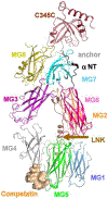
Crystal structures of C3 and C5. The domains of C3 (A) and C5 (B) are labeled. Figure and caption adapted from Janssen et al., 2005 (C3) and Fredslund et al., 2008 (C5) with permission of the copyright holder. Abbreviations: MG, macroglobulin; TED, thioester domain; ANA, anaphylatoxin; LNK, linker; CUB (complement C1r/C1s, Uegf, Bmp1).
Fig. 3.
Fig. 3.
Passage of complement component 9 (C9) across the blood retina barrier of nonexudative AMD (age-related macular degeneration) patients. Western analysis of post mortem neurosensory retina protein from macula (A) with corresponding pixel density graphs and from nasal periphery (B) with corresponding pixel density graphs. Tissue was obtained from the Minnesota Lions Eye Bank. Immunoblots were done with anti-C9 (R & D Systems, Minneapolis, MN) and α-tubulin (Sigma-Aldrich, Inc., St. Louis, MO). Loading control α-tubulin (50 kDa) bands are shown below each set of lanes. Graphs show band densitometry normalized to loading control calculated using Image J software. Numbers represent mean values (±SEM). Grading of eyes was done using the Minnesota Grading System (Decanini et al., 2007; Olsen and Feng, 2004). There were 5 normal eyes, and 5 intermediate-stage AMD eyes. Statistical analysis was performed using student’s two-tailed unpaired t-test. *p < 0.05, **p < 0.01. Normal and AMD eyes were matched for age and post-mortem interval. These findings were replicated with a second cohort of eyes from the Alabama Eye Bank. Figure and caption adapted from Schultz et al., 2019 with permission of the copyright holder.
Fig. 4.
Fig. 4.
Crystal structure of C3c bound to compstatin. Compstatin (analog 4W9A) binds to the C3c portion of C3 between MG4 and MG5. Compstatin sterically prevents C3 from interacting with C3 convertases. Figure and caption adapted from Janssen et al., 2007 with permission of the copyright holder. Abbreviations: MG, macroglobulin; LNK, linker.
Fig. 5.
Fig. 5.
Prolonged intraocular residence of Cp40-KKK. Pharmacokinetic profile of compstatin-based analogs Cp40-KK and Cp40-KKK in the vitreous of non-human primates. The intravitreal concentration of the lysine modified Cp40 analogs, Cp40-KK and Cp40-KKK, was determined by surface plasmon resonance-based quantification at various time points (i.e., 14, 28, 42, 56, 73, 90 days) following a single intravitreal injection of 500 μg of inhibitor in cynomolgus monkeys. A total of three eyes were used per treatment and each data point in the curve represents the mean value ± SD from N = 3 animals per group. Each curve represents successive measurements (intraocular inhibitor levels) from the same animal, averaged for a group of three treated eyes per inhibitor. Figure and caption adapted from Hughes et al., 2020 with permission of the copyright holder.
Fig. 6.
Fig. 6.
Retinal tissue distribution of Cp40-KKK and its co-localization with C3 in non-human primates. Non-human primates (NHPs) (6–7 years old) were treated with one dose of 500 μg of Cp40-KKK peptide via intravitreal Injection. One month later following the injection, the NHPs were euthanized. Both right and left eyes of NHPs were collected, fixed in 4% paraformaldehyde solution in PBS and held at 4 °C until dissection. The eyecup was carefully dissected into two halves; the eyecup half with the optic nerve head was cryopreserved overnight in 30% sucrose (prepared in PBS) at 4 °C. The following day, the sample was embedded in OCT, frozen in isopropanol (precooled in dry ice), and the tissue was sectioned at 12-μm thick sections and stored at −80 °C. The sections were then stained with both goat anti-human complement C3 (1:200 dilution) and anti-Cp40 antibody (final concentration of 2 μg/ml). The Cp40-KKK staining signal was amplified by using the Alexa Fluor™ 555 Tyramide SuperBoost kit (Thermo Fischer, B40923). The images were taken using a Nikon Eclipse Ti2 Confocal Microscope. Note: Control Retina tissue was from untreated NHPs. The middle (Cp40-KKK, 200X) and right panel (Cp40-KKK, 400X) of photomicrographs display retinal tissue sections from NHPs being treated with one dose of 500 μg of Cp40-KKK peptide at x200 and x400 magnification, respectively; the green-colored squares in the middle panel denote the magnified region of the retina. Red color: represents the C3 staining; Green color: represents the Cp40-KKK staining. Blue color: represents DAPI staining of nuclei. Yellow color: represents the co-localization of both C3 and Cp40-KKK. The retinal tissue consists of a total of 11 layers: 1. Internal limiting membrane; 2. Nerve fiber layer; 3. Ganglion cell layer; 4. Inner plexiform layer; 5. Inner nuclear layer; 6. Outer plexiform layer; 7. Outer nuclear layer; 8. Layer of rods and cones; 9. Retinal pigment epithelium; 10. Bruch’s membrane and choriocapillaris; 11. Choroid. Figure and caption adapted from Hughes et al., 2020 with permission of the copyright holder.
Fig. 7.
Fig. 7.
Complement focused model of major pathways in geographic atrophy pathophysiology. Several parallel pathways contribute to oxidative stress. Buildup of metabolic byproducts, oxidative stress, the impact of complement gene variants, and perhaps systemic complement deposition within the eye cause abnormally increased complement activity. This in turn contributes to several pathways including membrane attack complex activity, microglia/macrophage infiltration into the subretinal space, increased inflammation, and inflammasome activation. These potential pathways, acting in concert, contribute to cell death of photoreceptors, retinal pigment epithelium, and choriocapillaris. Abbreviations: AMD, age-related macular degeneration; MCP, monocyte chemoattractant protein; CCL-2, chemokine (C─C motif) ligand 2; RPE, retinal pigment epithelium.
Fig. 8.
Fig. 8.
Potential pathological consequences of deregulated complement activity in the AMD (age-related macular degeneration) retina. Several complement mediated mechanisms may act in parallel to bring about geographic atrophy. A. Increased membrane attack complex activity may cause cell lysis of retinal pigment epithelium, choriocapillaris, and photoreceptors. B. The anaphylatoxins C3a and C5a cause microglia/macrophages to migrate to the subretinal space. C. Complement dysregulation contributes to NLRP3 inflammasome activity within the RPE with secretion of IL-18 and IL-1β. D. The Y402H FH variant can competitively block TSP-1 mediated phagocyte clearance, leading to phagocyte accumulation, and subsequent inflammation (Calippe et al., 2017). There is also potential for phagocytosis of photoreceptors, RPE, and choroidal endothelium. E. Increased sublytic membrane attack complex activity overloads lysosomes leading to RPE dysfunction and sub-RPE deposits (Cerniauskas et al., 2020). Abbreviations: MAC, membrane attack complex; RPE, retinal pigment epithelium; BM, Bruch’s membrane; Mφ, macrophage; c3aR, C3a receptor; C5aR1, C5a receptor 1; NLRP3, NOD-, LRR- and pyrin domain-containing protein 3 inflammasome; FH(Y402H), Factor H Y402H variant; IAP, integrin associated protein; CR3, complement receptor 3; TSP-1, thrombospondin-1.

References

    1. Alcorlo M, Martínez-Barricarte R, Fernández FJ, Rodríguez-Gallego C, Round A, Vega MC, Harris CL, Rodríguez De Cordoba S, Llorca O, 2011. Unique structure of iC3b resolved at a resolution of 24 Å by 3D-electron microscopy. Proc. Natl. Acad. Sci. U. S. A 108, 13236–13240. 10.1073/pnas.1106746108. - DOI - PMC - PubMed
    1. Alcorlo M, Tortajada A, Rodriguez de Cordoba S, Llorca O, 2013. Structural basis for the stabilization of the complement alternative pathway C3 convertase by properdin. Proc. Natl. Acad. Sci. Unit. States Am 110, 13504–13509. 10.1073/pnas.1309618110. - DOI - PMC - PubMed
    1. Alcorlo M, López-Perrote A, Delgado S, Yébenes H, Subías M, Rodríguez-Gallego C, Rodríguez de Córdoba S, Llorca O, 2015. Structural insights on complement activation. FEBS J. 282, 3883–3891. 10.1111/febs.13399. - DOI - PubMed
    1. Amara U, Flierl MA, Rittirsch D, Klos A, Chen H, Acker B, Brückner UB, Nilsson B, Gebhard F, Lambris JD, Huber-Lang M, 2010. Molecular intercommunication between the complement and coagulation systems. J. Immunol 185 (9), 5628–5636. 10.4049/jimmunol.0903678. - DOI - PMC - PubMed
    1. Anderson DH, Radeke MJ, Gallo NB, Chapin EA, Johnson PT, Curletti CR, Hancox LS, Hu J, Ebright JN, Malek G, Hauser MA, Bowes Rickman C, Bok D, Hageman GS, Johnson LV, 2010. The pivotal role of the complement system in aging and age-related macular degeneration: hypothesis re-visited. Prog. Retin. Eye Res 29 (2), 95–112. - PMC - PubMed

Publication types