Review
. 2020 Dec 10;9(12):2650.
doi: 10.3390/cells9122650.

High Mobility Group Box-1 and Blood-Brain Barrier Disruption

Affiliations
Review

High Mobility Group Box-1 and Blood-Brain Barrier Disruption

Masahiro Nishibori et al. Cells. .

Abstract

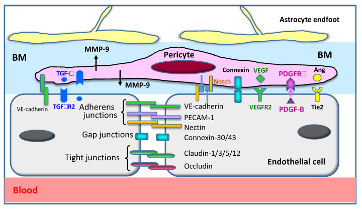
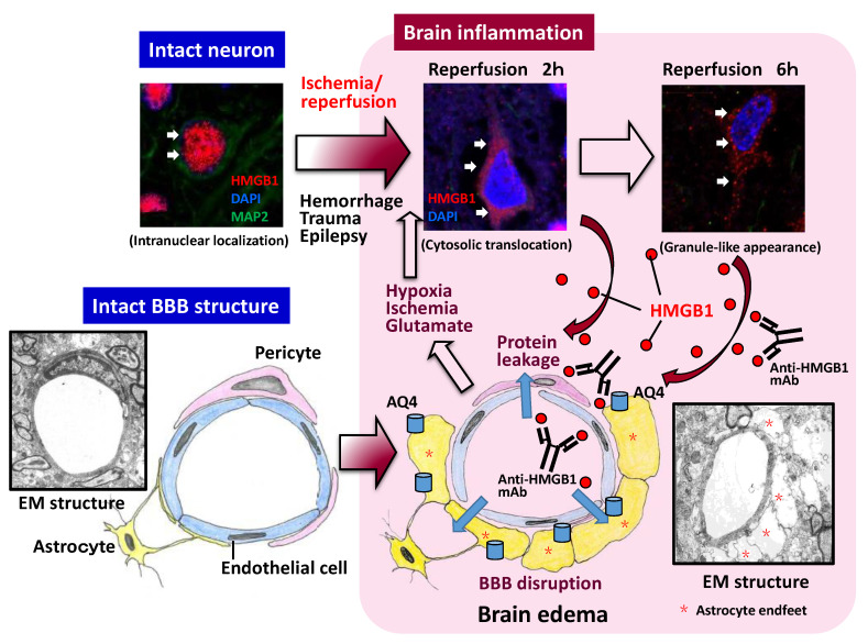
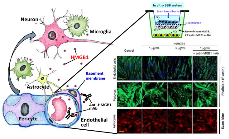
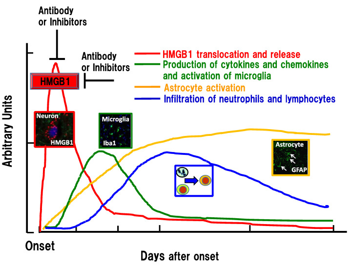
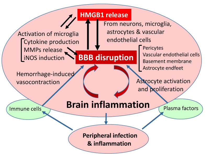
Increasing evidence suggests that inflammatory responses are involved in the progression of brain injuries induced by a diverse range of insults, including ischemia, hemorrhage, trauma, epilepsy, and degenerative diseases. During the processes of inflammation, disruption of the blood-brain barrier (BBB) may play a critical role in the enhancement of inflammatory responses and may initiate brain damage because the BBB constitutes an interface between the brain parenchyma and the bloodstream containing blood cells and plasma. The BBB has a distinct structure compared with those in peripheral tissues: it is composed of vascular endothelial cells with tight junctions, numerous pericytes surrounding endothelial cells, astrocytic endfeet, and a basement membrane structure. Under physiological conditions, the BBB should function as an important element in the neurovascular unit (NVU). High mobility group box-1 (HMGB1), a nonhistone nuclear protein, is ubiquitously expressed in almost all kinds of cells. HMGB1 plays important roles in the maintenance of chromatin structure, the regulation of transcription activity, and DNA repair in nuclei. On the other hand, HMGB1 is considered to be a representative damage-associated molecular pattern (DAMP) because it is translocated and released extracellularly from different types of brain cells, including neurons and glia, contributing to the pathophysiology of many diseases in the central nervous system (CNS). The regulation of HMGB1 release or the neutralization of extracellular HMGB1 produces beneficial effects on brain injuries induced by ischemia, hemorrhage, trauma, epilepsy, and Alzheimer's amyloidpathy in animal models and is associated with improvement of the neurological symptoms. In the present review, we focus on the dynamics of HMGB1 translocation in different disease conditions in the CNS and discuss the functional roles of extracellular HMGB1 in BBB disruption and brain inflammation. There might be common as well as distinct inflammatory processes for each CNS disease. This review will provide novel insights toward an improved understanding of a common pathophysiological process of CNS diseases, namely, BBB disruption mediated by HMGB1. It is proposed that HMGB1 might be an excellent target for the treatment of CNS diseases with BBB disruption.

Keywords: blood–brain barrier; high mobility group box-1; inflammation; monoclonal antibody; pericyte; stroke; trauma; vascular endothelial cell.


Conflict of interest statement

The authors declare no conflict of interest.

Figures

Figure 1
Figure 1
Structure of the blood–brain barrier (BBB). The BBB consists of vascular endothelial cells, pericytes, a basement membrane (BM), and astrocyte endfeet. Cellular components are integrated with many adhesive molecules and are functionally regulated by ligand–receptor systems. The major factors are drawn in this figure. Ang: angiopoietin; BM: basement membrane; MMP-9: matrix metallo proteinase-9; PECAM-1: platelet endothelial cell adhesion molecule-1; PDGF-B: platelet-derived growth factor-B; PDGFRb: platelet-derived growth factor receptor beta; TGF-b: transforming growth factor-b; TGFbR2: Transforming growth factor-b receptor 2; VE-cadherin: vascular endothelial cadherin; VEGF: vascular endothelial growth factor; VEGFR2: vascular endothelial growth factor receptor 2.
Figure 2
Figure 2
HMGB1 translocation and release after insults and its involvement in BBB disruption. In the upper three squares, typical HMGB1 translocation and release from the nucleus to extracellular space are shown after 2 h of occlusion/reperfusion of the middle cerebral artery. The released HMGB1 then affects the vascular endothelial cells and pericytes, leading to protein leakage and brain edema formation. BBB disruption further promotes HMGB1 translocation. There might be a positive feedback loop between BBB disruption and HMGB1 translocation. The existence of such a loop may be a reason why anti-HMGB1 therapy inhibited BBB disruption and HMGB1 translocation simultaneously. AQ4: aquaporin 4; BBB: blood–brain barrier; EM: electron microscopy; HMGB1: high mobility group box-1; mAb: monoclonal antibody.
Figure 3
Figure 3
Direct effects of HMGB1 on vascular endothelial cells and pericytes. A rat-reconstituted BBB system composed of vascular endothelial cells, pericytes, and astrocytes (upper right) was used. Recombinant HMGB1 at 1 or 5 mg/mL was added to the brain side (lower chamber), and the incubation was continued for 60 min. After 1 h of stimulation with HMGB1, the cells were fixed with paraformaldehyde. Endothelial cells and pericytes were labeled with Alexa-488-phalloidin, and the astrocytes were labeled with Evans blue. The contractile response was observed in endothelial cells and pericytes, which were inhibited by the presence of specific anti-HMGB1 mAb in the lower chamber (modified from Zhang et al., [15]). BBB: blood–brain barrier; HMGB1: high mobility group box-1; mAb: monoclonal antibody.
Figure 4
Figure 4
Time-course of inflammation-related events in the penumbra areas after ischemia, hemorrhage, and trauma. HMGB1 translocation and extracellular release form the initial peak to insults, among the known responses, which was followed in succession by microglia activation (cytokine/chemokine production), astrogliosis, and infiltration of immune cells. HMGB1 release may be a very early event that is common to several brain injuries. Inhibition of HMGB1 by the earlier intervention can lead to the diminution of the following inflammatory responses. GFAP: glial fibrillary acidic protein; HMGB1: high mobility group box-1; Iba1: ionized calcium-binding adapter molecule 1.
Figure 5
Figure 5
Flow diagram of relationship between HMGB1 release and BBB disruption. As described in the text, HMGB1 release is common to different kinds of injuries induced by ischemia/reperfusion, hemorrhage, trauma, and epilepsy, leading to BBB disruption through direct and indirect pathways. The peripheral inflammation caused by infection may exacerbate BBB disruption. Conversely, brain injuries may induce a vulnerability to infection in patients. BBB: blood–brain barrier; HMGB1: high mobility group box-1; iNOS: inducible nitric oxide synthase; MMPs: matrix metallo proteinases.

References

    1. Iadecola C. The pathobiology of vascular dementia. Neuron. 2013;80:844–866. doi: 10.1016/j.neuron.2013.10.008. - DOI - PMC - PubMed
    1. Ahmad A., Patel V., Xiao J., Khan M.M. The Role of Neurovascular System in Neurodegenerative Diseases. Mol. Neurobiol. 2020;57:4373–4393. doi: 10.1007/s12035-020-02023-z. - DOI - PubMed
    1. Sweeney M.D., Zhao Z., Montagne A., Nelson A.R., Zlokovic B.V. Blood-Brain Barrier: From Physiology to Disease and Back. Physiol. Rev. 2019;99:21–78. doi: 10.1152/physrev.00050.2017. - DOI - PMC - PubMed
    1. Abbott N.J., Patabendige A.A., Dolman D.E., Yusof S.R., Begley D.J. Structure and function of the blood-brain barrier. Neurobiol. Dis. 2010;37:13–25. doi: 10.1016/j.nbd.2009.07.030. - DOI - PubMed
    1. Shichita T., Sakaguchi R., Suzuki M., Yoshimura A. Post-ischemic inflammation in the brain. Front. Immunol. 2012;3:132. doi: 10.3389/fimmu.2012.00132. - DOI - PMC - PubMed

Publication types

LinkOut - more resources