Review
. 2020 Dec 10;9(12):2654.
doi: 10.3390/cells9122654.

The Role of Protein Disorder in Nuclear Transport and in Its Subversion by Viruses

Affiliations
Review

The Role of Protein Disorder in Nuclear Transport and in Its Subversion by Viruses

Jacinta M Wubben et al. Cells. .

Abstract

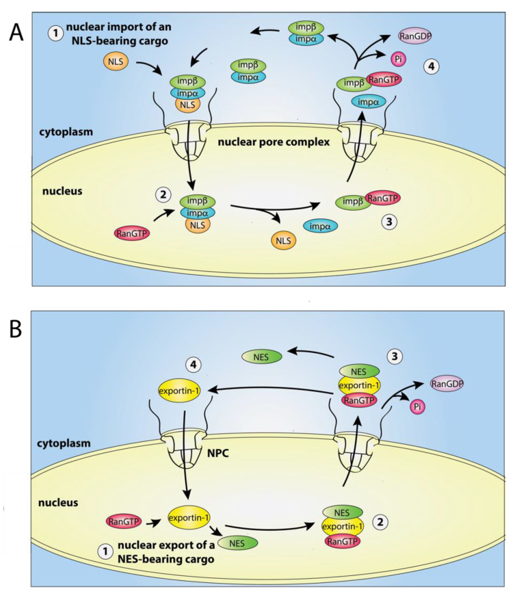
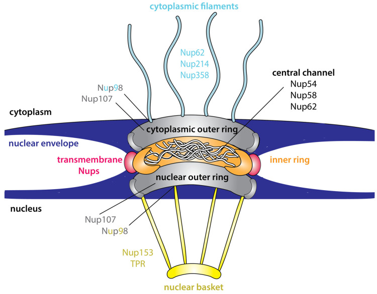
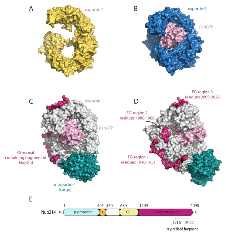
The transport of host proteins into and out of the nucleus is key to host function. However, nuclear transport is restricted by nuclear pores that perforate the nuclear envelope. Protein intrinsic disorder is an inherent feature of this selective transport barrier and is also a feature of the nuclear transport receptors that facilitate the active nuclear transport of cargo, and the nuclear transport signals on the cargo itself. Furthermore, intrinsic disorder is an inherent feature of viral proteins and viral strategies to disrupt host nucleocytoplasmic transport to benefit their replication. In this review, we highlight the role that intrinsic disorder plays in the nuclear transport of host and viral proteins. We also describe viral subversion mechanisms of the host nuclear transport machinery in which intrinsic disorder is a feature. Finally, we discuss nuclear import and export as therapeutic targets for viral infectious disease.

Keywords: nuclear export inhibitors; nuclear export sequence; nuclear import inhibitors; nuclear import sequence; nuclear transport receptors; nucleoporins; protein intrinsic disorder; viral infection.


Conflict of interest statement

The authors declare no conflict of interest. The funders had no role in the design of the study; in the collection, analyses, or interpretation of data; in the writing of the manuscript, or in the decision to publish the results.

Figures

Figure 1
Figure 1
The nuclear import–export cycle. (A) Nuclear import of a nuclear localisation signal (NLS)-bearing cargo (orange) by importin α/β (imp α/β). Protein cargo in the cytosol bind the nuclear import receptor importin α/β for transport through the nuclear pore complex (NPC) to the nucleus. Following import, guanosine triphosphate (GTP)-bound Ran (RanGTP) binding to importin-β causes the release of the cargo in the cytoplasm and the recycling of importin-α and importin-β-RanGTP. (B) Nuclear export of a nuclear export signal (NES)-bearing cargo (green) by exportin-1. RanGTP binds to exportin-1 in the nucleus to enable the exportin-1-RanGTP-cargo ternary complex to be formed and exported to the cytoplasm. The ternary complex is disassembled following the hydrolysis of RanGTP to RanGTP (guanosine diphosphate (GDP)-bound Ran).
Figure 2
Figure 2
Cross-section of the nuclear pore complex (NPC). The NPC is comprised of two outer rings (grey) and an inner ring which forms the central channel of the pore. The inner ring is formed from layers of nucleoporins (Nups) that are collectively depicted in orange. The phenylalanine-glycine (FG)-Nups are predominantly anchored within the central channel and contain long extensions intermittently spaced with FG-repeats which have properties characteristic of intrinsically disordered proteins (IDPs). The FG-Nups form the permeability barrier of the NPC. The transmembrane Nups (red) anchor the NPC to the nuclear envelope. Nups and NPC proteins that are referred to in this review are shown. TPR = translocated promoter region.
Figure 3
Figure 3
The nuclear export receptor exportin-1 adopts an extended or compact conformation and can bind to multiple partners. (AC) Surface representations of exportin-1 depicted in the same orientation. (A) The extended conformation of unbound exportin-1 from fungus. Figure generated from Protein Data Bank (PDB) ID 4FGV [54]. (B) The compact ring-like conformation of mouse exportin-1 (blue) bound to RanGTP (pink). Figure generated from Protein Data Bank (PDB) ID 3NC1 [58]. (C,D) The human exportin-1-RanGTP-snurportin-1-Nup214 complex, where D is a rotation of C along the Y-axis. RanGTP (pink) interacts with the inner surface of exportin-1 (grey). The nuclear export signal (NES) of the transport cargo, in this case snurportin-1 (teal), interacts with the NES binding groove on the outer surface of exportin-1. The receptor-cargo complex is transported through the nuclear pore complex (NPC) via its interactions with FG-Nups, such as Nup214 (crimson), of which a fragment (residues 1916-2026) containing three FG-regions (FG-region 1-3) is shown. Each of the three FG-regions contain multiple FG-repeats and serve as anchor points for binding hydrophobic surface pockets of exportin-1. Residues connecting the FG-repeat regions were not always clearly defined in the electron density. Although the Nup214 fragment was crystallised with an N-terminal maltose binding protein (MBP) fusion tag, MBP has been removed from the figure for clarity. Figure generated from PDB ID 5DIS [27]. (E) Schematic representation of the domain architecture of Nup214, with the crystallised fragment that is shown in C,D depicted. CTE = C-terminal extension of the seven-bladed β-propeller; CC = coiled-coil.

References

    1. Uversky V.N. Natively unfolded proteins: A point where biology waits for physics. Protein Sci. 2002;11:739–756. doi: 10.1110/ps.4210102. - DOI - PMC - PubMed
    1. Uversky V.N. What does it mean to be natively unfolded? Eur. J. Biochem. 2002;269:2–12. doi: 10.1046/j.0014-2956.2001.02649.x. - DOI - PubMed
    1. Croft S.N., Walker E.J., Ghildyal R. Human Rhinovirus 3C protease cleaves RIPK1, concurrent with caspase 8 activation. Sci. Rep. 2018;8:1569. doi: 10.1038/s41598-018-19839-4. - DOI - PMC - PubMed
    1. Dyson H.J., Wright P.E. Intrinsically unstructured proteins and their functions. Nat. Rev. Mol. Cell Biol. 2005;6:197–208. doi: 10.1038/nrm1589. - DOI - PubMed
    1. Wright P.E., Dyson H.J. Linking folding and binding. Curr. Opin. Struct. Biol. 2009;19:31–38. doi: 10.1016/j.sbi.2008.12.003. - DOI - PMC - PubMed

Publication types

LinkOut - more resources