Review
. 2020 Dec 10;21(24):9420.
doi: 10.3390/ijms21249420.

Congestive Hepatopathy

Affiliations
Review

Congestive Hepatopathy

José Ignacio Fortea et al. Int J Mol Sci. .

Abstract

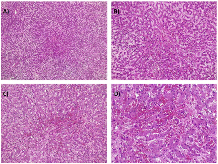
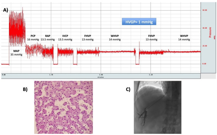
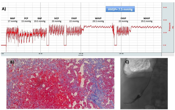
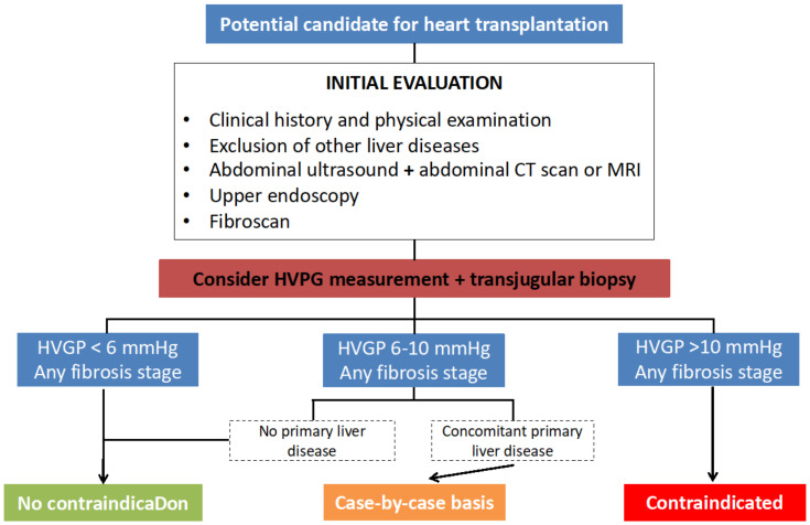
Liver disease resulting from heart failure (HF) has generally been referred as "cardiac hepatopathy". One of its main forms is congestive hepatopathy (CH), which results from passive venous congestion in the setting of chronic right-sided HF. The current spectrum of CH differs from earlier reports with HF, due to ischemic cardiomyopathy and congenital heart disease having surpassed rheumatic valvular disease. The chronic passive congestion leads to sinusoidal hypertension, centrilobular fibrosis, and ultimately, cirrhosis ("cardiac cirrhosis") and hepatocellular carcinoma after several decades of ongoing injury. Contrary to primary liver diseases, in CH, inflammation seems to play no role in the progression of liver fibrosis, bridging fibrosis occurs between central veins to produce a "reversed lobulation" pattern and the performance of non-invasive diagnostic tests of liver fibrosis is poor. Although the clinical picture and prognosis is usually dominated by the underlying heart condition, the improved long-term survival of cardiac patients due to advances in medical and surgical treatments are responsible for the increased number of liver complications in this setting. Eventually, liver disease could become as clinically relevant as cardiac disease and further complicate its management.

Keywords: cirrhosis; heart failure; heart transplantation; portal hypertension.


Conflict of interest statement

The authors declare no conflict of interest.

Figures

Figure 1
Figure 1
Laboratory parameters during the course of acute cardiogenic liver injury (ACLI) in a patient with respiratory failure due to drug overdose. Abbreviations: AST: aspartate aminotransferase; ALT: alanine aminotransferase; LDH: lactate dehydrogenase; Bb; bilirubin; INR: international normalized ratio.
Figure 2
Figure 2
(A) Cardiomegaly in a patient with ischemic cardiomyopathy. (B) Dilated suprahepatic vein in the same patient. (C) Doppler ultrasound in one of the dilated suprahepatic veins. (D) Hepatopetal flow in the portal vein highly modulated by the cardiac cycle.
Figure 3
Figure 3
(A) Idiopathic membranous inferior vena cava obstruction in a 44-year-old man. Magnetic resonance imaging shows a mildly nodular liver with altered parenchymal perfusion and dilatation of hepatic veins. (B) Severe tricuspid regurgitation in a 49-year-old man. Computed tomography scan shows dilatation of hepatic veins and reflux of contrast into the inferior vena cava and hepatic veins.
Figure 4
Figure 4
Idiopathic membranous inferior vena cava obstruction in a 44-year-old man. The image shows the dynamic phase of MRI. In addition to the significant hypertrophy of segment I, magnetic resonance imaging shows a mass (3.8 cm × 4.2 cm) that after administration of intravenous contrast presents a heterogeneous enhancement in the arterial phase with washout in the portal phase. Liver biopsy showed histological changes compatible with focal nodular hyperplasia.
Figure 5
Figure 5
(A) Postmortem example of the classical “nutmeg” liver with centrilobular congestion in congestive hepatopathy (CH). (B) Centrilobular regions show congestion and extravasation of red blood cells (4× objective). (C) Same findings as (B), with greater magnification (10× objective).
Figure 6
Figure 6
(AD). Postmortem example of a patient with sinusoidal obstruction syndrome. As in Figure 5, centrilobular regions show congestion and extravasation of red blood cells (increasing magnification from (A) to (D): 4×, 10×, 20×, and 40×; hematoxylin-eosin stain).
Figure 7
Figure 7
(A) Postmortem example of a liver with ischemic zones around centrilobular veins. (B) Centrilobular regions show congestion and coagulative necrosis (hematoxylin-eosin stain, 4× objective). (C) Same findings as 7B, with greater magnification (10× objective).
Figure 8
Figure 8
(A) A typical hemodynamic tracing of a patient with congestive hepatopathy due to cor pulmonale. The hepatic venous pressure gradient (HVPG) is calculated as the difference between wedged hepatic venous pressure (WHVP), and free hepatic venous pressure (FHVP). Both of them are elevated, but the HVPG is within the normal range. (B) Transjugular liver biopsy was performed and showed sinusoidal dilatation without significant fibrosis (hematoxylin-eosin stain, ×20 objective). (C) Occlusion of the hepatic vein with the balloon catheter. Abbreviations: MAP: mean pulmonary arterial pressure; PCP: pulmonary capillary pressure; RAP: right atrial pressure; IVCP: inferior vena cava pressure; FHVP: free hepatic venous pressure; WHVP: wedged hepatic venous pressure; HVPG: hepatic venous pressure gradient.
Figure 9
Figure 9
(A) A typical hemodynamic tracing of a patient with severe tricuspid regurgitation and concomitant hepatitis C. The HVPG is calculated as the difference between WHVP and FHVP. Both of them are elevated, and the HVPG is slightly elevated. (B) Transjugular liver biopsy was performed and showed significant fibrosis forming nodules (Masson stain, ×10 objective). (C) Occlusion of the hepatic vein with the balloon catheter. Abbreviations: MAP: mean pulmonary arterial pressure; PCP: pulmonary capillary pressure; RAP: right atrial pressure; IVCP: inferior vena cava pressure; FHVP: free hepatic venous pressure; WHVP: wedged hepatic venous pressure; HVPG: hepatic venous pressure gradient.
Figure 10
Figure 10
Protocol to determine the recommendation regarding liver disease in a potential candidate for a heart transplant when CH is suspected. We proceed to HVPG measurement and transjugular biopsy in those patients in whom advanced liver disease cannot be ruled out after the initial evaluation (e.g., nodular appearance of the liver). Our recommendation is hemodynamic-dependent, regardless of the fibrosis stage. In cases with a HVPG below 5 mmHg, there is no contraindication to perform an isolated heart transplant, whereas a HVPG > 10 mmHg discards it (no combined heart-liver transplantation has been performed so far in our hospital). In patients with a concomitant primary liver disease and a HVPG between 6–10 mmHg, the decision is patient-specific and relies mainly on the type of disease. If it is treatable (e.g., hepatitis C or B), we recommend proceeding with the heart transplant. The same recommendation is given in the absence of a primary liver disease and a HVPG between 6–10 mmHg. Abbreviations: CT: computed tomography; MRI: magnetic resonance imaging; HVPG: hepatic venous pressure gradient.

References

    1. Ford R.M., Book W., Spivey J.R. Liver disease related to the heart. Transpl. Rev. 2015;29:33–37. doi: 10.1016/j.trre.2014.11.003. - DOI - PubMed
    1. Fouad Y.M., Yehia R. Hepato-cardiac disorders. World J. Hepatol. 2014;6:41–54. doi: 10.4254/wjh.v6.i1.41. - DOI - PMC - PubMed
    1. Cagli K., Basar F.N., Tok D., Turak O., Basar O. How to interpret liver function tests in heart failure patients? Turk. J. Gastroenterol. Off. J. Turk. Soc. Gastroenterol. 2015;26:197–203. doi: 10.5152/tjg.2015.0086. - DOI - PubMed
    1. Moller S., Bernardi M. Interactions of the heart and the liver. Eur. Heart J. 2013;34:2804–2811. doi: 10.1093/eurheartj/eht246. - DOI - PubMed
    1. Henrion J. Hypoxic hepatitis. Liver Int. 2012;32:1039–1052. doi: 10.1111/j.1478-3231.2011.02655.x. - DOI - PubMed

LinkOut - more resources