. 2020 Dec 13;8(4):758.
doi: 10.3390/vaccines8040758.

Lactobacilli Expressing Broadly Neutralizing Nanobodies against HIV-1 as Potential Vectors for HIV-1 Prophylaxis?

Affiliations

Lactobacilli Expressing Broadly Neutralizing Nanobodies against HIV-1 as Potential Vectors for HIV-1 Prophylaxis?

Sarah Kalusche et al. Vaccines (Basel). .

Abstract

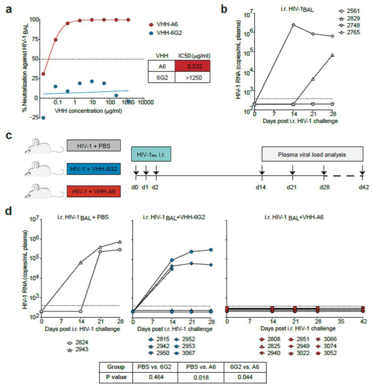
In the absence of an active prophylactic vaccine against HIV-1, passively administered, broadly neutralizing antibodies (bnAbs) identified in some chronically infected persons were shown to prevent HIV-1 infection in animal models. However, passive administration of bnAbs may not be suited to prevent sexual HIV-1 transmission in high-risk cohorts, as a continuous high level of active bnAbs may be difficult to achieve at the primary site of sexual transmission, the human vagina with its acidic pH. Therefore, we used Lactobacillus, a natural commensal in the healthy vaginal microbiome, to express bn nanobodies (VHH) against HIV-1 that we reported previously. After demonstrating that recombinant VHHA6 expressed in E. coli was able to protect humanized mice from mucosal infection by HIV-1Bal, we expressed VHHA6 in a soluble or in a cell-wall-anchored form in Lactobacillus rhamnosus DSM14870. This strain is already clinically applied for treatment of bacterial vaginosis. Both forms of VHHA6 neutralized a set of primary epidemiologically relevant HIV-1 strains in vitro. Furthermore, VHHA6 was still active at an acidic pH. Thus, lactobacilli expressing bn VHH potentially represent an attractive vector for the passive immunization of women in cohorts at high risk of HIV-1 transmission.

Keywords: HIV-1; Lactobacillus; VHH; humanized mouse model; nanobodies; neutralization; passive immunization; prophylactic vaccine; vector.


Conflict of interest statement

The authors declare no conflict of interest. HM is co-inventor and author of a patent related to the expression of antibody fragments in lactobacilli (Expression of antibody or a fragment thereof in Lactobacillus, US Patent Application 20130323819 A1)]. The funders had no role in the design of the study; in the collection, analyses, or interpretation of data; in the writing of the manuscript, or in the decision to publish the results.

Figures

Figure 1
Figure 1
VHHA6-mediated prevention of mucosal HIV-1 infection in humanized mice. (a) Neutralizing activity of anti-HIV-1 VHHA6 and control VHH6G2 against the HIV-1BAL infectious molecular clone (IMC) using TZM-bl cells. Dotted line represents the IC50. (b) Analysis of HIV-1 RNA levels in CD34T+ mice (N = 4) following high-dose intra-rectal (i.r.) HIV-1BAL challenges on two consecutive days. Dotted line represents the quantification limit of 384 HIV-1 RNA copies/mL of the qPCR assay. (c) Schematic representation of the experimental design used to test VHHA6–mediated HIV-1 prevention. CD34T+ humanized mice were given high-dose i.r. challenges with HIV-1BAL mixed with either PBS (N = 2), 50 µg control VHH6G2 (N = 6) or 50 µg anti-HIV-1 VHHA6 (N = 9) on three consecutive days and plasma viremia was tracked over time. (d) Analysis of HIV-1 RNA levels in the different CD34T+ mouse groups as described in (c). Dotted line represents the quantification limit of 384 HIV-1 RNA copies/mL of the qPCR assay. Statistical analysis in (d) was done using Fisher’s exact test.
Figure 2
Figure 2
Binding of purified VHHs to optC.664 gp140 SOSIP at acidic pH. Binding experiments were performed using 250 ng of VHHs 5, 9, 28 and A6 [29] and 200 ng immobilized optC.664 gp140 SOSIP at pH 4.2 (a) and pH 3.7 (b). Binding of VHHs was detected by ELISA via their C-terminal myc-tag. Binding efficiency of VHHs at acidic pH (blue/pink filled bars, respectively) was compared to binding at pH 7.4 (green bars). Full human mAb b12 was included for comparison (detection with α-human IgG-HRP). SOSIP pretreated at acidic pH for 1 h was used as control for antigen integrity (spotted bars). α-c myc and α-human IgG antibodies represent controls without addition of VHHs. Error bars represent SEM of duplicates from three experiments. Statistical differences in absorption between pH 7.4 and acidic pH were determined using t-test. ***: p < 0.001, **: p < 0.01; *: p < 0.05.
Figure 3
Figure 3
Expression of secreted VHHA6 from Lactobacillus rhamnosus DSM 14870 pAF100-VHHA6. (a) The expression cassette was cloned into the pIAV7 plasmid [50] between SalI and EcoRI restriction sites generating the plasmid pAF100 VHHA6. The expression cassette of VHHA6 is controlled by the apf promotor (P) and contains the signal peptide (SP) from the apf gene and a translational stop codon (pushpin). Plasmid pAF100-VHHA6 allows production and secretion of soluble VHHA6 with a fused C-tag. (b) Soluble VHH (14.4 kDa) was detected in the supernatant from L. rhamnosus DSM 14870 pAF100-VHHA6 by Western blot using a biotinylated camelid anti C-tag antibody and streptavidin-HRP via ECL. pIA7: empty vector control, SN: supernatant, CE: cell extract (c) Neutralizing activity of soluble VHHA6 expressed in E. coli or L. rhamnosus pAF100-VHHA6 using a panel of HIV-1 pseudoviruses. Standardized TZM-bl assays were performed to determine IC50 values [μg/mL]. The panel of pseudoviruses includes the most common subtypes B and C and patient relevant Tier 2 and 3 neutralization sensitivities. IC50 values are colour-coded (>50 μg/mL: white, 10–50 μg/mL: yellow, 1–10 μg/mL: orange, 0.1–1 μg/mL: red, <0.1 μg/mL: dark red) and were determined from the respective neutralization curves using GraphPad Prism.
Figure 4
Figure 4
Expression of cell-associated VHHA6 from Lactobacillus rhamnosus DSM 14870-pAF900-VHHA6. (a) The expression cassette was cloned into the pIAV7 plasmid [50] between SalI and EcoRI restriction sites generating the plasmid pAF900-VHHA6. The expression cassette of VHHA6 is controlled by the apf promotor (P) and contains the signal peptide (SP) from the apf gene and a translational stop codon (pushpin). Plasmid pAF900-VHHA6 allows production of cell wall attached VHHA6 with a fused E-tag. Cell wall anchoring is mediated via a prtP anchor (ank) region fused to the VHH. (b) VHHA6 (40 kDa) was detected by Western Blot in the cell extract using an anti-E-tag antibody (arrow). pIAV7: empty vector control, SN: supernatant, CE: cell extract. (c,d) Surface display of VHHA6 was shown by flow cytometry. L. rhamnosus DSM 14870 were stained with Thiazole Orange (TO), whereas VHHA6 was detected via its E-tag with rabbit α-E tag and goat α-rabbit IgG-BV421 (BV421). The dark blue population is double positive for TO and the E-tag associated to VHH. Background signal with wildtype bacteria was 0.1% (c, right upper panel), while 41% of pAF900-VHHA6 bacteria showed VHHA6 expression on the surface (d, right upper panel). Light blue population represents unstained bacteria (for TO), whereas non-bacterial particles are shown in grey.
Figure 5
Figure 5
Reduction in background binding of GFP-labeled HIV-1 virus particles lacking Env to Lactobacilli by Methyl-α-D-mannopyranoside (MaMP) analyzed by flow cytometry. The addition of MaMP (0.05 M, right panel) reduces background binding of GFP-labeled HIV-1 virus particles lacking Env (pCHIV − Env) to L. rhamnosus DSM14870 expressing VHHA6 on the surface (pAF900-VHHA6, blue) or not (pIAV7, purple). MaMP has no effect on specific binding of NL4-3 viral particles with Env (pCHIV + Env) to lactobacilli expressing VHHA6 (blue: 12.4% compared to 11.9%) and only marginally increases binding to wildtype Lactobacilli (purple: 2.3% compared to 4.1%). Error bars represent SEM of two experiments.

References

    1. UNAIDS Fact Sheet. [(accessed on 10 November 2020)];2020 Available online: https://www.unaids.org/en/resources/fact-sheet.
    1. Cohen M.S., Baden L.R. Preexposure prophylaxis for HIV--where do we go from here? N. Engl. J. Med. 2012;367:459–461. doi: 10.1056/NEJMe1207438. - DOI - PubMed
    1. Stephenson K.E., Wagh K., Korber B., Barouch D.H. Vaccines and Broadly Neutralizing Antibodies for HIV-1 Prevention. Annu. Rev. Immunol. 2020;38:673–703. doi: 10.1146/annurev-immunol-080219-023629. - DOI - PMC - PubMed
    1. Bar K.J., Sneller M.C., Harrison L.J., Justement J.S., Overton E.T., Petrone M.E., Salantes D.B., Seamon C.A., Scheinfeld B., Kwan R.W., et al. Effect of HIV Antibody VRC01 on Viral Rebound after Treatment Interruption. N. Engl. J. Med. 2016;375:2037–2050. doi: 10.1056/NEJMoa1608243. - DOI - PMC - PubMed
    1. Caskey M., Klein F., Lorenzi J.C., Seaman M.S., West A.P., Jr., Buckley N., Kremer G., Nogueira L., Braunschweig M., Scheid J.F., et al. Viraemia suppressed in HIV-1-infected humans by broadly neutralizing antibody 3BNC117. Nature. 2015;522:487–491. doi: 10.1038/nature14411. - DOI - PMC - PubMed

LinkOut - more resources